Potential Diagnostic Biomarkers in Natural Killer (NK) Cells of Patients with Chronic Fatigue Syndro

IrisRV

Well-Known Member
That's good, I'd trust INIM with that but personally I would not trust Quest to do the same.
I'm with you there. :)
Their main focus appears to be pathogen testing and treatment, so it wouldn't change their treatment plan if you had low or normal NK cell function. They would give antivirals/abx regardless and that is their main approach.
True, but OMI and INIM also focus on pathogen testing and treatment. In additon, they look at immune dysfunction and treat where there is some possibliity of doing so. IVIG for low IgG, Imunovir or inosine for low NK cell function, and so on. I can spend longer off AVs when I get some immune treatment, which is easier on my liver and kidneys.

Not all groups test and treat the same since there's no established protocol yet, but since the direction of research is definitely suggesting immune implications in this neuroimmune illness, I'm surprised a university clinic isn't looking as the immune side. Oh well, the top folks are doing the best they can in the environment they're in. We can't fairly ask for more.

I really wish that all new ME research would take into account duration of illness in their cohort.
Me, too. We need to know a lot more about the progression of this illness. It's clear a lot of things are changing significantly over time.

Oddly, my NK cell function and cytokines look like those of a young short-term patient rather than the old, long-term patient that I am. I wonder if that has anything to do with getting a lot of pathogen and immune treatments. Perhaps my immune system is getting enough support from those treatments that it's no longer exhausted and is back in fight mode. o_O
 

weyland

Well-Known Member
Oddly, my NK cell function and cytokines look like those of a young short-term patient rather than the old, long-term patient that I am. I wonder if that has anything to do with getting a lot of pathogen and immune treatments. Perhaps my immune system is getting enough support from those treatments that it's no longer exhausted and is back in fight mode.
I'm guessing that has a lot to do with it. We know that antivirals like Valcyte and even herbals like Equilibrant can measurably modify cytokine levels. The AHCC in Equilibrant will have an effect on NK cell function.
 
Last edited:

weyland

Well-Known Member
Transfection of primary NK cells with pre-miR-99b compared with transfected nonsense control identified 37 genes with differential expression passing the inclusion criteria (P≤0.05 fold change ≥1.5). Twenty one genes exhibited down regulation, with fold changes between 1.5 and 1.8. Sixteen genes demonstrated increased expression between 1.5 and 2.6. Interferon gamma (IFN-Y), which is a key mediator of NK cell activation, was the most highly induced gene (2.6 fold). Granzyme B (GZMB), which encodes a component of the cytotoxic granules that NK cells release upon recognition of target cells, was induced 1.6 fold. Genes involved in cellular activation, vesicle formation, motility and modulation of the actin cytoskeleton were also up regulated (KLRF1, STOM, S100A4, FEZ1, ARPC3 and TPM3P5). Cathepsin L (CTSL) was the most down regulated gene by hsa-miR-99b. This protein is required for cleavage and activation of perforin & granzyme B within lytic vesicles [32]. CD6 and IL-8 (interleukin-8) were also down regulated (1.6 and 1.5 fold respectively). Reduced expression of these genes is associated with NK cell maturation following activation [33, 34]. Reduced expression was also identified in genes involved in the autophagy, WNT and AKT signalling pathways (ULK1, ATG2A, PMEPA1, DKK3, PDCD4).

One consistent finding in CFS/ME patients is the reduction in the cytotoxic activity of NK cells [11, 13, 14], frequently associated with an alteration in the level of components of cytotoxic granules notably granzyme and perforin [13, 14]. In this study Granzyme B (GZMB) was significantly up regulated by hsa-miR-99b suggesting an enhanced cytotoxic capacity. However CTSL (Cathepsin L) exhibited the greatest degree of down regulation by hsa-miR-99b. This enzyme is required to process lytic granule components, granzyme and perforin from precursor to active forms suggesting an explanation for the increased GZMB mRNA seen here and in the literature leading to reduced effector function. This mechanism suggests an accumulation of precursor proteins for GZMB and perforin in lytic vesicles with reduced cytotoxic function which is supported by recent data which identified reduced NK cell cytotoxicity in CFS/ME patients without an associated impairment in degranulation [78]. Over expression of hsa-miR-99b and hsa-miR-330-3p induced a gene expression pattern consistent with NK cell activation increasing IFN- γ expression and genes involved in cellular motility, vesicle formation and modulation of the actin cytoskeleton (KLRF1, STOM, S100A4, FEZ1, ARPC3 and TPM3P5), confirmed by over representation analysis. Activated NK cells in the peripheral blood of CFS/ME patients have been postulated to result from incomplete resolution and control of viral infection which is the most commonly cited trigger for onset of CFS/ME symptoms. This data also demonstrates a molecular basis for the reduction in NK cytotoxicity seen in CFS/ME patients mediated by miRNA, via direct down regulation of enzymes required for processing cytotoxic vesicle components and down regulation of genes within Autophagy, WNT and AKT pathways (ULK1, ATG2A, PMEPA1, DKK3, PDCD4) required for normal proliferation and maturation of activated NK cells.

These are the most interesting parts to me so far. A possible explanation for the exact way in which the NK cell function is being affected. And it would appear to be a regulatory cause.
 

weyland

Well-Known Member
Are they inconsistent or just different? Inconsistent would be unfortunate. That would need to be explained before either group could make a claim of having a reliable biomarker. Different, however, could just mean they're looking at somewhat different aspects and both could be right (or wrong :p).
This is just getting bloody confusing now. I haven't been following this very closely because it seemed too sensational, but there seems to be massive confusion regarding exactly what findings the Australians will be using for their biomarker test. According to several articles on the subject, they will be using their ion channel SNP findings, but this hasn't been officially confirmed. They're not talking much either, I assume because a commercial test is in the works.

Back on the topic of the thread, I was able to dig up a 2014 paper from the Aussies that covers their miRNA findings. You were correct, they were looking a different way. Kerr's group was looking at miRNAs expressed in isolated PBMCs. The Aussies were looking at circulating miRNAs in plasma. A good explanation for why they found different things. That's what I get for trusting someone else to interpret this stuff for me.
 

Seanko

Well-Known Member
ME Research UK have written a layman's guide to the paper

Layman's guide

[bimg=700|no-lightbox]http://www.meresearch.org.uk/wp-content/uploads/2016/03/MiRNA.jpg[/bimg]

Comment by ME Research UK

Ribonucleic acid (RNA) is found in all living cells, and controls the manufacture of the proteins needed for all essential functions of life, from hormones to immunological responses. It comes in different forms, and the best known is messenger RNA which passes information from our DNA to the sites where proteins are actually made. In recent years, however, another fascinating form has been identified – microRNA or miRNA. While other types of RNA have a ‘positive’ role in the creation of proteins, miRNAs tend to prevent things happening – they ‘silence’ mRNA molecules by cleaving them, destabilising them, or interfering with their work.

A lot of effort has gone into discovering whether miRNAs are involved in human disease, and a role in cancer was first reported in 2002 in chronic lymphocytic leukaemia (read more). Since then, a large array of different miRNA molecules have been linked with different diseases and, in fact, there is a specific database containing information about these links (read more). While it’s now clear that miRNAs are involved in regulating blood cell formation and moderating immune cascades, they probably have a role in constraining or subduing the workings of many, if not most, biochemical pathways in the body.

Dr Robert Petty and Dr Jonathan Kerr (Queen Mary University of London, and St George’s University of London & Universidad del Rosario, Colombia)
have just published a paper in the journal PlosOne (read more) on the role of miRNAs in ME/CFS. The work was undertaken during Dr Petty’s PhD project, and the research group received funding from ME Research UK and the CFS Research Foundation among others over the years, producing a range of scientific reports (read more).

The team examined miRNA expression in mononuclear white blood cells (T-cells, B-cells, natural killer cells and monocytes involved in defence and immunity) in Fukuda and Canadian Consensus-defined ME/CFS patients and healthy controls. There were three stages to the experimental work. The first step was to determine whether miRNA expression (i.e. its activity) was different between one set of 15 ME/CFS patients and 30 healthy control individuals. Next, the researchers undertook a replication stage, attempting to repeat their findings in another independent group of 20 ME/CFS patients and 20 controls, and to identify the particular types of mononuclear cells involved. The final step involved a separate experiment to see if the ME/CFS-associated miRNAs could be linked with particular genes by ‘transfecting’ them into primary natural killer cells and observing which genes were produced.

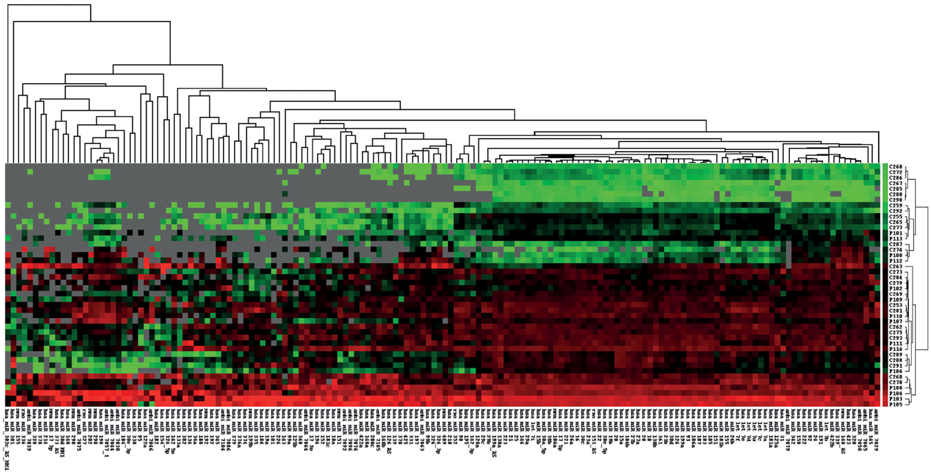
Unsupervised cluster analysis of miRNA microarray data. Each column represents one sample, each row a miRNA. miRNA expression is represented in red for high expression, green for low expression and grey for data excluded at normalization.

In essence, the analysis identified ‘differential expression’ or activation of 34 miRNAs, all of which were up-regulated. Using quantitative PCR to validate the findings, expression changes were confirmed in four of these miRNAs – hsa-miR-99b, hsa-miR-330-3p, hsa-miR-30c and hsa-miR-126 – which, in addition, were found to be suitable for further investigation as potential biomarkers for ME/CFS as their specificities ranged from 0.71 to 0.78. The two miRNAs which showed the greatest degree of over expression were hsa-miR-99b and hsa-miR-330-3p, which are also know to be important in other illnesses. For instance, hsa-miR-99b has a distinctive pattern of microRNA expression in primary muscular disorders (read more), while hsa-miR-330 signatures are associated with certain lymphomas (read more).

At the replication analysis, changes in miRNA expression were found in the four types of white blood cells, with the most significant abnormalities occurring in natural killer cells. ‘Transfection’ of natural killer cells with hsa-miR-99b or has-miR-330-3p led to gene expression changes, including in genes involved in the activation of cellular processes and immunity. The researchers concluded that the natural killer cells were ‘activated’ but with reduced functioning, consistent with what we know already about the low activity of these cells in ME/CFS (read more).

Overall, the researchers found that four miRNAs expressed in mononuclear white blood cells have potential as biomarkers in ME/CFS, particularly hsa-miR-99b and hsa-miR-330-3p which may also be involved in the natural killer cell dysfunction characteristic of the illness. As the authors point out, the results are particularly interesting since the messenger RNAs regulated by hsa-miR-99b and hsa-miR-330-3p have a large degree of overlap with the messenger RNAs found to be upregulated in previous work by the Kerr group, strengthening the case for the role of abnormalities in the innate immune system in the pathogenesis of ME/CFS.

Because miRNAs are ‘protected’ and stable in various body fluids and tissues, there is now an enormous literature on their use as possible biomarkers, mainly in cancer but also in diseases like epilepsy, malarial infection and multiple sclerosis. In all these illnesses, there is a need for non-invasive, easily detected, sensitive biomarkers, and ME/CFS is no different. For that reason, these interesting findings deserve to be taken much further in large validation studies, as well as in investigations to elucidate the particular role played by immune system abnormalities in the pathogenesis of the disease.
 

Merida

Well-Known Member
Think it is important to appreciate that spinal cord injury/myelopathies ( ie disease/ disorder of the spinal cord) also
Cause reduced NK cell counts, depressed NK function, depressed lymphocyte transformation, depressed T cell function/activation, elevated interleukins 6/2r, and more. " Deficits in immune function and the presence of a chronic inflammatory state have been documented in the acute and chronic phases of spinal cord injury."
( Spinal Cord Medicine :,Principles and Practice, editor-in chief Vetnon W. Linn, MD, PhD, 2003)

This might be a good group to compare with. It is known that Epstein Barr, herpes simplex, cytomegalovirus, adenovirus, enterovirus, Coxsackie B , HHV6, HIV can cause myelopathies. ( SH Wong et al. Myelopathy but Normal MRI: where next? Practical Neurology 2008; 8: 90-103. ) Also, there is research in spinal cord injury that demonstrates multiple hormone imbalances, autonomic problems, sleep disorders, dysfunction of the thalamus !!!!
And fatigue ! Plus more.

Think is is important to remember that spinal cord injury can come in many forms - overt trauma, virus, incorrect spinal fluid flow causing syringomyelia or myelomalacia (Hard to dx), vascular ( new disorder : thoracic epidural arteriovenous malformation - neurosurgeon Robert Bray), genetic, autoimmune, toxins, spirochete disease, fungal, more.

Gee, thanks to everyone who is posting. We are all struggling to get our lives back. May the answer(s) present to the few great researchers who are trying to help. I do believe that thoughts have power. Let's put it out there!
 

weyland

Well-Known Member
For that reason, these interesting findings deserve to be taken much further in large validation studies, as well as in investigations to elucidate the particular role played by immune system abnormalities in the pathogenesis of the disease.
Yes please. Sooner rather than later, thanks.
 

Get Our Free ME/CFS and FM Blog!


Forum Tips

Support Our Work

DO IT MONTHLY

HEALTH RISING IS NOT A 501 (c) 3 NON-PROFIT

Shopping on Amazon.com For HR

Latest Resources

Top