


Geoff’s Translation
The GIST
This is part one of a series of blogs on the Mast Cell Masterminds Conference, which took place in September 2024 in Fort Hood, Oregon. Thanks to Dr. Kaufman and the conference organizers for making it possible for me to view the presentations.

Alexis Cutchins MD spoke on connective tissue and venous disease.
One thing to note is that a mast cell conference will never solely be about mast cells. Mast cell activation can affect the body in so many ways that any mast cell conference will include presentations on a wide range of disorders.
 Health Rising’s Quickie Summer Donation Drive is On!
Take connective tissues. They’re loaded with mast cells! When mast cells go off, they can damage the connective tissues, and the connective tissues (skin, blood vessels, spinal cord, gut lining, ligaments) are everywhere.
Some of the 3 1/2-day Mast Cell Masterminds conference presentations addressed connective tissue issues, and this overview will focus on a specific type of connective tissue: the blood vessels.
This blog will cover the following presentations:
- Alexis Cutchins – Emory University – “It’s Not Just POTS: The Role of Connective Tissue Disorders, Venous Disease, and Mast Cells in Postural Orthostatic Tachycardia Syndrome“
- Meredith McDermott, MD (Minimally Invasive Procedure Specialists, South Denver) – “A Clinical Approach to the Diagnosis and Treatment of Pelvic Venous Disease“
Some information not in the presentations is included in the blog.
The GIST
-

Collateral veins (left) disappear after a stent.
This is part one of a series of blogs on the 2024 Mast Cell Masterminds Conference focused on diseases of the pelvic veins.
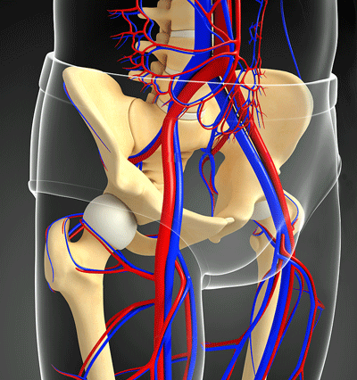
- While connective tissue damage can occur in many areas of the body, the high collagen levels found in the veins puts them at special risk. There are several places in the pelvic area where the weaker veins are at increased risk of being compressed by the stronger, more muscular arteries.
- This compression (or congestion) can block blood flows resulting in swelling and pain in the abdominal/pelvic area and/or legs and the development of non-pelvic symptoms like dizziness, brain fog, fatigue, migraines, IBS, etc. Collateral veins that develop to enhance blood flow can also impact the nerves, causing pain.
- As with POTS and hypermobile Ehlers-Danlos (hEDS), most people with the signature finding of pelvic venous diseases, “venous insufficiency” (i.e., reduced blood flows in the pelvic veins), do not experience symptoms. While it’s not clear why some people experience symptoms while others do not, it may be that the severity of the blockage or compression, or other factors play a role.
- With up to 80% of POTS patients having a diagnosis of pelvic venous disease (as well as mast cell activation syndrome), it’s clear that pelvic venous diseases are part of the ME/CFS/FM/POTS/IBS/long-COVID suite of diseases. The chief problem – obstructed blood flows – fits very nicely with the idea that problems with blood flows – whether from the heart, through the veins, or in the microcirculation – play a key role in these diseases.The pelvis appears to be simply another place where connective tissue damage/problems with blood flows can occur.
- The two main syndromes – May-Thurner Syndrome and Nutrcracker Syndrome – feature compression of the veins in the pelvic area.
- The primary issue McDermott sees is left iliac vein compression, also known as May-Thurner Syndrome. In May-Thurner Syndrome, a thick, muscular artery, sitting on top of the weaker, less robust left Iliac vein, compresses it, reducing blood flows from the legs to the upper body.
- Symptoms can include left leg swelling and heaviness that worsen late in the day or with prolonged standing/sitting, and which improves when the leg is elevated, feelings of pressure and pain during walking that eases when lying supine, red or purplish-brown skin, or visible spider or varicose veins, dysmenorrhea (menstrual pain), fatigue, dizziness, orthostatic intolerance, gut bloating/pain, pain during or after intercourse (dysparenuria).
- In both cases, stents to decompress the veins has proven to be “a safe, effective, and minimally invasive” treatment. In more severe cases, vascular surgery can reposition the veins so that they are no longer compressed.
- In pelvic congestion syndrome (PCS), the valves in the veins fail, resulting in engorged, dilated veins. PCS can produce all the symptoms of venous insufficiency plus urinary tract symptoms, increased urinary frequency, pain with urination, irritable bladder symptoms, palpitations, varicose veins, and orthostatic intolerance. As with venous insufficiency, all of these symptoms get better when lying down.
- PCS treatment involves embolizing (shutting down) using a “minimally invasive” procedure (coils, embolizing agents) that stops the abnormal blood flow, thereby relieving pressure.
- While we need more studies, the evidence to date suggests that treating pelvic venous syndromes may help alleviate orthostatic intolerance, brain fog, and fatigue in some people. People with POTS who have not responded to POTS treatments and who experience pelvic/leg pain might particularly take note of some of the pelvic venous syndrome connections.
Learned Something Helpful? Support Health Rising and Keep the Information Flowing!
HEALTH RISING IS NOT A 501 (c) 3 NON-PROFIT
Connective Tissues and Postural Orthostatic Intolerance Syndrome (POTS)

Meredith McDermott MD spoke on diagnosing and treating pelvic venous syndromes.
In her POTS presentation, Dr. Cutchins noted that she doesn’t just look for the signature finding in POTS – an increased heart rate during standing – but for signs that the connective tissues have been damaged; i.e.. things like hypermobility, elastic skin, abnormal stretch marks, flushing, rash, dermatographism (raised marks after scratching/rubbing the skin), evidence of venous pooling, and edema (swelling, color changes in the feet).
As we’ll see, if the connective tissues on the outside show signs of damage, it’s possible that damage on the inside – in the pelvic veins, for instance – has occurred as well.
A Locus of Damage – the Veins
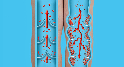
While connective tissue damage can occur in many areas of the body, the veins appear to be at special risk. The beefy, muscular arteries that send oxygenated blood flowing to the tissues are not so affected, but the weaker, more flaccid veins that return the used-up blood to the heart/lungs are.
They may be less robust, but because the venous system holds 70% of our blood volume and regulates blood flows to the heart, the venous system plays a major role in cardiac output. (David Systrom has found that preload failure – the failure to deliver normal amounts of blood to the heart and reduced cardiac output is common in ME/CFS and long COVID.)
Since the venous system relies on collagen and elastin, two components of connective tissue, to regulate blood flow, and because veins contain more collagen than arteries, connective tissue problems affecting collagen are more likely to occur in the veins.
Collagen damage leads to “stretchy veins” which don’t close properly, allowing blood to accumulate in the abdomen and lower body when we stand, and potentially causing problems like orthostatic intolerance, varicose veins, skull-based venous compression syndromes and pelvic vein compression.
Venous Insufficiency
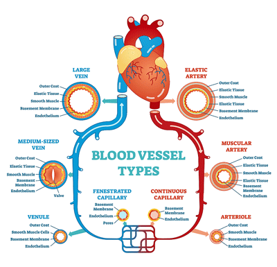
Under high pressure, muscular arteries provide oxygenated blood to the tissues. The weaker veins bring blood back to the heart and contain 70% of our blood volume.
Venous Insufficiency refers to the inability of the veins to deliver normal amounts of blood, a seemingly serious but underdiagnosed problem. Most vascular surgeons, on the lookout for clots, often do not recognize the symptoms or signs of venous insufficiency. Only in the last five years or so has it become clear that venous insufficiency/venous compression can cause an array of ME/CFS-like symptoms.
This is one of those puzzlingly tricky diagnoses. Twenty to 40% of people have some compression of the veins in the pelvic area and are asymptomatic. One study found that almost 70% of POTS patients and 40% of healthy controls exhibited some compression of the left iliac vein.
The same thing is true with postural orthostatic tachycardia syndrome (POTS) and hypermobile Ehlers-Danlos Syndrome (hEDS). Most people who fit the heart rate criteria for POTS or who are hypermobile are completely healthy, and up to 90% of people who meet the hEDS criteria have no symptoms.
A number of factors can turn asymptomatic venous compression in the pelvic region pathological. A high degree of narrowing, the buildup of scar tissue, the development of collateral veins that impact the nerves, lesions that cause symptoms only when put under load such as when standing, the presence of other factors (Leiden V, antiphospholid antibodies, estrogen exposures, pregnancy, obesity, immobility) and probably other factors can turn an asymptomatic case of venous compression or insufficiency into a symptomatic problem.
Please note that ultrasounds are not sufficient for diagnosing these conditions. MRVs (magnetic resonance venographies) are needed to accurately assess blood flows and can also pick up other pelvic problems.
Beyond the Pelvis

Most people with hypermobility, who meet the heart rate criteria for POTS, or have some degree of venous compression are asymptomatic.
If pelvic venous syndromes were just about pelvic pain, this blog would not exist. The real surprise is that they’re also linked to and may be contributing to many ME/CFS/FM/long-COVID symptoms. Check out the list of symptoms they can cause: achy and heavy feeling legs, tingling sensations in the legs, pelvic pain, flank pain, cramping muscles in the legs – particularly at night, restless legs, swollen and discolored feet and ankles, pain that is worse when you stand and gets better when you lie down, dizziness, orthostatic intolerance, anxiety and fatigue.
Smith and Rowe showed there was more to pelvic venous symptoms than the usual pelvic and leg pain doctors look out for. Their 2022 survey showed that people with pelvic congestion – which is caused by a blockage of blood flows in the pelvic veins – often experienced symptoms not associated with the pelvis such as severe fatigue (72%), dizziness (63%), IBS symptoms (61%), brain fog (33%), migraines (49%), polyuria or dysuria (41%), excessive sweating (31%), TMJ pain (31%), and loose skin or lax joints (18%).
In a paper submitted for publication, Cutchins estimated 80% of POTS patients had signs of pelvic venous disease; 73% were diagnosed with MCAS or had suspected MCAS, 24% had hypermobile EDS, 26% polycystic ovary syndrome (pCOS), and 53% experienced symptom worsening or had their symptoms triggered by COVID-19.
The pelvis appears to be another place where connective tissue and circulatory problems can occur.
The high incidence of pelvic venous disease in POTS is likely due to blood pooling in the abdomen/pelvis/legs, which reduces blood flows to the heart, thus triggering the baroreceptors to increase the heart rate in an attempt to improve them.
While pelvic venous syndromes have not been studied in long COVID, an increased risk swelling, varicose veins, clot risk, impaired blood vessel functioning, and hypercoagulation suggests that pelvic vein problems may be present.
The takeaway: Pelvic venous diseases are part of the ME/CFS/FM/POTS/IBS/long-COVID suite of diseases. If you have symptoms associated with them and ME/CFS-like symptoms, these syndromes might be something to check out. The chief problem – obstructed blood flows – fits very nicely with the idea that problems with reduced blood flows – whether from the heart, through the veins, or in the microcirculation – play a key role in these diseases.
The connective tissue connection potentially fits with the spinal and blood vessel issues, Peter Rowe’s neuromuscular strain, the pathological hypermobility that is sometimes present, the pain hypersensitivity, and even the gut problems sometimes seen in these diseases. The pelvis appears to be simply another place where connective tissue damage/problems with blood flows can occur.
Collateral Damage
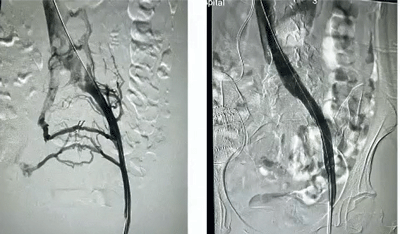
Collateral veins (left) disappear after a stent.
When a vein becomes compressed or blocked in the pelvis (or other areas), the body produces a tangle of small collateral veins in an attempt to maintain blood flow. While collateral veins can often help move blood and thereby relieve symptoms, they can also press on nerves or other structures, causing pain.
Pelvic Venous Syndromes
Note that most of these syndromes are considered by the medical profession to be “compression syndromes,” not connective tissue diseases. At some point, that may change, but for now, note that they are also associated with connective tissue problems that can weaken the veins, making them more susceptible to being compressed. Patients can have a variety of issues that may require both stenting and emobolization.
May-Thurner Syndrome
The primary issue McDermott sees is left iliac vein compression, also known as May-Thurner Syndrome. In May-Thurner Syndrome, a thick, muscular artery, sitting on top of the weaker, less robust left Iliac vein, compresses it, reducing blood flows from the legs to the upper body.
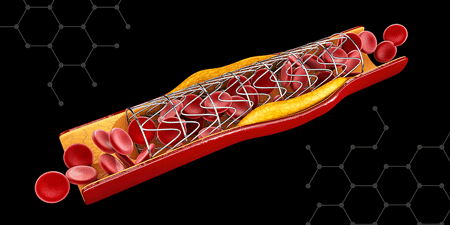
Stenting is a minimally invasive procedure commonly used in several pelvic venous syndromes.
Symptoms can include left leg swelling and heaviness that worsen late in the day or with prolonged standing/sitting, and which improves when the leg is elevated. There may also be feelings of pressure and pain during walking that eases when lying supine, as well as red or purplish-brown skin, or visible spider or varicose veins. Other ME/CFS symptoms may be present.
Treatment – Conservative options include leg elevation, compression stockings, and monitoring to determine if the problem resolves. The standard of care for relieving vein compression, however, is stenting. First, a balloon widens the narrowed vein, and then a stent is permanently placed to hold it open, restoring normal blood flow. This often dramatically reduces pain, swelling, and risk of further thrombosis or venous insufficiency. More severe cases may involve relocating the right iliac artery to prevent it from compressing the vein.
Nutcracker Syndrome
In Nutcracker Syndrome, the left renal vein gets trapped between the abdominal aorta and the mesenteric artery.
Symptoms can include blood in the urine (hematuria – can be microscopic and intermittent), orthostatic proteinuria (increased protein levels in urine when standing), left flank and/or abdominal pain, varicose veins in the pelvic area, feelings of pelvic congestion, dysmenorrhea (menstrual pain), fatigue, dizziness, orthostatic intolerance, gut bloating/pain, pain during or after intercourse (dysparenuria).
Treatment – Treatment is similar to May-Thurner. Anticoagulants may be tried. Putting in stents to decompress veins has proven to be “a safe, effective, and ‘minmally invasive’ treatment”. In more severe cases, vascular surgery can reposition the vein so that it is no longer compressed.
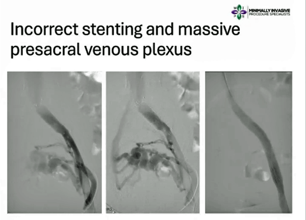
A short stent doesn’t work (middle image), but when a longer stent was put in (right image) and blood flow was restored and the collateral veins disappeared, the patient’s pain disappeared.
Pelvic Congestion Syndrome
PCS occurs when the valves in the veins fail, resulting in engorged, dilated veins. It can produce all the symptoms of venous insufficiency plus pelvic pain, low back and/or hip pain, pain after intercourse, urinary tract symptoms, increased urinary frequency, pain with urination, irritable bladder symptoms, palpitations, varicose veins, and orthostatic intolerance. As with venous insufficiency, all of these symptoms get better when lying down.
Treatment – Embolization is a “minimally invasive” procedure that stops the abnormal blood flow, thereby relieving pressure. It involves implanting tiny metallic coils or a liquid agent to block the dilated and damaged veins. The veins then shrink over time, reducing swelling, pain, and congestion. Depending on how many veins are being treated, the procedure usually takes 30 minutes to a few hours, and the patient can go home on the same day. It reportedly has a high success rate.
Valve Trouble – Varicose Pelvic Veins
When the valves in the veins break down, the veins become engorged. Embolizing agents can close these veins down.
Varicose veins can occur in all these syndromes. They occur when malfunctioning valves in the veins allow the blood to pool and stretch the veins, resulting in bulging, twisted, and, at times, visible veins. One cause is weakened blood vessel walls – possibly due to mast cell attacks. While they often cause only cosmetic concerns in the legs, they can also lead to pain, heaviness, swelling, cramping, skin changes, and ulcers, particularly in the areas around the ankles.
Treatment – Compression stockings, leg elevation, weight management, and increased exercise may provide relief for people with mild cases. Using ultrasound to target the source of the varicose veins, and then closing them down with embolizing agents (coils, plugs, or liquid agents) can reduce pressure and help alleviate interstitial cystitis and vulvodynia, as well as pelvic pain. It appears to be quite effective. Sclerotherapy may work for some types of pelvic veins.
Variococeles
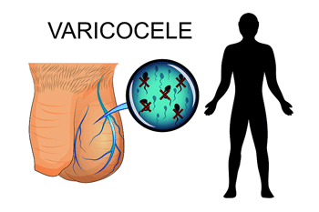
Variococeles are a pelvic venous issue that occur only in men. They are usually not a problem.
The pelvic venous syndromes occur more often in women, but varicoceles occur only in men. Varicoceles (enlarged veins in the scrotum) on the left side in men are caused by valve or anatomical problems.
Like the pelvic venous syndromes, connective tissue problems can increase the risk of having a varicocele. (Both my twin and I have several connective tissue issues (varicocele, pectus carinatum). I also have a hiatal hernia and plantar fasciitis. He does not have ME/CFS…)
The ME/CFS/POTS Connection

A clear overlap exists between pelvic venous syndromes and the POTS/ME/CFS suite of diseases.
While we need more studies, the evidence to date suggests that treating pelvic venous syndromes may help alleviate orthostatic intolerance, brain fog, and fatigue in some people. People with POTS who have not responded to POTS treatments and who experience pelvic/leg pain might particularly take note of the pelvic venous syndrome connection.
Smith and Rowe found that treatments for pelvic venous problems resulted in significant decreases in symptom scores assessing pelvic pain and dysautonomia.
Several case studies have featured POTS or EDS patients who presented with pelvic and/or lower extremity pain. A 16-year-old with POTS and EDS experienced palpitations, concentration difficulties, dizziness, left leg swelling, and pelvic pain. Propranolol (10 mg) twice daily had not helped, and she was taking 0.1 mg of fludrocortisone once a day.
Her pelvic venography showed “severe obstruction of the left common iliac vein with near-complete flow arrest” and the development of numerous collateral veins. Stenting resulted in a significant reduction of her orthostatic symptoms as well as her pain, leg swelling, etc.
Similar results were seen in a case series of three female patients with POTS, Ehlers-Danlos syndrome (EDS), and May-Thurner syndrome (MTS). One woman with hypermobile EDS and POTS experienced left leg swelling and pelvic pain, brain fog, dizziness, and difficulty staying upright. Neither propanolol nor fludrocortisone had helped her POTS symptoms.
A pelvic ultrasound was normal, but an MRV found “severe compression” of both the left iliac and left renal veins, indicating that she had both May-Thurner syndrome and Nutcracker syndrome. Stenting of her left iliac vein resulted in a complete resolution of her POTS and concentration symptoms.
Another woman with EDS and POTS had it all. She experienced chronic abdominal pain, weakness, severe concentration difficulties, lightheadedness, palpitations, pelvic pain, left leg swelling, and lower abdominal bloating that would worsen during menses. An MRV showed narrowing of the left iliac vein (May-Thurner), blockage in the left renal vein (Nutcracker), and retrograde (backwords) flow in the left ovarian vein (pelvic congestion syndrome). The authors wrote:
“These findings are consistent with severe venous insufficiency, which is a common feature of POTS.”
A stent in her left iliac vein resolved her left leg pain and swelling, but she continued to experience abdominal pain and bloating. An embolization of the left gonadal vein resulted in a significant improvement in POTS symptoms, decreased abdominal bloating and leg pain, and an improvement in her ability to remain upright. To address occasional episodes of dizziness, she continued to take 2.5 mg of midodrine 3× daily.
Conclusion
If you experience unresolved abdominal/pelvic/leg pain and the symptoms of ME/CFS/POTS/long COVID you might want to investigate pelvic venous syndromes. From what I can tell, you’ll need a venography (MRV) (hopefully with contrast?) to tell if the veins in your pelvic area are being compressed or congested. If they are, the treatment options mostly include “minimally invasive” procedures such as stenting and/or emobolization.
Learned Something Helpful? Support Health Rising and Keep the Information Flowing!
HEALTH RISING IS NOT A 501 (c) 3 NON-PROFIT


I feel like the percentage in the “doesn’t show symptoms” category could be misleading because despite having clear compression issues on imaging, I’ve been told by countless doctors that they don’t cause symptoms. (And the symptoms many of us experience while being told that are the exact ones listed as potential symptoms.)
I’m thrilled with this research. I hope they can keep making these connections!
My HR peaks randomly, and only on some days, but is always improved by lying down and usually by box breathing, even when walking.
I’ve also developed a bladder prolapse, which I manage with hypopressive exercises.
Do you think that may be related to the condition you describe?
I don’t have pelvic pain, but some right buttock discomfort and occasional sharp pain in the front of my right thigh and more frequent and extended flares of my long-standing IBS since LC. Ask frequent leg heaviness after eating, especially large meals
My rather mild long covid PEM response tends to be more related to excessive social stress than to reasonable levels of physical exertion. Interestingly, my daughter who has been diagnosed with ME CFS has a similar pattern. Thanks for any insights.
Are you hypermobile at all. The prolapse speaks to weaker areas of tissue in some places in the body. Theose places in my opinion are more prone to disease
Interesting, Maxine, to hear about your sharp pain in your right thigh. I too had sharp stabbing pains in my front right thigh, with icy wet feelings. After about 6 months, my right thigh is now numb – apparently in the subcutaneous area. Could this be a part of venous pelvic syndrome? I’ve also been experiencing increased urgency, and recently very painful kidneys that took me to hospital to get them checked out, with not much result.
Thanks Cort for all that you do – including using your spoons to let us know what’s happening out there in Research World. Much obliged.
Thanks! 🙂
Hello Cort,
Thank you for your insightful blog post. I found it particularly valuable, as it aligns with my belief in the importance of identifying the cause, understanding the effect, and pursuing an effective cure.
The vascular system—comprising arteries and veins—serves as the essential lifeline for all living organisms. When a blockage occurs, neither medication nor supplementation can effectively reach the areas beyond the obstruction. As a result, the affected tissues—whether muscular or part of vital organs—are deprived of oxygen and nutrients, ultimately leading to degeneration and cell death.
For this reason, I have consistently advocated for an approach focused on identifying the underlying cause and addressing it directly.
Unfortunately, this is often where the greatest challenge lies. In my own case:
A 2007 MRI revealed moderate calcified aortic and branch vessel atherosclerotic plaque.
Prior to that, a 2001 CT scan identified compression of blood vessels at the thoracic outlet near the clavicle.
However, no action was taken to eliminate the issue.
While identifying the root cause is essential, it is only the first step. Proactive intervention is necessary to stop the progression and prevent further damage.
Thank you once again for your work and dedication.
Sincerely,
Sieglinde
The underlying cause, in my opinion, is the integrity if the tissue.
We all have subclinical eds, or eds that is not aligned to the beighton tear.
Eds just comes up all the time in this suspense, in autoimmunity etc. It’s highly heritable.
Just my belief but I’m glad it’s getting recognition
Thank you Cort. What a great article.
Now I know what it was called-Nutcracker Syndrome. In 1980 after 3 years of debilitating pain and recurrent UTIs I had surgery to realign my L renal vein. No-one back then understood it and I was told by one kidney specialist to “stand on my head” which actually could have been useful if not so impractical.
Then after being diagnosed with hypermobility disorder and suffering from severe varicose veins and iliac valve incompetence again surgery improved my life.
What surgery helped in the end, Maggie?
Varicose vein surgery. And yes connective tissue issues for many of us
Left-sided varicocele can also be a consequence of May-Thurner compression.
Connective tissue .. AGAIN!
I keep saying tgis us the ground zero from which the disease is creates.
I particularly fit in this category of pelvic instruction.
Of course when I suggest tgis to my doctor.. nothing!
We need crispr and stem cells to treat these veins
Yes, Ehlers-Danlos Syndrome (EDS) is a genetic condition, typically caused by mutations in specific genes, and can often be identified through genetic testing.
Classical EDS is associated with mutations in the COL5A1 or COL5A2 genes.
Vascular EDS is linked to mutations in the COL3A1 gene.
Hypermobile EDS (hEDS), however, does not yet have a known genetic marker or confirmed genetic cause.
I do have COL5A1 but not COL5A2.
So what’s next? CRISPR-Cas9 gene editing could offer a potential future solution—but as of now, there are no known or published clinical applications of CRISPR for EDS. It remains an experimental possibility, likely out of reach for most, both technically and financially.
I agree with you that it’s out of reach for us nowbut i think it’s the cure.
Telemorase therapy is a potential scatter gun approach.
it’s like we ve aged faster than normal people. I can’t think of any other way around it.
Granted i’m no scientists but it’s always been my gut feeling that it starts in the connective tissue. I see it time and time again in m.e.cfs, icluding jen brea, who says she didn’t classify as an eds patient, but had weak connective tissue in the ligaments i her neck.
I think its that route that reaslly needs looking at. regenerative medicine and pulling on the strings of the eds puzzle itself as theres not enough research
Would you consider a differential diagnosis?
Example:
Mast Cell Activation Syndrome (MCAS) is a systemic condition that often goes unrecognized, especially when it overlaps with chronic illnesses such as fibromyalgia and Ehlers-Danlos Syndrome (EDS). Its broad spectrum of symptoms—particularly those involving the skin—make it a complex and frequently misdiagnosed disorder.
I’m totally open to that idea.
It seems to me that hypermobility disorders breed mast cell disorder tho.
My thinking is that eds is not understood enough at the moment.
They are still coming up with new categories of eds.
I think the hyperexcitability in the tissues is a result of complex interaction between the tissue and outside factors.
But that tissue type needs to be there.
That’s my gut feeling.
Connective tissue problems run through family lines.
Stress type humans, phenotypes, sensitive, whatever you want to call that propensity to cascades which start with trauma of any kind( biological, emotional, chrmicjal) sensitise mast cells. It’s been proven psychological trauma for example sensitises mast cells. Any kind of trauma will do this.
But the question is. Why do they become sensitised when others don’t?
We see this very explicity in autistic people. Why do their healing cycles shut down?
Why Is their cell danger response so switched on and non resilient?
Why are people with m.e. so more prone to overwhelm of the allostatic load.
Depending on the individual, you can see damage and sensitise accumulate through childhood.
Some people are resilient enough to grt into their 50s and
Suddenly the accumulation becomes too much to handle.
I believe eds is a spectrum disease and so much very low end eds is being missed.
So to me, at present, mast cells and mast cell activation syndrome are a result of a sensitive organism developing less and less resistance to assault and more and more response from the body to try and fight tgis. Mast cell degranulation. Glial cell activation. Inflammation are compensatory mechanisms to try and fight the triggering the cell danger response and also try to shut the organism down and stop it from dying.
That’s the birth place of tgis illness for me.
The anti purigenic therapies, mast cell stabilisers, telomerase and down the line crisor and gene editing once genetic markers are fully known will be the treatment.
When I take your sentence literally, –
“Depending on the individual, you can see damage and sensitise accumulate through childhood. Some people are resilient enough to grt into their 50s and Suddenly the accumulation becomes too much to handle.”
I see two possible connections in terms of thematic or indirect relevance — particularly if you’re exploring immune hypersensitivity, inflammatory load, or general systemic overload in chronic conditions like acromegaly and Cushing syndrome.
Meaning: acromegaly
Organomegaly
Metabolic changes (insulin resistance, etc.)
Joint pain, headaches
Other systemic complications
Or:
Conceptually relate to Cushing syndrome:
A sensitive organism (implying vulnerability or dysregulation)
Decreasing resistance to stressors (“assault”)
Increased response from the body
Both acromegaly and Cushing syndrome may contribute to mast cell activation or degranulation as part of broader immune dysregulation. They are fundamentally hormonal disorders, and both can be traced to dysfunction in the hypothalamic-pituitary-endocrine axis.
It would be worth investigating further, since in both cases, symptoms could be misinterpreted while visual signs missed or not understood.
I forgot to mention the possibility of a neoplasm — a new and abnormal growth of tissue in a part of the body, which is not necessarily cancerous.
It’s definitely a very complex interplay of many things.
My sister has pcos ( and I suspect but never say, eds). Insulin resistance is common in pcos.
Just those connective tissue problems rearing their head again to me.
I assume the mitochondrial membranes are also not strong in eds and more prone to shut down etc.
I was using the accumulative nature of damage literally.
Bob naviauxs work makes most sense to me. Basically a series of insults that force the body into shutdown.
I need to go back and refresh my view of his work.
It seems intuitively correct to me. And he has made a lot of progress in understanding tye metabolic signatures that occur during cell danger response activation.
Of course crushing syndrome etc are all really useful and valuable things tease apart.
I think the fact that m.e. runs through family lines means there is smthg genetically heritable.
My guess is it’s eds as I’ve pointed out too many times now!!
Then that heritage reacts with the world and complex webs of illness are created.
Also common with MCS is SIBO.
Oliver…speaking of aging faster…
In the early days of my onset, I was put in hospital for a herniated disc that refused to heal…once in hospital I had zero appetite or desire to eat so after 5 days of no food my herniated disc healed I pulled the pic line out of my arm myself and walked out under my own power…I could feel my immune system start to function as it should.
Upon eating once again, my skin began to bleed in several places and in one week the once bleeding spots all turned into moles along with a very strange looking set of smaller moles confined to an area the size of a 25 cent piece
After once again fasting for 8 straight days nothing happened.its as if my immune system is now jambed and stuck permanently. That was way back in 1993
That’s reminiscent of smthg that happened to me. I too have a herniated disc and a similar kind of ‘ eating of tissue’ it past fairly quickly thankfully. Your experience sounds rough.
Mast cells are known to destabilised and damage tissue would be my poor guess.
Reacting to food
Pelvic venous insufficiency and Nutcracker Syndrome (called Nutcracker Physiology or NCP when patients are asymptomatic) can cause pain well beyond the legs and abdomen. Although I have been diagnosed with fibromyalgia since 2006, my first symptom was a permanent occipital headache that I woke up with in June 2004 when I was just 19 yo – it has never gone away, not even for a single minute. This headache was eventually diagnosed as New Daily Persistent Headache (NDPH) by Mayo Clinic Jacksonville, who are at the forefront of chronic headaches correlated with Nutcracker Syndrome.
Cort, you reason that symptomatic pts will “need a venography (MRV) (hopefully with contrast?) to tell if the veins in your pelvic area are being compressed or congested.” You’re not far off- I personally had a slightly different experience firsthand. As part of the team of neurologists and vascular surgeons studying Nutcracker at Mayo Clinic, after telling me that Mayo has started. to identify a correlation between NDPH patients and Nutcracker, and that is could also explain my myriad other symptoms (GI, tachycardia, IBS, ED, etc), my neurologist first ordered a Doppler Ultrasound which shows the rate of speed that blood is flowing through the LRV (left renal vein). After that report came back consistent with Nutcracker syndrome (my LRV peak velocity was almost 2.5x the normal rate), they then ordered a special 4D MRI called “MRA with TWIST (Magnetic Resonance Angiography with Time-resolved angiography With Stochastic Trajectories), a 4D dynamic MRA technique — essentially a rapid series of contrast bolus images that let the radiologist watch flow direction over time. I had to hold my breath repeatedly for 10 seconds about 30 times in the machine. That report stated that my imaging showed “abnormal reflux from the left L2 lumbar vein into the spinal EVP, causing venous engorgement of that plexus.” I am apparently a candidate for vein embolization surgery, which you mentioned in your piece. Thanks for shedding light on this new frontier of medicine. But I had to fly to Mayo Florida to find a doctor aware of this condition- that’s how new this area of medicine is.
On this site I’ve seen a lot of different blog posts on alternate/missed diagnoses, some which might cause ME, others which often go along with ME
Might be useful to have these articles collected in one place so patients can go over many such blog posts to make sure they haven’t missed an important condition they have.
Perhaps there could also be created a tag for such posts, or whatever you call it when a post has associated categories listed under the name of the article, like how this one has: “cardiovascular, connective tissue, Ehlers Danlos Syndrome, etc”
Just a thought, dunno how important such a move would be or if it has already been done some other way
All the best
Not for this blog, but the Bateman Horne Center has a list such as you describe, and the ME/CFS Roadmap, put together by a patient, does too.
https://mecfsroadmap.altervista.org/
(Sorry, I don’t have the link for the Bateman Horne Center document and I’m a bit too tired to track it down right now. Shouldn’t be too hard to find on the website.)
I agree it would be handy as a tag for Cort to add as well.
“Fasting to Heal Autoimmune Disease | GreenMedInfo | Blog entry” https://greenmedinfo.com/blog/fasting-heal-autoimmune-disease-1
On fasting and the cell danger response seethe above article
Yes Wallace…fasting goes as far back as biblical times. I think its mentioned in the Bible some 160 (give or take)times
I hope this is not a completely uneducated question but can a repair surgery for an nguinal hernia cause a compression injury to the pelvic area? Does this scenario potentially fit here?
Thank you
Via scar tissue, maybe? Or some injury or process during surgery? I can see why you are asking.
Thanks for taking the time to respond.
Sure. 🙂
“The standard of care for relieving vein compression, however, is stenting. First, a balloon widens the narrowed vein, and then a stent is permanently placed to hold it open, restoring normal blood flow. This often dramatically reduces pain, swelling, and risk of further thrombosis or venous insufficiency.”
Does this mean the compression is mostly on a short track of this long vein? If so, that sounds a lot like what mechanically is called buckling. That is a well known problem in mechanics when a tube that has too thin and / or too elastic walls has lateral (along the length) pressure on it.
If there is no sideways pressure, the tube will tend to buckle in the middle. With some sideways pressure, it’ll most often buckle at the position of sideways pressure.
See the picture in https://www.researchgate.net/figure/shows-the-schematic-of-the-buckling-failure_fig28_352019226
That would fit the view of connective tissue disease. In the body, the veins are often not supposed to go from point A to point B in a straight line. Connective tissue “guides” and supports the vein / tube to follow a desired track.
With weaker supporting connective tissue, the vein / tube would follow the desired track less precise. It would sort of “take the inside corners of a bend”. That would make it’s trajectory from A to B a little bit shorter then when going through the supposed route.
If that happenend over the years and the vein remained the same length, it would be a little bit too long for the path it takes. And buckling happens very easily if a flexible thin walled tube is a bit longer then it should be. The same would happen if people shorten with age as they often do. Is pelvic compression common among elderly people?
It’s gratifying to see people reading our research! I would offer a more detailed lecture on the entire picture at RenegadeResearch’s TouTube channel. A lecture by me (Steven J Smith MD) and another by Peter C. Rowe MD. I would suggest Peter’s first, then mine. A link to mine is:
https://youtu.be/J_WRh8c8mDE
I hope that works and I am happy to answer any questions if I can.
Steven J. Smith MD
You guys just keep moving the field forward 🙂 Looking forward to checking out the presentations.
Thank you for visiting and offering to answer questions. It’s much appreciated, as is your research.