




Geoff’s Narration
The GIST
Jarred Younger’s face lit up in a recent YouTube video when he talked about a recent study long-COVID study: “Reduced Adenosine Triphosphate-to-Phosphocreatine Ratios in Neuropsychiatric Post-COVID Condition: Evidence From 31P Magnetic Resonance Spectroscopy“.

This is the first long-COVID study to actually measure energy availability in the brain. No similar ME/CFS or FM studies have been done.
The study used, for the first time in long COVID (or ME/CFS), a technique that measures energy availability in the brains of long-COVID patients.
Where did this paper come from? Not the U.S.! No, No! It came from Germany – which has become a seedbed of innovation.

It’s potentially a big deal. The brain is easily the most energy-intensive organ in the body. Clocking in at about 2% of body weight, it consumes about 20% of the body’s total energy. Even at rest, it still consumes more energy than any other organ of the body.
Reduced brain energy production in long COVID and ME/CFS has been inferred using a variety of techniques that measure cerebral blood flows (MRI, SPECT, transcranial Doppler ultrasound), metabolites (proton magnetic resonance spectroscopy, glucose uptake (FDG-PET), and oxygen uptake (MRI; near-infrared spectroscopy; oxygen-15 PET).
The phosphorus-31 magnetic resonance spectroscopic imaging (31P—MRS) technique used doesn’t tell us how much ATP the mitochondria are producing or if the brain is consuming more ATP than normal. Its superpower is its ability to determine how much actual energy is available to the brain.
It does this by measuring the phosphorus metabolites that play a major role in energy production. ATP (adenosine triphosphate) – the premier energy producer of the cell – contains a base (adenine), a sugar (ribose), and three phosphate groups. The energy in ATP lies in the bonds between the phosphate groups. When those bonds are broken, energy is released.
The authors focused on something called the ATP/phosphocreatine ratio (ATP/PCr) to measure energy availability. When ATP is used up, it gets depleted to ADP (adenosine diphosphate). ATP is revived when phosphocreatine donates a phosphate to ADP and creates ATP (adenosine triphosphate).
ATP doesn’t efficiently move about in the cell, though. Particularly in high energy cells like the brain and the muscles, something called the creatine kinase (CK) shuttle is needed to quickly get ATP to where it’s needed.
THE GIST
-

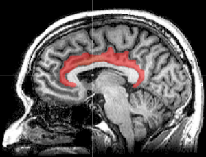
A large energy deficit was found in the cingulate cortex. Jarred Younger believes that part of the brain may play a key role in these disorders.
In a recent video, Jarred Younger’s face lit up when he described a long COVID study that measured the energy levels available in the brain. (See the video in the blog).
- While many studies have indirectly suggested that energy deficits exist in the brains of people with long COVID, ME/CFS, etc., no one has directly measured them until now.
- The German group used a technique that can assess the phosphorus and its metabolites that make up ATP. ATP refers to adenosine triphosphate. The three phosphates in ATP are where ATP’s energy is stored.
- The study particularly looked at the ATP/phosphocreatine ratio. While ATP provides most of the energy for our cells, it’s difficult to move ATP from the mitochondria to the place in the cell that needs it.
- To overcome this problem, an enzyme called creatine kinase adds creatine to ATP, turning it into phosphocreatine. Phosphocreatine (PCr) then transports ATP via something called the creatine kinase shuttle to where it’s needed in the cell. PCr is then converted back into ATP.
- The authors focused on phosphocreatine and ATP (PCr/ATP) concentrations. In a normal brain, they should be roughly equal, but they were significantly reduced in a large part of the brain called the cingulate cortex in the long COVID patients.
- It might seem that the reduced ATP concentrations found (relative to phosphocreatine) would indicate mitochondria were having trouble producing ATP. While the authors acknowledged that the mitochondria might be having some problems, they concluded that the main problem involved the creatine kinase shuttle.
- Phosphocreatine concentrations were increased relative to ATP because it was not being used up. ATP was being created, but once it was converted into phosphocreatine and then shuttled to another part of the cell, something went wrong. In other words, a transport problem existed.
- That the cingulate cortex was the hub of the energy problems in the brain was more than intriguing. The cingulate cortex – particularly the anterior cingulate cortex – has shown up in ME/CFS brain imaging studies many times.Jarred Younger has said that if he could affect any region in the brain, it would be the cingulate cortex. Problems with the cingulate cortex could make everything more effortful.
- Bigger studies are clearly needed, and Jarred Younger is getting the tools needed to do one.
- If these findings are validated, creatine supplementation is an interesting and apparently safe option. Creatine, via phosphocreatine, plays a major role in getting ATP from the mitochondria to where it’s needed in the cell.
- If the creatine kinase shuttle is lacking sufficient creatine, then creatine supplementation could help. That said, other problems that don’t involve creatine could be contributing to the CK shuttle issue.
- Still, some studies have found low brain creatine levels, and one ME/CFS study found them in the same region of the brain.
- A small Oxford 2024 ME/CFS creatine supplementation study found creatine supplementation (16 g creatine monohydrate for six weeks) increased brain creatine in cingulate/prefrontal regions and improved fatigue and some cognitive measures.
- A small Japanese study also found that creatine and glucose (8 g of creatine monohydrate and 3 g of glucose per day) for 8 weeks reduced several long COVID symptoms (including body aches, breathing problems, difficulties concentrating, headache, and general malaise).
- Creatine supplementation isn’t going to be the be-all and end-all of any of these diseases, but it might help. If you’re interested in creatine supplementation, check out Courtney Craig’s Health Rising blog here.
Support Health Rising and Keep the Information Flowing!
Health Rising is not a 501 c (3) non-profit
The CK shuttle takes ATP from the mitochondria and then adds creatine to it to create phosphocreatine (PCr), which moves around the cell much more efficiently. Once phosphocreatine gets to the part of the cell that needs more ATP, the CK shuttle adds the phosphocreatine to ADP that’s floating around, and voilà, the cell has ATP. (More about the shuttle later.)
Phosphocreatine, then, is kind of the secret sauce that keeps the energy flows in the cell moving. Without it, ATP has a hard time getting from the mitochondria to where it’s needed in the cell, and things bog down.
Small Blood Vessel Focus
In their introduction, the authors focused on the very small blood vessels, or microvasculature, as they made the case for their decision to assess the energy availability in the brain.
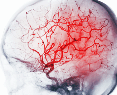
The authors proposed that damaged small blood vessels and clotting could be contributing to energy deficits in the brain.
They proposed that inflammation, increased clotting (hypercoagulation), and autonomic nervous system problems were damaging the small blood vessels, causing low oxygen levels (hypoxia), and hurting the mitochondria. Plus, they noted evidence that a leaky brain barrier may be allowing pro-inflammatory cytokines into the brain.
They referenced a VanElzakker/Loggia long-COVID study which found that blood vessel dysfunction (fibrinogen, sL-selectin) was correlated with neuroinflammation. (Fibrinogen can actually trigger immune cells in the blood to enter the brain.) The study found evidence of neuroinflammation “across a wide swath of brain regions including midcingulate and anterior cingulate cortex, corpus callosum, thalamus, basal ganglia, and at the boundaries of ventricles.”
In conclusion, there are many ways that the fragile but oh-so-important brain may be getting tweaked in long COVID, and in ME/CFS (which has similar findings).
Results
“We present the first direct evidence of deficient high-energy phosphate metabolism in the central nervous system of patients with PCCo (long COVID).” the authors
The sample size was not large (29 people with long COVID; 29 people who had fully recovered from COVID-19) but was larger than many brain imaging studies.
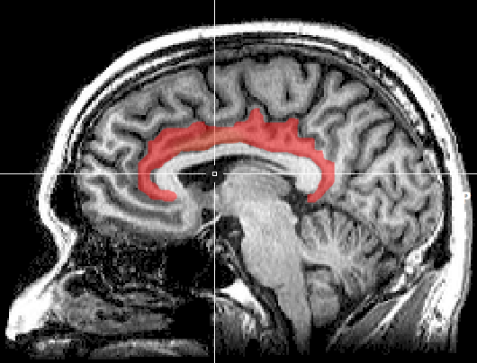
A large energy deficit was found in the cingulate cortex. Jarred Younger believes it plays a key role in these disorders.
The study found a “large, significant cluster of reduced ATP/PCr concentration…centered on the cingulate cortex” in the long-COVID patients. This indicated that this area of the brain was experiencing a significant energy deficit.
The nice high probability value (P < .001) indicated that the finding was solid; i.e., the long-COVID and the recovered COVID-19 patients had very different ratios.
The researchers dug deeper and assessed the phosphorus levels across the three different regions of the cingulate cortex and found reduced levels across all of them.
Lower ATP/PCr ratios in the anterior cingulate cortex were significantly correlated with poorer performance on a cognitive test called MOCA. That suggested that the energy deficit in the ACC may be contributing to the brain fog in long COVID.
Core Brain Region?
Jarred Younger has said that if he could affect any region in the brain in these diseases it would be the cingulate cortex. I asked him in our talk (upcoming) if he thought the cingulate cortex could be a core hub in the brain which causes dysfunctions in other areas of the brain, and he said yes. Problems with the cingulate cortex could make everything more effortful, and the anterior cingulate cortex, in particular, has popped up again and again in ME/CFS and fibromyalgia.
Reduced energy availability in the cingulate cortex was the main finding. The authors did report, though, that reduced ATP levels were found in other areas of the brain.
The Cause – Not the Mitochondria After All
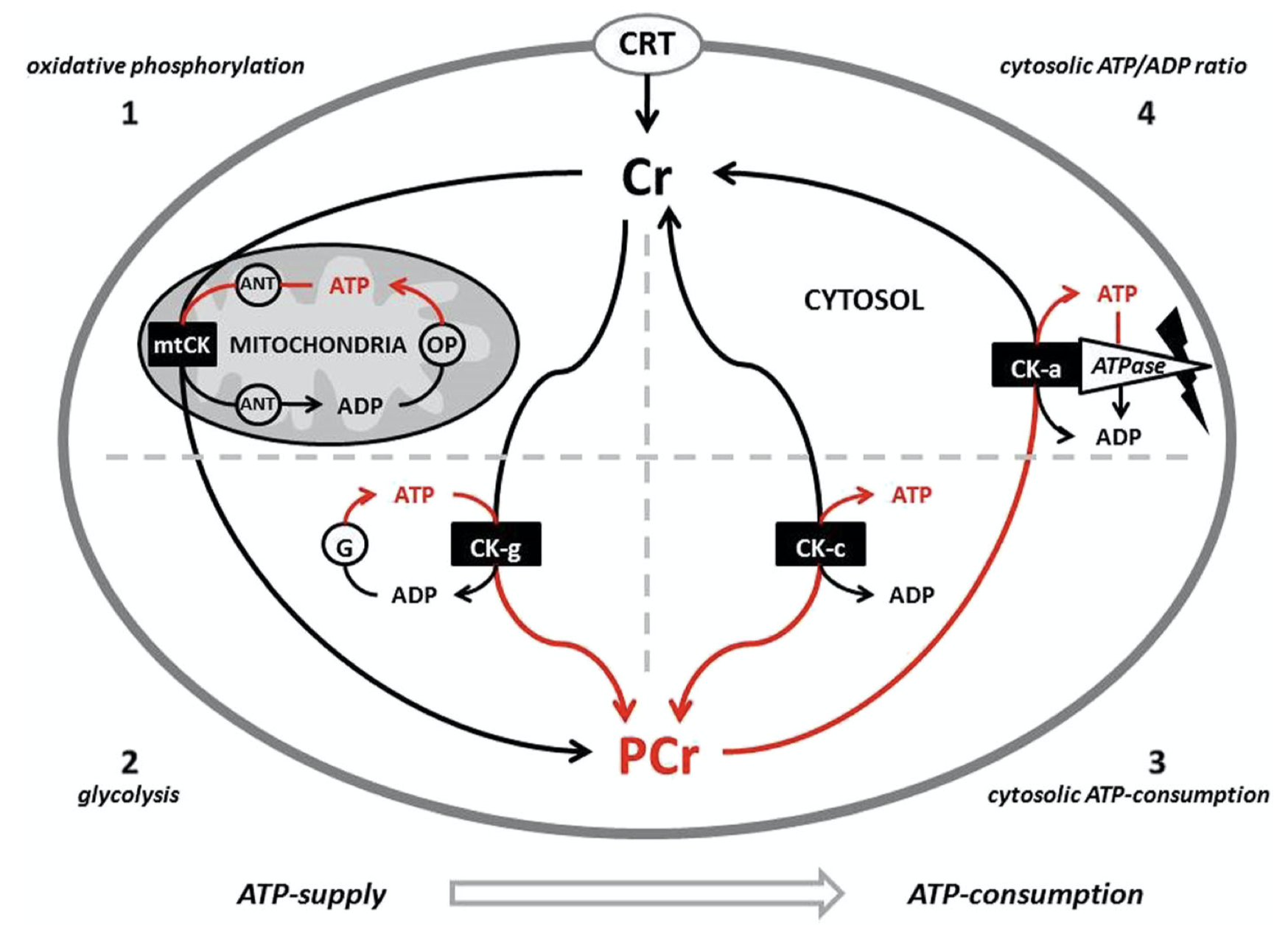
The authors believe that a transport mechanism in the cell called the CK shuttle is failing to deliver energy across the cell. (Image – Richard Krieider, Jun 17, 2017. J Int Soc Sports Nutr)
The authors came up with an intriguing cause for the energy availability problems seen. Their analyses suggested that the reduced ATP/PCr ratio found in the long-COVID patients probably resulted from reduced ATP production.
They didn’t believe the reduced ATP production was mainly caused by the mitochondrial problems, however. (That would be too simple (lol).)
If mitochondrial ATP production was simply low, then PCr levels – because it’s derived from ATP – should have been low too, but PCR was increased relative to ATP. That suggests that ATP is being produced, but it’s not making it to the part of the cell that needs it.
A low ATP/PCR reading indicated that phosphocreatine is present but is not being readily turned into ATP, suggested an energy transport problem is present. (Other studies have suggested that an energy transport problem may exist with the carnitines).
They fingered the creatine kinase (CK) shuttle, which moves ATP from where it is produced (the mitochondria) to where the cell needs it, as the chief problem.
A number of things could have gone wrong. The creatine kinase enzyme may be less active, the transport system inside the cell may be impaired, the cell may not be using phosphocreatine properly, creatine may not be efficiently getting into the cells, and mitochondrial ATP production may be impaired (to some extent).
Core Physiological Factor?
We’re always searching for core problems, and the key role the CK shuttle plays in cellular energy production certainly fits that bill. Because the shuttle is present in cells across the body, it’s possible that it could be causing problems elsewhere. If it is, it might particularly show up in high-energy areas such as the brain and the muscles.
Because different forms or isoforms of the CK shuttle exist in different parts of the body, some areas might be affected while others are not.
You have to go all the way back to 1992 to find the first skeletal muscle 31P NMR study in ME/CFS, which found rapid exhaustion and reduced intracellular concentrations of ATP. In 1993, Barnes found evidence of reduced phosphocreatine utilization in about a quarter of ME/CFS patients. McCully tried three times, on the other hand, and did not find evidence of prolonged phosphocreatine recovery after exercise. A 2023 NMRS study, which did not assess phosphocreatine (i.e. energy production), nevertheless found reduced creatine levels in the same part of the brain – the anterior cingulate cortex – as the long-COVID paper.
In fibromyalgia, phosphocreatine skeletal muscle abnormalities were not found in three small 1994 studIes, or a small 2000 study, but abnormalities were found in a small 1998 study, a larger 2000 study, in the anterior cingulate of the brain in 2021, and in 2023 in a small study in the thalamus. Interestingly, in the last study, reduced blood creatine levels were associated with neuroinflammation, and the authors proposed that “Low creatinine levels may be the prime peripheral biomarker of neuroinflammation in FM patients”.
On that note, in 2019, Nacul found lower serum creatine kinase levels in people with severe ME/CFS compared to healthy controls.
A small 2014 Gulf War Illness study found it took abnormally long to replenish PCr levels in the muscles after exercise. A larger 2023 long-COVID study found slower phosphocreatine recovery after exercise in calf muscle.
The evidence for skeletal muscle phosphocreatine abnormalities in these diseases is mixed, with some studies finding them and others not. At least three studies, however, have found reduced phosphocreatine levels in the brain.
Treatment Possibilities

A small ME/CFS study suggested that creatine may help.
This study’s findings need to be validated, but creatine supplementation is an interesting and apparently safe option. Creatine, via phosphocreatine, plays a major role in getting ATP from the mitochondria to where it’s needed in the cell.
Whether creatine helps or not depends on where the problem is in the CK shuttle. If the CK shuttle lacks the creatine, it needs to operate properly, then creatine supplementation could help. If the primary problem lies elsewhere (CK enzyme problems including loss, inflammation, low oxygen levels, reduced mitochondrial ATP production), then creatine supplementation might not help.
The long-COVID study’s finding made a small Oxford 2024 ME/CFS creatine supplementation study all the more interesting. While the study did not measure ATP/PCr levels directly, it did find that creatine supplementation (16 g creatine monohydrate) increased brain creatine in cingulate/prefrontal regions and improved fatigue and some cognitive measures.
A small Japanese study also found that creatine and glucose (8 g of creatine monohydrate and 3 g of glucose per day) for 8 weeks reduced several long COVID symptoms (including body aches, breathing problems, difficulties concentrating, headache, and general malaise).
Check out Courtney Craig’s blog on creatine supplementation in ME/CFS, long COVID, fibromyalgia, etc.
Like any supplement, creatine is probably not going to be the answer. The answer likely requires addressing fundamental factors like inflammation, blood vessel problems, and autonomic nervous system dysregulation.
It might be helpful, however.
Next Steps?

Hopefully, this study will spawn larger, more comprehensive ones. Jarred Younger is getting the tools needed to do one.
Note that Jarred Younger is getting the equipment needed to duplicate this experiment. When I talked to him, I believe he was about a month away from getting the last piece.
I asked Perplexity AI what it recommended for follow-up studies that could: a) determine if this finding is valid and, if it is; b) try to get at the cause.
(1) A larger study that separated out ME/CFS-like patients from other long-COVID patients and used the same technique but added more complete cognitive and symptom assessment to see if the findings track with brain fog, orthostatic intolerance, fatigue, PEM, etc. would be very helpful. Do exertional challenges to see if the ATP/PCr ratio worsens.
(2) Pair the brain 31P-MRS testing with similar assays of cells found in the body such as PBMCs, fibroblasts, or muscle to see if the same energy availability problems are present. Besides assessing CK activity, check out creatine transport, mitochondrial respiration, and coupling efficiency. (3) Do root-cause studies to determine whether the abnormality is driven by inflammation, persistent immune activation, microvascular dysfunction, altered pH regulation, autonomic dysregulation, or, interestingly, sleep disruption.
(4) Measure brain or systemic acid-base handling, lactate/pyruvate balance, and markers of redox stress alongside CK-related endpoints to help determine whether the problem lies in low ATP production or transport problems.
Good for Germany for opening up this intriguing area of research! Brain research is blowing up right now.
- Coming up – David Goldstein’s ME/CFS and long-COVID brain norepinephrine paper





That sounds like it could be a Ca2+/Mg2+ balance thing. Cytosol creatine kinase (the enzyme that will convert a lot of phosphocreatine plus adp to creatine plus atp inside cells / outside mitochondria) seems to be sensitive to / needing magnesium. Calcium resembles magnesium a lot (same colomn on Mendeleev’s table). Due to that similarity, both ions can bind to many same enzymes. When the ‘wrong’ ion is bound to the enzyme however, it risks to become deactivated.
After neuronal firing (each and every single time a neuron ‘works’), there is a large calcium influx in the neuron (ref1). Too much calcium in the cytosol can become toxic and create things like neuronal ROS, mitochondria death up to neuronal cell death (and neurons are rather hard to replace!). When it comes to creatine kinase, Ca2+ is the only ion that can displace Mg2+ at the binding site of the enzyme in a way that the enzyme is inhibited (ref2).
That could be reversed to some extend by increasing Mg2+ (if the neurons would take it up). That then increases the Mg2+ versus Ca2+ ratio and therefor activates the creatine kinase enzyme a bit more. Desired effect: more ATP available for neuronal firing. Side effect: even more excessive neuronal cytosol Ca2+ levels with more risk for ROS, mitochondrial death and neuronal death.
=> Basically, it could short term help if the body isn’t wise enough to counteract that attempt. In reality, it’s most likely trying to override an important safety mechanism trying to prevent even worse health over time.
ref1:
https://pmc.ncbi.nlm.nih.gov/articles/PMC3048837/
Title “Neuronal calcium sensor proteins: generating diversity in neuronal Ca2+ signalling”
citates:
“High intensity Ca2+ signaling necessitates high ATP consumption to restore basal (low) intracellular Ca2+ levels after Ca2+ influx through plasma membrane receptor and voltage-dependent ion channels.”
“it leads to a significant increase in [Ca2+]cyt after depolarization”
(my note: each neuronal firing (neuronal work) causes such depolarization, creating a Ca2+ influx, requiring a lot of ATP to flush it out and we don’t seem to have enough of ATP = problem!)
ref2:
https://academic.oup.com/clinchem/article-abstract/26/8/1137/5664372
Title “Effect of cations on the human creatine kinase isoenzymes.”
citations:
“We report our findings on the effect of Ca2+, K+, Na+, Fe2+, Mn2+, Zn2+, and Cu2+ on the activity of creatine kinase isoenzymes derived from human tissues. The only inhibitory cation was Ca2+, and Dixon plots of the data indicate the inhibition to be competitive for the Mg2+ in the reaction assay mixture. This effect of Ca2+ on all three isoenzymes is completely reversed by including Mg2+ (10 mmol/L) and ethylenediaminetetraacetate (2 mmol/L) in the assay mixture.”
=> My take: IF it is this at play, THEN it is not the cause of a problem but an essential safety mechanism that is best not fidled with in order to prevent quick neuronal degeneration
In simpler words:
* Too rapid firing of neurons can create massive neuronal damage.
* Each time a neuron fires, Ca2+ (calcium ions / cations) are increased inside the neuron.
* The neuron itself needs time and energy (ATP) to recharge.
* Removing the excess calcium from the inside of the neurons also needs time and energy.
* So long there is too much calcium inside the neurons, creatine kinase will be slowed and with it slow both (and synchronously!) the ‘recharging’ of the neuron as well as ‘clearing the calcium safety break’.
Therefor: if the body manages things well, it knows when it is safe to fire the neuron again. It can do that by making sure that the excess calcium is removed a bit slower then that the neuron is ‘recharged again’.
This effects become bigger over a long series of neuronal firing (long and intense brain work). Calcium then accumulates (ref2). So it is a sort of ‘memory’ telling the neuron how hard it worked over the recent past and how exhausted it is.
That stems with our experiences: when our brains get exhausted more and more, our thinking becomes slow and full of mistakes.
important citation here from (ref2) above:
“Because proton pumping creates an inside-negative membrane potential, Ca2+ as a cation will tend to accumulate in the mitochondrial matrix”
(my note: mitochondria will also try to remove exess calcium from the cytosol, but not by pumping it out of the cells but ‘temporarily swallow’ it inside themselves)
Other important parts of (ref2) for this comment:
“Therefore, Ca2+ signaling and excitability mutually regulate each other such that an optimal neuronal activity level is achieved for a given metabolic capacity.”
and
“As will be described below, neuronal Ca2+ homeostasis is altered during aging and in neurodegenerative conditions, and uncontrolled Ca2+ signaling may also actively contribute to neurodegenerative processes.”
(read uncontrolled here also as ‘the brake is not working well” or “someone tried to remove the brake because the brain worked to poorly after exhaustion”)
Calcium again! How interesting. I must say, DeJurgen the research lost a lot when you came down with ME/CFS. We, on the other hand, gained a lot 🙂
any thoughts on how Prusty’s showing mitochondrial fragmentation would factor into this?
Hi Djergen, is there anyway you can private message me at 2017399410. Thank you
I think you’re going to like this study too Cort 🙂
https://www.nature.com/articles/s41467-026-75725-y
Rob Wust is doing great work in the Netherlands! Thanks 🙂
@sunie on August 2, 2026 at 11:51 pm
any thoughts on how Prusty’s showing mitochondrial fragmentation would factor into this?
———————————————————————————————————————————–
Yes, and it’s rather complex, also for me. I had to dive in deeper to get the details right. I’ll try and give an overview while ommitting complex details.
With intense exertion, cells see a strong increase in calcium in their cytosol. That quickly becomes toxic and needs to be kept under control. The simplest way of doing that, is by ‘pumping calcium out’ of the cells as quickly as possible. The problem is that that consumes quite a bit of ATP. During peak loads, that ATP may be unavailable. Brain cells firing too quickly without enough ATP can kill them.
An alternative is that the mitochondria can ‘swallow’ calcium. By *temporary* doing so, they increase their internal calcium (Ca2+) content quickly. They need to handle that later, but the cells can avoid the direct / ‘need energy now’ ATP cost of actually pumping the Ca2+ out of the cell. (ref1) for example shows that by doing so, cells can produce more peak ATP under conditions of oxygen shortage. For brain cells that is a nice potential saver! The Ca2+ ions however need to be pumped out again and that will cost a bit more then what was initially saved. The benefit is however that this can be done later at resting time.
(ref2) shows that mitochondria actually ‘deliberately’ initiate fragmentation during injury, with the specific purpose to be able to further increase the ability to take in Ca2+ in order to temporary avoid the ATP cost of pumping Ca2+ out of the cells. Fragmentation changes the morphology so that / in order to increase this uptake of Ca2+ from the cytosol. The main goal from that is to temporarily increase peak output beyond what the mitochondria would be able to do so (without fragmentation) with the limited amount of oxygen they can get their hands on.
Now saving cells and mitochondria from an accute energy shortage is important, as that can be devastating to them both. There are multiple payback problems with that however:
Mitochondria can use this only up till a certain concentration of *free* Ca2+ ions. Then this ability stops. But this limit can be bypassed by forming calciumphosphate solids inside mitochondria. Those consume phosphate, removing phosphor from the ATP / ADP / AMP pool (as is seen by research in ME/CFS), decreasing the ATP /ADP pool. Also, calcium solids are a *lot* more difficult to remove from the mitochondria again then Ca2+ ions. They need to be broken down in solution bit by bit so that calcium solids slowly transform again to Ca2+ ions that can be removed from the mitochondria. So mitochondrial Ca2+ buffering due to minutes of overexhaustion can cost (need to check numbers, too tired now) hours of time to clear Ca2+ / calciumphosphate from mitochondria. Having less phopsphate due to calcium binding to it in solids doesn’t help either, as that depletes the ATP / ADP pool needed to provide the energy needed to clean this mess up… :-(.
The other alternative is to split mitochondria into good parts and bad parts and fill the bad part with calcium deposits and break it down via mitophagy. In ME/CFS the mitophagy pathway however is observed to be slow / crippled too. So forget this potentially quick way to deal with the problem.
Summery:
Exertion increases calcium buildup in cells.
Keeping calcium concentration inside cells within limits is vital.
Directly removing excess calcium costs plenty of ATP.
During ATP shortage this is infeasible, an alternative is needed. (pacing would be even a lot better!)
Mitochondrial calcium buffering can come to the rescue.
If needs are even more dire, mitochondria can fragment to increase their rate of calcium buffering.
Recovering from that (short lasting overexertion) is however a costly process.
It takes a lot more time and in many cases will leave the cells full of fragmented mitochondria that are filled with problematic calcium deposits (especially if it happens every other day).
So concerning to ME/CFS: the less spare energy one has, the smaller the exertion is needed to activate this calcium buffering emergency mechanism with all the trouble clearing the damage from it out. That costs time and energy. With low baseline energy (due to poor quality mitochondria piling up) that recovery process takes longer then in healthy people too. That aligns well with ME/CFS versus healthy controls.
See (ref2) for pictures on mitochondrial fragmentation found in this research.
(ref1): https://pmc.ncbi.nlm.nih.gov/articles/PMC3048837/
Title “Neuronal Calcium Homeostasis and Dysregulation”
citations:
“The following consideration outlines why mitochondrial buffering can be regarded as a mechanism that limits the maximal rate of O2 consumption required to maintain Ca2+ homeostasis. Pumping out one Ca2+ ion from the cytoplasm across the plasma membrane or the to the endoplasmic reticulum (ER) membrane costs one ATP molecule, either directly via activity of the plasma membrane Ca2+ ATPase (PMCA) or the sarcoplasmic endoplasmic reticulum Ca2+ ATPase (SERCA), or indirectly by activity of the Na+–Ca2+ exchanger (NCX) which exchanges three Na+ for one Ca2+ and then the three Na+ are pumped out by the Na+/K+ ATPase at the expense of one ATP. To restore the ATP level, three protons must be pumped by the ETC. If Ca2+ enters the mitochondrial matrix, this comes at the expense of only two protons (i.e., 0.67 ATP molecules). Thus, mitochondrial Ca2+ buffering requires fewer protons to be pumped and accordingly less O2 needs to be consumed.”
(ref2)
https://rupress.org/jcb/article/219/5/e201909154/151605/Mitochondrial-fragmentation-enables-localized
Title “Mitochondrial fragmentation enables localized signaling required for cell repair”
“Fission of injury-proximal mitochondria allows for greater amplitude and duration of calcium increase in these mitochondria, allowing them to generate local redox signaling required for plasma membrane repair.”
We also get this one ‘free’ with mitochondrial fragmentation: extra ROS (oxidative stress)
“Increase in [Ca2+]m promotes mROS production”
=> So this fragmentation seems to be another ‘tool’ for preventing something even worse (massive cellular / neuron death due to accute ATP shortage), even if itself has plenty of side effects.
Rather the ‘fixing’ the amount of fragmentation by trying to block it, we may need better ways to prevent the need of this ‘tool’.
Would intense exertion be walking non stop for 15 minutes for an ME person especially if in late 70s?
The question isn’t clear but let me respond on mitochondrial calcium buffering (that has related implications like mitochondrial division and fragmentation) in skeletal muscle.
Arm and leg muscles are skeletal muscles.
https://www.cell.com/biophysj/fulltext/S0006-3495(16)31755-6
Title: “Mitochondrial Calcium Uptake and Matrix Calcium Buffering in Skeletal Muscle”
Saying “To elucidate the role of mitochondrial Ca2+ uptake in skeletal muscle (SM) function we ablated MICU1, the Ca2+ sensing regulator of mtCU in mouse. MICU1 ablation in SM resulted in impaired gatekeeping and attenuated Ca2+ uptake and resulted in less endurance exercise performance when challenged with fatigue protocols.”
In more simple language: if they block the ability of mitochondria to buffer calcium (a mechanism that can prolong short term strong exertion at a significant cost in the long term if the mitochondria and cells can’t recover enough after it), then test mouse had poorer exercise endurance.
In the free available abstract they give numbers. There they also blocked something very much related to this calcium buffering so it will influence numbers. Still, the outcome is clear: “When challenged in an incremental exercise test, PiC-ablated mice could only run for 8.5±1.7 min, whereas control mice ran for the full 20 min, showing impaired performance.”
Trying to block this mechanism decreases endurance a lot. I can only guess something similar happens when this mechanism hasn’t been able to recover and as a result mitochondria start to falter. The most important insight here is however that muscle cells also have similar problems with calcium buffering and overload as neurons do. If they do so, increased calcium concentrations in the cytosol have a reasonable chance to inhibit Creatine Kinase there too.
This is interesting! What about those of us who also have endometriosis and were told that creatine can make those endometrial cells worse/re-produce better….. Have you or anyone run into this conundrum where creatine might be helpful for me/cfs but might hurt for endo?
Just wanted to mention that creatine-glucose study you mentioned briefly actually was not Japanese, it just was published in a Japanese journal. The authors are in Serbia, Canada, Hungary and Norway. Some of the Serbian researchers had a previous brief publication on creatine as well (“Creatine supplementation combined with breathing exercises reduces respiratory discomfort and improves creatine status in patients with long-COVID” by Slankamenac et al.)
As it happens, I’ve recently experienced some LC symptom improvements when taking Creatine and by coincidence using sugar-based cough drops and candy for a respiratory infection. So I’m so glad to learn about this research!
Updating my comment, I later saw that the Serbian research group has published multiple creatine paper, including one mentioned in Courtney Craig’s blog post on creatine. That paper is “Effects of six- month creatine supplementation on patient- and clinician- reported outcomes, and tissue creatine levels in patients with post- COVID- 19 fatigue syndrome.”
The researchers appear to be based in the Applied Bioenergetics Lab, Faculty of Sport and PE, University of Novi Sad, Novi Sad, Serbia — seems worth keeping an eye on this team.
🙂 Thanks for clearing that up and for the tip!
Thanks this article. Appreciated.
Creatine supplementation did nothing for me.
I have LC for 2.5 yrs.
However, I can feel my brain warming up if I exert too much.
Especially certain types of cognition. Then my brain feels like it is
simply shutting down. But if I take a 2-3 hour break the heat goes
away.
Very interesting. The search for an intrinsic (primary) mitochondrial problem has been going on for a while and not turned up a conclusive picture. I´d rather follow the vanElzakker route and try to explain the major PCC/ME/CFS findings together: endothelial dysfunction –> neuroinflammation (possibly including viral reactivation) –> mitochondrial dysfunction (i.e. as a secondary factor). The finding of neuroinflammation in the lining of the ventricles fits with previous findings of enlarged chorioid plexi in PCC (Diez-Cirarda, 2025) and could indicate that a leaky blood brain barrier is centerpiece in all this (would be interesting to know if markers of BBB leak correlate with PEM).
For you, Cort: would be great to discuss with Jarred Younger how he interprets the findings of this study?
There is a problem with the supply of oxygen (and nutrients) because ME patients have fewer capillaries and a different muscle distribution. This is basically how I understand it in short. It affects the whole body and brain. An oxygen shortage could then explain the fight-or-flight response as a compensation. And the dysfunction of various systems.
HR-P, I seem to remember you being vaccinated with BCG perhaps a couple of years ago. Did you experience any improvement in your symptoms?
I think so, I have seen an overall improvement over the last years. But do I know if it is better than the natural course? No. I have had 9 or 10 BCG vaccinations so far – whenever my baseline declines I repeat. Is this the best regimen? I don´t know!
I’m more of the vascular – inflammation- mitochondrial dysfunction mind as well. I did talk briefly with Jarred about it but had not read the study beforehand. He’s definitely excited by but unfortunately we did not get into the weeds about the CK shuttle. In other words, I was not prepared! (lol)
Beste Cort, ong een maand geleden ben ik begonnen met Creatine in de hoop op wat verbetering (lig 24-24 op bed) maar integendeel ik kreeg nog meer last van uitputting en ben om die reden gestopt. Vreemd zou je zeggen omdat het tegenstrijdig is.
Hartelijke groet
Google translate to English:
Dear Cort, about a month ago I started taking Creatine hoping for some improvement (I am in bed 24/7), but on the contrary, I suffered even more from exhaustion and stopped for that reason. Strange, you might say, because it is contradictory.
Kind regards
Sorry that happened. It was certainly worth a try. We’re just a heterogenous group. Something that helps one person hurts another. It happens all too often.
Subsets can’t come too quickly!
Hi Cort, it was Lenny who tried the creatine, I was just translating from their Dutch, I let them know you replied
Cort has replied to you but to my reply, please see above
Thank you, Germany!!!! And, as always, thank you, Cort!
creatine does not cross blood-brain barrier, it’s too big of a molecule
but creatine is not curing anything or even treating, it’s just “overclocking” a malfunction system, like a 4-cylinder car with only 2 working cylinders that spins 10% faster to at least move somewhat better
not everyone responds to ribose but it’s worth a try for another kind of ATP production encouragement but of course also won’t be a cure
this is like HIV/AIDS research, they are going to go in circles for decades with useless observational studies and then 40 years later finally make some progress
unfortunately most of us won’t survive another decade forget 40 years
“Overclocking”, well said! 🙂 I feel like my system is overclocking all the time – used to say “ME/CFS car’s stuck in first gear, and pushing oneself equals pushing the gas pedal hard (= a state of nervous system over-activation) so the car can at least jump a little bit forward, but then the nervous system gets stuck in that overactivated state like a gas pedal getting stuck”. Funny how patients often come up with similar analogies for ME/CFS.
What you write corresponds to what I wrote about my experience with supplements below.
I waited much longer than it took for them to understand and have a treatment for HIV and AID.
It took about 6 years to get the first FDA-approved medication (AZT in 1987) after the HIV virus was identified in 1983, and roughly 15 years to reach truly effective life-saving therapy (combination therapy or “the cocktail” in 1996) that turned the disease into a manageable chronic condition.
Just 15 years for HIV and AIDS …and they’re not even close to understanding this illness!
Because people were dying they spent money on it. If they spent that kind of money on ME/CFS/FM we would have answers given the technology we have now.
Forty years ago, Dr. Cheney memorably said something like the old guard is going to have to disappear before they move on this illness. Well, the old guard was replaced by the new guard, and things are still the same – at least regards ME/CFS and FM.
Yes, we are moving forward on virtually every front – except for federal funding…which adjusted for inflation may not have moved an inch in 40 years.
And just a warning again…. if you areover 50yo I’d make sure you’ve had your kidney function checked by routine exam in the year prior to starting creatine. Otherwise you could be looking at more exams, after taking it, if your kidney eGFR #s tank and creatinine goes up. Your Dr might not believe you when you say you’ve been supplementing. Mine ordered a MRI! (which I refused.) I stopped taking it, made them redo the blood test a couple weeks later and it was back to it’s usual #. Ps it did nothing for my energy or brain functioning in the month I took it.
Cort, I’m grateful that you’re giving this research the momentum it deserves.
Here are my thoughts:
Mitochondrial toxicity?
Mitochondrial ATP production was discussed as early as 2019, with the proposal that proton (H⁺) leak and mitochondrial uncoupling are dynamically regulated according to cellular ATP demand and the rate of ADP/ATP exchange. See references in the link I included.
This brings me back to an important question: Could phytanic acid alter intracellular pH?
Experimental studies have shown that elevated phytanic acid concentrations can:
Inhibit mitochondrial respiration
Reduce ATP production
Increase reactive oxygen species (ROS)
A reduction in ATP would limit the energy available for ATP-dependent ion transporters, including proton pumps and other mechanisms that maintain intracellular pH homeostasis.
In addition, phytanic acid is an amphipathic molecule, possessing both hydrophilic and lipophilic properties, which allows it to interact with biological membranes and potentially influence membrane function.
Taken together, a plausible mechanistic pathway is:
Elevated phytanic acid → mitochondrial dysfunction → reduced ATP production → impaired ATP-dependent ion transport and proton handling → altered intracellular pH homeostasis.
Whether phytanic acid also affects intracellular pH through more direct effects on membrane properties or proton flux remains an important question for further investigation.
As I laid out here: https://swaresearch.blogspot.com/2023/10/mitochondria-function.html
More interesting research re: the brain:
https://www.frontiersin.org/journals/medicine/articles/10.3389/fmed.2026.1824498/full?utm_source=substack&utm_medium=email
Did all the patients in this study have the same strain of Covid? I had Omicron in 2023. Following a prescription for Paxlovid and several negative Covid tests, I thought I was okay. One month later, I started to have high blood pressure and significant heart irregularity. I found an excellent cardiologist and after a few months I thought it was okay to fly to Maui to see our daughter. I got Covid again 8 months later in May 2024, a different strain. This time I took a different anti-viral and once again tested negative. One month later, I began to have great trouble with balance and walking, but not the heart and blood pressure problems from before. I am still stuck in gait and balance problems and have had some serious falls. I broke my wrist on the last one. Covid is constantly mutating. This is why having it doesn’t protect you from getting it again. I don’t see how any serious study can be made of Covid unless all the participants had the same strain.
Sorry to hear about that and wishing you all the best! Always appreciate your comments.
If feels ironic that on an article on the brain – something I have fervently believed to be central to ME/CFS – I post a link to very credible research just published on viral reactivation and long covid. As I have always said, I am happy to be proven wrong, and especially if that results in effective treatment. This research is fascinating work on viral reactivation, and implies a viral family I have never heard of:
https://www.nature.com/articles/s41586-026-10740-z
To stay up to date on latent viruses in LC and ME/CFS, keep an eye on Liisa Selin and her collaborators—particularly Ayano Kohlgruber, whose current study builds directly on Selin’s foundational work. Kohlgruber’s work should give us concrete answers regarding the specific targets driving T-cell exhaustion in ME/CFS.
Alongside Akiko Iwasaki’s current research on herpesvirus reactivation, the PolyBio network is stacked with early-career researchers mapping T-cell targets. At the recent PolyBio spring conference, one researcher showed the first direct proof of T cells targeting EBV and VZV in Long COVID, while Michela Locci is zeroing in on B-cell pathology. Locci’s ongoing study analyzes fine-needle aspirates taken directly from lymph nodes, where she’s already identified significant B-cell impairment. She’s now expanded her study to try to determine the role of EBV in this.
On top of that, the PolyBio network has started small clinical trials testing antivirals like Valtrex and Truvada in Long COVID to target early-phase EBV reactivation (led by Putrino and Iwasaki). That’s a great step, though they really should look into Famvir as well—I’m on it, I greatly benefit, and its side effect and toxicity profile is so much better than both Valtrex and Truvada.
Cort Johnson has done a great job breaking all of this down in recent months.
Perhaps ‘The Answer’ will lie somewhere between our key realms of interest (the brain/virology).
I thought this recent Japanese research on long covid, viral reactivation and the brain was interesting:
https://mainichi.jp/english/articles/20260623/p2a/00m/0li/018000c
And that an anti-dementia drug might help. Yet, according to this theory, so might antivirals. It made me wonder if anyone with ME/CFS has benefitted from Huperzine A, which acts on the same acetycholine pathways like the anti-dementia drug. I’ve never tried it, maybe I will be a human guinea pig again and try…
Interested to know which particular symptoms you greatly benefit from with Famvir? Thanks, M
How has Famvir been of help? I have had EBV reactivate 9 times in the last 10 years. I am desperate for answers. Thank you.
How has Famvir been of help?
Does it help prevent EBV reactivation?
Valacyclovir/famciclovir suppress my ME/CFS episodes fully. When I am acute they suppress all the symptoms within one hour (I fulffilled the CCC when I was at my worst between 2021-2024). When I am not acute I can do significantly more, e.g. my baseline (level of exertion intolerance) improves quite drastically. When I go off these drugs I am as sick as before. They are no cure. The most helpful change that these meds have brought to me is that I could let go of the constant fear of relapses that could get out of control and a deterioration of my health level. I am mild/moderate and in the first years of being ill when I learned to manage it with pacing without these drugs the fear of becoming housebound or bedbound was looming in the background all the time.
Both these drugs (an Truvada) have good-very good activity against early-phase EBV relplication. Which is why Dr. Lerner, the first doctor who developed valacyclovir protocols with his patients came up with the idea that ME/CFS might be caused by a tissue-specific, early-phase abortive reactivation. All these drugs are useless in a standard lytic EBV reactivation where the virus is replicated and a cytokine storm has been unleashed.
Bateman and Putrino have run small, prospective open-label studies into valacyclovir in the last years in LC. You can there learn more about dosage.
If you have further questions, I am happy to help.
Hi Lina. I’m glad that the valaciclovir works for you.
I have just finished a course of it for suspected shingles reactivation (test said negative) and was hoping it might have some useful side-effects, but unfortunately I’m feeling worse than I have in a while, and will be watching out for signs of improvement now I’m finished.
As Cort says, we’re a heterogeneous bunch.
Yes, it’s known that it doesn’t work for everyone. Theoretically, one clear reason for that would be EBV negativity. Do you know your status regarding that virus?
I’m really sorry to hear that you’re feeling worse right now.
Before trying or continuing any antiviral protocols, the most essential first step is to verify baseline antibody status—specifically checking whether you are EBV-positive (seropositive) in the first place. Antivirals targeted at herpesviruses only have a theoretical mechanism if the body actually harbors the virus.
Establishing serostatus (such as checking VCA IgG and EBNA-1 IgG) provides the baseline data needed to know whether that specific pathway is even relevant before considering any targeted treatments.
I agree Lina! I believe Liisa Selin probably has the best theory for the cause of ME/CFS & Long Covid! There is T Cell Exhaustion taking place & probably a genetic weakness in the immune response that keeps the immune system from being able to effectively neutralize or suppress certain viruses. Manuel Ruiz has a similar theory & people should follow his work. I follow him on Twitter/X. I think Mathias will eventually come around to the root cause of ME/CFS being immune dysfunction/viral, even though many of the symptoms manifest in the brain.
Thanks for making me aware of Manuel Ruiz’s advocacy. I’m not surprised that he, just like Selin, is a patient-researcher. If you have ME/CFS and take your lived experience seriously, you recognize that this feels infectious—and because symptoms flare every time you overexert, your mind naturally turns to latent viruses.
That was my immediate intuition during my first flare, which led me to convince my doctor to try acyclovir on a hunch. That was lucky, of course, given how many herpesviruses exist and that acyclovir only targets a subset of them.
What infuriates me now is how hard the medical establishment pushed back against patients who had the right instincts as far back as the 1980s, when acyclovir was new and patients realized it suppressed their flares.
For decades, we’ve been told that ME/CFS is so impossibly complex that it took half a century just to gain basic insights. But the reality looks quite different. The core pathomechanism may be straightforward enough that attentive patients identified it early on as smoldering EBV reactivation—especially since in vitro studies had already shown acyclovir’s activity against early-phase EBV reactivation by 1980. They began calling it Chronic EBV, only for the medical establishment in the 1980s to pivot, rename it “Chronic Fatigue Syndrome,” and dismiss those patients as ill-informed or hysterical.
While we don’t have absolute proof yet that ME/CFS is fundamentally a smoldering chronic EBV infection, the framework and studies are finally aligned to bring that proof to light. What this history really reveals isn’t the complexity of the disease, but how dogmatic and misogynist the medical establishment has been in writing off patient intuition.
I have had ME for years, but suddenly it got worse. My IgG and IgA antibodies were falling and B cells also low. T cells were alright (no testing on B & T cells since last year).
I started on IVIG infusions with horrible side effects, but actually a couple good days, still fatigued but mobile. Then every month I had more good days, kind of where I was over a year before.
My immunologist approved me to do genetic testing and I’m waiting for results from Finland.
Thanks for sharing, Mojo. Good luck with the IVIG and please let us know if the genetic testing reveals anything helpful. We should be getting results from the RECOVER IVIG clinical trial soon.
Creatine (Megamax brand) did nothing for me at the dosage recommended on the bottle.
Might try again to observe if any effects on brain fog, but someone else said in the comments that creatine is too big a molecule to cross blood-brain-barrier.
Two thoughts:
1) Whatever brain processes limit functionality in ME/CFS – might they actually be protective to prevent from running into ME/CFS exertion limits elsewhere in the bpdy?
2) It may not be as easy as just corecting an imbalance with supplements:
Again and again I’ve made the experience that supplements can temporarily increase energy/ability to be active through one pathway in the body, while ME/CFS exertion limits STILL exist ELSEWHERE in the body, so that the supplement will cause a temporary increase of functionality followed by an ME/CFS crash (in particular when – but not limited to – trying to fill up the deficit too fast). Happened with all kinds of stimulating or functionality-increasing supplements including magnesia and inositol. Happened only recently when trying to quickly fill up a lab-proven omega-3 deficit gave me a hyperfocus-like brain activity high and overstimulation sequeing into a crash.
It is as if ME/CFS exertion intolerance does not tolerate the higher functionality made possible by the supplement. Filling up a deficit is NOT easy with severe ME/CFS in my case.
This makes makes a lot of sense, that supplements may allow us to overdrive the ME/CFS body, resulting in a crash.
I got very energised when my daughter saw very significant improvements on creatine+ a high absorption iron supplement last year.
In terms of energy, she went from 3 out of 10 to maybe 6 out of 10
Although she’s a little better than a 3 now, she couldn’t sustain the improvement at a 6
It was a bit disappointing, even if it did give me some hope that surely ‘something’ can and will help this condition
I doubt it was placebo. She’ s tried many supplements with no benefits
I wonder if ‘something’ in the creatine+iron mix worked on some sort of key pathway, even if that wasn’t sustained. And whether that is a clue…
I thought this 2024 Australian study was VERY interesting at the time. I have gone back to it, and it still is! I think they are ‘warm’.
Interesting linkage between kynurenine pathway and hepcidin (iron).
Perhaps the iron superficially helped my daughter, temporarily, and the help it gave her points to the Hepcidin issue? And therefore kynurenine, and inflammation…..
https://pubmed.ncbi.nlm.nih.gov/38015302/
What are vaccines designed to do?
Correct…cause the immune system to create an immune response/reaction🚀
Brain/dopamine issues. Makes sense of the findings of Nath et al ???
https://www.sciencealert.com/long-covid-linked-to-lasting-damage-in-the-brains-dopamine-system
Hello Cort,
I don’t have anything profound or thought-provoking to put in a comment, but I just wanted to let you know that I’m still reading!
Cheers,
Sarah T
(Australia)
Good to hear, Sarah! Hang in there. Lots of stuff is coming. 🙂
FIP is the most lethal corona virus illness. It occurs in cats and was 100% fatal when we lost our precious Ragdoll kitten nearly 10 years ago. Even at that time, the company making Remdesivir knew it could be a cure, but they wouldn’t let vets have it because the company hoped to use it in humans and they did in hospitalized patients.
Now Remdesivir and two other antivirals are approved for FIP in cats and the cure rate is almost 100%.
Why aren’t these antivirals being studied in Long Covid? I could find very little to suggest that this kind of research is happening.
Great read on such an important study!
I completely agree with the idea that the brain and muscles lose functionality primarily because of their high energy demand, rather than some unique tissue characteristic.
I was so happy to see you dig deeper into this study to test that connection, and even happier to learn the authors found this exact issue showing up wherever energy demand in the brain is highest. Since my own technical skills here are limited, I really appreciate you doing the deep dive to confirm it!
Thanks so much, Lina 🙂