

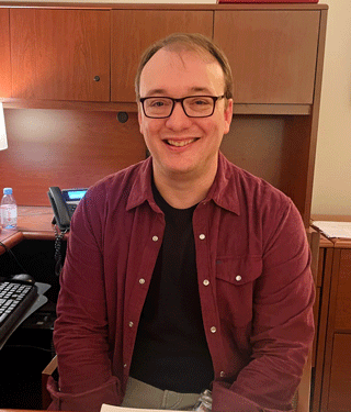
Jarred Younger, founder of the Neuroinflammation, Pain and Fatigue Laboratory, talked about what’s exciting him in these diseases
Jarred Younger pioneered the first LDN trials in FM, helped develop a new, less invasive method for measuring brain inflammation, and a way to determine whether immune cells from the body are getting into the brain. He’s assessed the effects of dextromethorphan and botanical agents, is currently assessing the effects of psilocybin in fibromyalgia, and is about to begin a major NIH-funded LDN study.
I call him the “neuroinflammation man”. He’s proposed that neuroinflammation plays a major role in diseases like ME/CFS and fibromyalgia for over 15 years, and more recently, long COVID, and he’s never been more convinced pf that than he is now.
Besides ME/CFS and fibromyalgia, he’s also studied Gulf War Illness, rheumatoid arthritis, traumatic brain injury, TMJ, and others. He established the Neuroinflammation, Pain and Fatigue lab at UAB in Birmingham in 2014. I got to talk with him in a Zoom interview last month.
Jarred Younger Talks
Jarred Younger looked decidedly under the weather in our talk, but no worries – the area around his eyes has simply swelled after an eye test. After the ceiling in his office collapsed, he also temporarily ended up in a dreary space with weird yellow paint. He’ll be moving into a beautiful new building soon.

I wanted to go over what’s exciting Jarred in the ME/CFS, fibromyalgia, and long COVID fields right now. It turned out there was quite a lot.
Measuring ATP in the Brain
First we talked about a “beautiful” long COVID study which prompted him to get a new device. The researchers used a magnetic resonance spectroscopy (MRS) device to measure energy availability in the brain.
He noted that studies using this technique in ME/CFS found reduced energy availability in the muscles but no one had applied it to the brain, which he thought was a shame because a) the brain must be involved, b) it generates all our experiences, and c) and because it generates its own ATP you can’t tell diddly (my term) about energy production in the brain by looking at the body.
There’s a reason no one has used this technique on the brain in ME/CFS, though. It won’t work with standard MRIs; specialized coils that are tuned to detect phosphorus are needed; hence the name “phosphorus-31 magnetic resonance spectroscopic imaging (31P—MRS).

Finally, a way to directly measure energy availability in the brain
Jarred explained that these coils have usually been produced in-house by a team of physicists and engineers. Unless your academic institution had that kind of capability, you were just out of luck. This is how academic institutions often work. They engineer a hot new technique and then keep it to themselves. Several groups are now producing them, and they are becoming available.
They’re not cheap (@120K), but they’re nothing like an MRI machine. When we talked he was waiting on the last coil to arrive. A nice thing about this technique is that should his ME/CFS study be successful, other groups should be able to get the coils and validate and expand on his results.
Regarding ATP (adenosine triphosphate). The premier energy producer of the cell – ATP contains a base (adenine), a sugar (ribose), and three phosphate groups. The energy in ATP lies in the bonds between the phosphate groups. When those bonds are broken, energy is released.
The study didn’t suggest that the mitochondria were necessarily impaired. Instead, it pointed an arrow at a possible energy transport problem. ATP produced by the mitochondria must be broken down and transported to parts of the cell that need it. The transport mechanism called the CK shuttle, which uses creatine, appeared to have broken down.
While Jarred noted that the study assessed people with long COVID, it’s findings – the ATP decrease and the location of that decrease – made perfect sense with what we know about ME/CFS.
The Anterior Cingulate Cortex – the Brain Hub in ME/CFS/FM and Long COVID?
This long COVID study zeroed in on the anterior cingulate cortex. That was a fascinating finding, given that Younger had said in an earlier video that if he could impact one area of the brain in ME/CFS, it would be this part.
He explained that the cingulate cortex is made up of three different parts. The part that he’s most interested in in ME/CFS is the anterior mid-cingulate which takes information from many parts of the brain to determine if we have the resources to do something.
Say, a smoke detector on the second floor of a house starts beeping. You probably need to bring a ladder or get a chair or something to help you get to the detector, then you have to remove the cover (without falling :)), find out what kind of batteries to get, get those batteries, put them in, and move the ladder or chair back to where you got it.
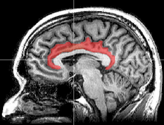
The cingulate cortex. Jarred believes the anterior part of the cortex may be making effort and movement difficult in ME/CFS and long COVID.
It’s a relatively simple task for a healthy human being, but simply getting up to the second floor could be too much for someone with ME/CFS. Your anterior cingulate might decide that you don’t have the resources and slam on the effort brake. I related how early on I would walk up the block and decide to go down another block. I would tell myself O”k, go,” and it felt like there was a force field holding me back.
Jarred said, “The worst thing you can do is try something and run out of energy for your basic processes, so it’s designed to be extremely powerful and to stop you, unless it determines that you have the resources; it will not let you do that thing. It’s like you said, like a force field.”
It’s not a psychological thing, he said, it’s a conserved mechanism in mammals that goes way back. Younger didn’t know what was causing energy levels to plunge in the cingulate but pockets of inflammation could be the cause.
There are parts of the brain where localized inflammation would go almost unnoticed but not so with the anterior mid-cingulate. He did note that a systemic, chronic problem would require cutting down on our energy expenditure, and that low ATP and inflammation might be how it manifests.
We didn’t discuss this in the interview, but studies that have used endotoxins or vaccines to trigger immune activation in the brain indicate that the anterior cingulate cortex is affected. It’s one of a group of brain regions (basal ganglia/ventral striatum, insula, amygdala, hypothalamus) that seem particularly driven by cytokine induced inflammation.
As such, low energy production in the ACC could make it the locus of the “effort” factor in “sickness behavior,” wherein the brain makes everything effortful in order to keep us in bed, conserving our energy, and not spreading an infection when we have a cold.
Note that the anterior mid-cingulate – the effort part of the cingulate cortex – influences large parts of the brain – most of which have been found affected in these diseases. One of the more interesting parts, given recent ME/CFS and long COVID studies, is those that involve movement. It turns out the anterior cingulate cortex affects five brain areas involved in generating movement: the supplementary motor area, premotor cortex, primary motor cortex, basal ganglia, and cerebellar-motor networks (!).
Problems in the ACC, then, could be affecting our ability to move. It’s no wonder that Jarred Younger believes it may be a kind of brain hub for ME/CFS and long COVID dysfunction.
The basal ganglia connection is fascinating because they are the “reward/motivation” areas of the brain and, again, play a role in “effort allocation”. Without a sufficient “reward,” the brain will not gear up to take on a task.
It’s possible that, when it comes to effort, people with ME/CFS and long COVID may be getting hit every which way: low energy levels in the anterior mid-cingulate make things seem very effortful and may also block motor cortex activity (muscle recruitment). Meanwhile, a battered basal ganglia clamps down on a crucial element of activity: reward/motivation.
It makes sense that so many brain regions may be involved: it must take a lot, after all, to produce one of the most functionally disabling conditions known to man.
Fibromyalgia, the ACC, and the Misery Index
When I asked about fibromyalgia, Jarred reported that a more frontal part of the anterior cingulate cortex is abnormally activated. This part of the cingulate cortex determines how painful a pain sensation is. It adds in the emotional or suffering aspect of pain. If pain is making you particularly miserable, this part of the brain is likely responsible.

The part of the ACC affected in fibromyalgia amps up the misery component of pain.
Jarred noted that the ACC plays an important role in helping us heal from an injury by preventing us from moving the affected limb, but in fibromyalgia it restricts everything. Once again, we have a part of the brain whose role it is to restrict movement and activity, and once again, Jarred suspected neuroinflammation; i.e., microglial activation was responsible.
The Neuroinflammation Man
It’s the same process: inflammation consumes tons of resources, including ATP, lowering the brain’s energy levels. So what about neuroinflammation, I asked Jarred? What is going on with that?
His heat-mapping and metabolite studies have identified neuroinflammation, and PET studies have as well. The elevated lactate levels he’s found indicate that something has gone wrong with aerobic energy production in the brain, causing it to switch to an alternative energy source.
Jarred, though, is dealing with a similar resources issue to the one he had last time we talked: not enough graduate students to write up the papers. This does not seem to be an uncommon issue. Ron Davis has talked about how difficult it is to keep good post-docs around. They get hired by industry or have the opportunity to get a tenure track at another university or whatever. In Jarred’s case, his graduate students have completed their degrees and moved on.
Jarred said he has six papers that clearly show brain inflammation. It’ll be good to get those done because, for all the talk about neuroinflammation, we still lack the large studies that validate it, and his papers should do that. The neuroinflammation news was encouraging (it’s there!) and frustrating (help needed to write it up) at the same time, but for real frustration, the brain invasion study takes the cake.
At the Last Minute the Brain Invasion Study Goes “Poof”!
“That was the most, without a doubt, the most disappointing event in my academic career” Jarred Younger
What an exciting project this was. In fact, of all the projects underway, this may have been the most exciting. Why? Because it could have revolutionized the field.
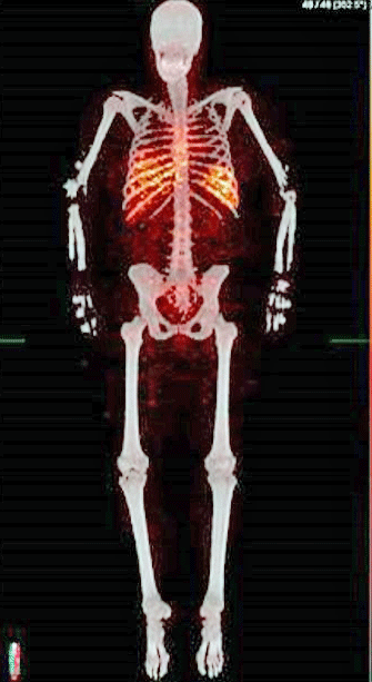
The tests had been done and Jarred was ready to start assessing ME/CFS patients when the radiochemist/radiologists pulled out….
About ten years ago, ME Research UK (MERUK), to its everlasting credit, funded a high-risk/high-gain study to determine whether immune cells from the body were invading the brain in ME/CFS. Incontrovertibly, determining this would slam the door shut on whether ME/CFS was a real and serious disorder. (Given their funding levels, the NIH clearly doesn’t believe this). It would undoubtedly have sparked a lot of interest. Jarred said he’d been working on this new technique for about ten years.
The thing is that the brain has its own unique set of immune cells, and it doesn’t play well at all with the body’s immune cells. Unless the brain has no other options, it will always keep the body’s immune cells out. If it lets them, or if they just find a way in on their own, there’s going to be damage.
The study was to use a radiotracer to determine whether T-cells were entering the brain. It’s relatively easy to assess T-cells in the body because they spread quickly, but they can take about 3 days to reach the brain. Until the radiochemists he was working with developed a way to track them for longer periods, there was no way to watch them long enough to see they’re entering the brain.
The back-and-forth with the FDA over the use of a new technique to assess T-cells in the brain proved enormously complicated. At one point, they had to start over because the FDA disallowed a reagent they’d been using.
Last summer, he reported that initial tests on healthy controls indicated the new technique was successful and he was ready to begin testing people with ME/CFS. At some point, the radiochemist and radiologist suddenly departed for Canada, and the project collapsed.
Unless he can find another radiochemist, the project is over. (He mentioned Michelle James at Stanford (???). One of the heroes of the story has been MERUK, which funded an extraordinary project in the first place and has shown remarkable patience.
A similar thing happened with the nanoneedle in ME/CFS when the inventor left for another university, which offered him a tenure-track position but did not allow him to continue his nanoneedle work.
The Centrality of Fatigue
Speaking of neuroinflammation or the lack of it. I asked Jarred about his neuroinflammatory study, which found widespread neuroinflammation in rheumatoid arthritis patients with fatigue, but only localized inflammation in those without fatigue.
RA provides an interesting test case of the widespread neuroinflammation/ME/CFS/FM/Long COVID hypothesis because a subset of people with RA (or any chronic pain condition) come down with fatigue, pain, sleep, and cognitive problems that look very much like ME/CFS/FM. (A recent study found that over 16 years, 15% of female ME/CFS patients also developed RA.)
Younger believes, and his study showed, that in these patients, the dramatic inflammation they experienced in their joints had spread to their brain.
While RA-only patients now have a wide swath of drugs available to them, none of those drugs help with the fatigue, widespread pain, etc. that the “RA+” patients have. This is why Daniel Clauw, a prominent rheumatologist, chronic pain doctor, and researcher, doesn’t believe people with ME/CFS/FM have an autoimmune disease.
Younger believes the classic autoimmune drugs don’t help dispel the fatigue, pain, etc. in RA+ patients, because these drugs can’t make it past the blood-brain barrier. The RA+ example suggests that effective treatment of these conditions may require drugs that can pass the blood-brain barrier.
(As always, there are exceptions. We know that some people can respond well to peripheral-acting drugs. It’s possible that knocking down inflammation in the body (and possibly tightening the blood-brain barrier (BBB) at the same time?) could reduce neuroinflammation in the brain. Rinvoq, for instance, does not cross the blood-brain barrier but returned a very ill person with ME/CFS to health. IVIG and Mestinon unquestionably help some people, but neither crosses the BBB.)
Still, a drug that crosses the BBB may have the best chance of succeeding. It’s not always clear which drugs do or do not cross the BBB, but low-dose naltrexone (LDN), baricitinib, GLP-1 agonists, and maraviroc all appear to cross it to at least some extent.
Younger’s Big Search
“That’s my number one issue, how do you get… promising agents…through the blood-brain barrier?
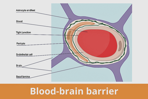
Jarred Younger’s big search: find neuroinflammation reducing drugs that can make it past the blood brain barrier.
Jarred noted, “If you have central involvement, it’s going to be much more complicated, and you have to have different medications to handle that, and that’s what I work on. There aren’t a lot of centrally active (brain) anti-inflammatories…That’s my number one issue, how do you get stuff… we’ve got tons of promising agents, but how do you get them effectively through the blood-brain barrier? And that’s why I’m looking at a lot of different interventions”
His focus is on drugs that can tame the microglia. Every day, he sees possibilities crop up on PubMed that do exactly what he wants them to. They simply need to be tested. He mentioned two recent possibilities: a secretion called temporin from a frog and a peptide (sedanolide) derived from celery.
It’s an exciting time, given how much interest there is in neuroinflammatory treatments, and a frustrating one, given how poor funding remains for diseases like ME/CFS and FM in the U.S.
Neuromodulatory Approaches
That brought up neuromodulatory tools that use ultrasound or magnets to alter the brains functioning. Repetitive transcranial magnetic stimulation (rTMS) appears to have some positive effects, but Jarred pointed out that because it can only reach a couple of centimeters into the brain, it can’t come close to affecting what may be the hub organ in the brain – the anterior cingulate cortex.
A new technique called tFUS ultrasound is another story. It’s the only neuromodulatory approach that can reach into the deeper parts of the brain. It’s a powerful tool, though, and that can cut both ways. Younger is excited about its potential, but given the “Wild West” nature of the tFUS field right now, he’s being cautious and hasn’t made a choice yet.
The Big NIH-Funded LDN Trial
We will finally learn what LDN is doing in the brain.
Younger just got a rare NIH grant to do a major clinical LDN trial. LDN trials have suddenly become a kind of hot topic, with several underway in ME/CFS and long COVID.
Younger’s, though, is different. The best LDN dose has always been a question, and he’ll assess multiple doses in the first, lengthy preliminary phase of the trial to determine it. Because it will mostly be a remote trial, people who are bedbound will be able to participate.
He’s also going to use extensive brain scans – and that means assessing ATP production, doing thermal mapping and assessing metabolites – in a subset of patients to try and finally get clear on what LDN is doing when it works. (The brain scans by themselves will be very informative.)
Jarred pointed out that determining why LDN works when it does is important not just for understanding LDN better but also for understanding what’s happening in ME/CFS. If, as he expects, LDN is tamping down inflammation, this study will indicate that we need to prioritize therapies that fight inflammation in the brain.
Dextro-Naltrexone
The big NIH LDN trial and the dextro-naltrexone saga show that NIH can support clinical trials but highlights its inherently conservative nature. It’s perhaps no surprise that the NIH would begin supporting LDN trials for these diseases after other groups began doing the same.
Younger tried to get the NIH to fund an exploratory trial of a potentially much more potent form of naltrexone, called dextro-naltrexone, but it was, not surprisingly, a bridge too far for the NIH.
Dextro-naltrexone’s superpower is its potential for higher dosing and therefore more effectiveness. LDN’s dosing is rate-limited because at a certain point it starts to knock out the endogenous opioid system – nothing that anyone with a chronic pain condition would ever want to contemplate. Among other things, that can produce “deep unhappiness”.
Jarred believes the side effects that start to kick in for some people are caused by this process. Because dextro-naltrexone doesn’t inhibit the endogenous opioid system, it’s possible that much higher doses – and that potentially means – a greater reduction in neuroinflammation could result.
Stating, “I’ve been talking about this forever, you know?”, Jarred noted that he’s been talking up dextro-naltrexone for 15 years. Despite its potential, it’s never been trialed in humans. In fact, if I have it right, it didn’t even physically exist; i.e., no one was producing it.
About 8 months ago, Jarred took the bull by the horns and said: “If no one else is going to do this, I will find a way.” He put out a video, got funding from “lots of private donors”, and then got an Australian company to produce enough to do preliminary testing. He expects to have it in a couple of months.
The State of Things
“The sheer amount of new stuff(coming out) ..is so exciting”
When I mentioned that things seem to be moving forward on a technological level, in particular, he agreed and referenced all the new players in the ME/CFS field. Ten years ago he could look at the title of a paper and knew who had written it because the field was so small.
Now he regularly sees papers on ME/CFS popping out by people he’s never heard of. He said a lot of new expertise and ideas are flooding into these fields, and that “the sheer amount of new stuff(coming out) while it’s daunting, and… (and is) almost impossible to stay on top of, is so exciting.
Still, interest in ME/CFS and post-infectious diseases has blossomed in Europe to the extent that he expected that, if things keep going the way they are, Europe would end up more productive than the US.
Lastly, the Sex Hormones
Then we moved back in time to a 2018 paper where he found that progesterone and testosterone play a protective effect in fibromyalgia. Nancy Klimas’s research suggests something similar, and some doctors are treating their FM patients with testosterone.
The gender issue; i.e., the female predominance issue, suggests that the sex hormones probably are involved, yet research into the sex hormones has been paltry.
Jarred noted that the sex hormones also impact immune functioning and that not only are women more vulnerable to immune events like infections at particular times during their menstrual cycle, but this also includes the primary immune cells of the brain – the microglia.

Did some women get an infection at just the wrong time? (Image by Sydney Fought, CC BY-SA 4.0 via Wikimedia Commons)
One hypothesis is that high estradiol levels, which occur about 1/2 way through the menstrual cycle, may chronically sensitize microglia in some people with these diseases.
Younger wondered whether a genetic predisposition to microglial activation could come into play. Someone sneezes into the face of a female with a predisposition to microglial activation at a time in their menstrual cycle when their microglia were already buzzing away, and the rest could be history.
He noted that it’s been clear for decades that lower testosterone is associated with an increased risk of pain disorders in men.
Coming Up
To sum up, Jarred Younger has a lot coming up! He’s got 1/2 dozen papers to write, which will cement the proposition that widespread neuroinflammation is present in ME/CFS. He has psilocybin/LDN/dextronaltrexone trials either underway or in process; he’ll be applying for a grant to use the new technique to assess ATP availability in the brain, and he’s checking out tFUS instruments that can reach deep enough in the brain to get to where he believes the real action is in ME/CFS.
Plus, he’s excited about the new interest in these fields. Yes, it always takes longer than we would want, but as former SolveME head Carole Head said, the arc is upward.