


An inflammatory process (neuroinflammation) almost certainly plays a major role in the development of many neurological diseases. Andrew Lloyd has pointed out that the symptoms in chronic fatigue syndrome (and fibromyalgia) are “brain symptoms”‘; i.e. they are flu-like symptoms produced by the brain when we get ill. That suggests, of course, that neuroinflammatory processes are at work.
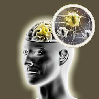
The search for inflammation in ME/CFS and FM may strike gold in the brain
The search for the inflammatory basis of fibromyalgia never really got off first base, but the search for the inflammatory component that many felt must be in chronic fatigue syndrome (ME/CFS), has never died. The tie-in between the infectious onset that many people experience and the flu-like symptoms that have dogged many for decades has simply been too strong to give up on the idea that the immune system plays a huge role in ME/CFS.
Now, technology is giving researchers a deeper look at inflammation. The Lipkin/Hornig cytokine study was comprehensive enough and large enough to uncover a strong inflammatory component occurring earlier in the disease. The Stanford ME/CFS Initiative will soon be publishing gene expression results that strongly implicate inflammation. In Jarred Younger’s intense Good Day/Bad Day study, leptin – an immune factor able to sensitize the microglia in the brain – jumped to the front of the line.
The ability to finally uncover strong evidence of inflammation in the body is gratifying, but it may pale next to what researchers will find when they get into the most heavily guarded area of the body – the brain.

The ability to “see’ the immune activity in the brain is coming and it’s coming fast. The barriers to accessing the brain are falling.
Neuroinflammation in Chronic Fatigue Syndrome and Fibromyalgia
Thus far one neuroinflammation ME/CFS study has been done. The small Nakatomi study found evidence of neuroinflammation in the cingulate cortex, hippocampus, amygdala, thalamus, midbrain, and pons regions of the brain. The fact that the degree of the widespread neuroinflammation was strongly correlated with the severity of the fatigue, pain, cognitive problems and depression found in the ME/CFS group was encouraging.
The small Japanese study found evidence of widespread inflammation in the brains of ME/CFS patients.
The areas seemed to fit what we know about ME/CFS. One inflamed area regulates the reticular activating system which determines how awake or aroused one is. Several others are involved in pain sensitivity, cognitive impairment and mood.
Nakatomi proposed two possible causes for the neuroinflammation.
- (1) damage to one of part of brain is causing other parts of the brain to overwork leading to N-methyl-D-aspartate receptor overload and the production of factors that produce inflammation
- (2) the immune response to an initial infection remains active
Other causes are surely possible. Younger has proposed that infection, stress, diet, trauma, etc. plus other factors have caused the microglial cells/astrocytes to become hyperactive to stimulus.
Assessing Inflammation in the Brain
The sophisticated brain imaging techniques that have been developed can do many things. Until recently, though, they’ve never been aimed at assessing the level of immune activity in the brain.
The recognition that the microglia and the inflammation they cause may play a major role in many neurological disorders changed that. We now know that the microglial cells produce dozens of immune factors and other mediators that can produce pain, fatigue, sleeplessness, mood changes and other symptoms.
That understanding prompted the development – years in the making now – of scanning techniques that can assess microglial activity/inflammation in the brain. Those scanning techniques are coming to fruition now. For neurologists 2015 may be the year they finally start getting a good, close look at the levels of inflammation in the brain.
The Race Is On
This is a competitive area of research. Whoever develops the best scanning technique is going to not only get big chops in the research community, but will, if they’re allied with a company, see the cash rolling in. Being able to better understand the biggies, or track treatment efficacy in the biggies, like Multiple Sclerosis (MS), Alzheimer’s, Parkinson’s, etc. is a big deal. That means the race is on. Dozens of competitors are involved.
The remarkable variety of techniques being developed is all to the better. Neuroinflammation is a complex topic that effects numerous aspects of the brain. It appears that someone, somewhere is attempting to image virtually every aspect of the brain – from immune cells to oxidative stress to neuron health – involved in neuroinflammation.
The rich broth of techniques that will result should allow researchers to better analyze and understand neuroinflammation. That’s very good news for anyone with a neurological disorder. This is what it looks like when the research community gets excited about a topic.
A Dec. 2014 review article (possibly already outdated) looked at and assessed the various technologies emerging. Sit back and check out the rather wild world of neuroinflammation imaging.
Imaging Neuroinflammation – from Bench to Bedside Benjamin Pulli and John W Chen* Center for Systems Biology and Department of Radiology, Massachusetts General Hospital and Harvard Medical School, 185 Cambridge Street, Boston, MA 02114, USA. J Clin Cell Immunol. 2014 ; 5: . doi:10.4172/2155-9899.1000226
Imaging Inflammation in the Brain
Attempts have been made to adapt major scanning techniques: MRI, positron emission tomography (PET), single photon emission computed tomography (SPECT) and optical imaging to assess inflammation in the brain.
Targeting Immune Cells and Immune Activity
Adhesion molecules such as VCAM-1 and CD62 open the doors, so to speak, that allow immune cells to get into inflamed area. Studies suggest that magnetic resonance (MR) techniques that track adhesion molecule activity are subtle enough to detect small levels of neuroinflammation. Another technique tracks the levels of myeloperoxidase – a proinflammatory enzyme secreted by neutrophils and monocytes in inflamed tissues. Another approach is looking at COX-1 and 2 enzyme levels. These are enzymes that convert arachidonic acid to prostaglandins – a major player in inflammation.
Markers of Neuronal Damage

Some techniques look for markers of neuronal damage
Matrix metalloproteinases (MMP’s) are substances that damage tissues in response to microglial and other immune activity. They’re associated with excitotoxicity, nerve damage and gaps in the blood-brain barrier. At least four different markers of MMP activity are being assessed.
Translocator protein (TSPO)
Translocator Proteins have been the most extensively studied neuroinflammation imaging technique. This technique targets a receptor that appears on activated microglial and astrocyte cells. Watanable used the PET ligand 11C-PK11195 to find evidence of neuroinflammation in ME/CFS.
In one small study a TSPO scan picked up increased microglial activity 17 years after a group of patients had experienced a traumatic brain injury. In multiple sclerosis this technique picked up evidence of increased microglial activation in areas of the brain that appeared normal otherwise. It’s been used to assess neuroinflammation in a variety of animal models and in humans.
The 11C-PK11195 ligand that was first developed, however, had several drawbacks. It’s not particularly specific (precise) and has a poor signal-to-noise ratio and its very short half-life (20 minutes) restricts its use.
Second generation TSPO markers are more precise. One called 11C-DAA1106 appears to be five times more specific than 11C-PK11195. Two others (11C-CLINME, PBR111) appear to have higher binding potential and at the time the article was written were undergoing tests in humans. At least two other ligands for the TSPO are being tested.
Others
Other markers of neuroinflammation that have been developed include scanning for cannabinoid receptor, radioactively labeling white blood cells, tagging cells with iron oxide, tracking cells that take in iron oxide, fluorine 19F-MR imaging and imaging specific immune cells. Some of thse techniques have received multiple studies and are viable alternatives.
Tracking Demyelination

Others look for burnt out and dead neurons…
MRI’s have been used to identify areas where inflammation has broken down the myelin covering of the nerves. This technique, however, has been hampered by low specificity. Probes are being developed now though, that are much more precise. At least eight different compounds have been developed for use with MR, PET or FRI scans.
Tracking Neuronal Death
The most severe consequences of neuroinflammation are lots of dead neurons. Inevitable disease progression and/or irreversible damage appears to be associated with the death of neurons in MS and stroke. Techniques to scan seven different factors associated with neuronal death (receptors, proteins) are being tested.
The Future
It’s not clear what the future will bring but it will certainly bring many more ways of assessing and ultimately understanding the immune status of the brain. Whether they are directly tracking micoglial and astrocyte activity, neuron damage or death, oxidative stress, or accumulations of immune cells or cytokines, or other factors, methods are being developed that will tell us much.
Already researchers recognize microglial activation, and more subtle forms of damage can take place in parts of the brain far removed from the center of damage. (A recent autopsy study of traumatic brain injury patients found that virtually their entire brains had been affected.) Expect many more revelations as this technology matures.
In the last month studies using new PET imaging techniques examining people with epilepsy, Alzheimer’s, schizophrenia and dementia have been published and the efficacy of three new inflammation tracers have been reported on.
Given the strong sense among many that ME/CFS and FM are neuroinflammatory disorders the development of this new technology is good news indeed. At least three neuroinflammation studies (Nakatomi followup, Andrew Lloyd, Jarred Younger) are underway in ME/CFS.


Your entire viral infection history from a single drop of blood
June 8, 2015
Systematic viral epitope scanning (VirScan). This method allows comprehensive analysis of antiviral antibodies in human sera. VirScan combines DNA microarray synthesis and bacteriophage display to create a uniform, synthetic representation of peptide epitopes comprising the human virome. Immunoprecipitation and high-throughput DNA sequencing reveal the peptides recognized by antibodies in the sample. The color of each cell in the heatmap depicts the relative number of antigenic epitopes detected for a virus (rows) in each sample (columns). (credit: George J. Xu et al./Science).
New technology called called VirScan developed by Howard Hughes Medical Institute (HHMI) researchers makes it possible to test for current and past infections with any known human virus by analyzing a single drop of a person’s blood.
With VirScan, scientists can run a single test to determine which viruses have infected an individual, rather than limiting their analysis to particular viruses. That unbiased approach could uncover unexpected factors affecting individual patients’ health, and also expands opportunities to analyze and compare viral infections in large populations.
The comprehensive analysis can be performed for about $25 per blood sample, but the test is currently being used only as a research tool and is not yet commercially available.
Stephen J. Elledge, an HHMI investigator at Brigham and Women’s Hospital, led the development of VirScan. He and his colleagues have already used VirScan to screen the blood of 569 people in the United States, South Africa, Thailand, and Peru. The scientists described the new technology and reported their findings in the June 5, 2015, issue of the journal Science.
Virus antibodies: clues that last decades
Bacteriophage (credit: Wikimedia Commons)
VirScan works by screening the blood for antibodies against any of the 206 species of viruses known to infect humans*. The immune system ramps up production of pathogen-specific antibodies when it encounters a virus for the first time, and it can continue to produce those antibodies for years or decades after it clears an infection.
That means VirScan not only identifies viral infections that the immune system is actively fighting, but also provides a history of an individual’s past infections.
To develop the new test, Elledge and his colleagues synthesized more than 93,000 short pieces of DNA encoding different segments of viral proteins. They introduced those pieces of DNA into bacteria-infecting viruses called bacteriophage.
Each bacteriophage manufactured one of the protein segments — known as a peptide — and displayed the peptide on its surface. As a group, the bacteriophage displayed all of the protein sequences found in the more than 1,000 known strains of human viruses.
To test the method, the team used it to analyze blood samples from patients known to be infected with particular viruses, including HIV and hepatitis C. “It turns out that it works really well,” Elledge says. “We were in the sensitivity range of 95 to 100 percent for those, and the specificity was good — we didn’t falsely identify people who were negative. That gave us confidence that we could detect other viruses, and when we did see them we would know they were real.”
Harvard Medical School | Viral History in a Drop of Blood
International study
Elledge and his colleagues used VirScan to analyze the antibodies in 569 people from four countries, examining about 100 million potential antibody/epitope interactions. They found that on average, each person had antibodies to ten different species of viruses. As expected, antibodies against certain viruses were common among adults but not in children, suggesting that children had not yet been exposed to those viruses. Individuals residing South Africa, Peru, and Thailand, tended to have antibodies against more viruses than people in the United States. The researchers also found that people infected with HIV had antibodies against many more viruses than did people without HIV.
Elledge says the team was surprised to find that antibody responses against specific viruses were surprisingly similar between individuals, with different people’s antibodies recognizing identical amino acids in the viral peptides. “In this paper alone we identified more antibody/peptide interactions to viral proteins than had been identified in the previous history of all viral exploration,” he says. The surprising reproducibility of those interactions allowed the team to refine their analysis and improve the sensitivity of VirScan, and Elledge says the method will continue to improve as his team analyzes more samples. Their findings on viral epitopes may also have important implications for vaccine design.
Elledge says the approach his team has developed is not limited to antiviral antibodies. His own lab is also using it to look for antibodies that attack a body’s own tissue in certain autoimmune diseases that are associated with cancer. A similar approach could also be used to screen for antibodies against other types of pathogens.
* Antibodies in the blood find their viral targets by recognizing unique features known as epitopes that are embedded in proteins on the virus surface. To perform the VirScan analysis, all of the peptide-displaying bacteriophage are allowed to mingle with a blood sample. Antiviral antibodies in the blood find and bind to their target epitopes within the displayed peptides. The scientists then retrieve the antibodies and wash away everything except for the few bacteriophage that cling to them. By sequencing the DNA of those bacteriophage, they can identify which viral protein pieces were grabbed onto by antibodies in the blood sample. That tells the scientists which viruses a person’s immune system has previously encountered, either through infection or through vaccination. Elledge estimates it would take about 2-3 days to process 100 samples, assuming sequencing is working optimally. He is optimistic the speed of the assay will increase with further development.
Abstract of Comprehensive serological profiling of human populations using a synthetic human virome
The human virome plays important roles in health and immunity. However, current methods for detecting viral infections and antiviral responses have limited throughput and coverage. Here, we present VirScan, a high-throughput method to comprehensively analyze antiviral antibodies using immunoprecipitation and massively parallel DNA sequencing of a bacteriophage library displaying proteome-wide peptides from all human viruses. We assayed over 108 antibody-peptide interactions in 569 humans across four continents, nearly doubling the number of previously established viral epitopes. We detected antibodies to an average of 10 viral species per person and 84 species in at least two individuals. Although rates of specific virus exposure were heterogeneous across populations, antibody responses targeted strongly conserved “public epitopes” for each virus, suggesting that they may elicit highly similar antibodies. VirScan is a powerful approach for studying interactions between the virome and the immune system.
references:
George J. Xu, Tomasz Kula, Qikai Xu, Mamie Z. Li, Suzanne D. Vernon, Thumbi Ndung’u, Kiat Ruxrungtham, Jorge Sanchez, Christian Brander, Raymond T. Chung, Kevin C. O’Connor, Bruce Walker, H. Benjamin Larman, Stephen J. Elledge. Comprehensive serological profiling of human populations using a synthetic human virome. Science 2015 DOI: 10.1126/science.aaa0698
related:
Your Viral Infection History from a Single Drop of Blood
Topics: Biotech
share | e-mail print
Comments (2)
June 9, 2015
by tim the realist
I think this analysis should be expanded and performed across animal populations also. Especially those animals who are immunologically resistant to human diseases. Perhaps this technique could be modified and applied to blood samples from humans who have survived or are more resistant to various types of cancers? Maybe new drug targets culd be found to enhance the body’s immune response, or at least measure the response to existing pharmocological treatments?
May also be usefull to research organ rejections?
Log in to Reply
June 9, 2015
by intelligent design
This is the direction that future medicine will be taking. Local community based testing in this fashion will enable a better and more accurate treatment process.
Log in to Reply
Please log in to post a comment.
Exciting stuff..
Would Neurofeedback be an appropriate treatment to consider for chronic pain, fatigue caused by fibromyalgia, chronic fatigue, small fiber neuropathy, Sjogren’s?
Cort,
Thank you again for a valuable research summary and report. I hope we can all appreciate that spinal cord injury can cause brain inflammation. Most everyone thinks of spinal cord injury as related to some major spine trauma. However, spinal cord injury can result from a number of conditions: tethered cord, syringomyelia, Lyme disease, spinal arteriovenous malformations, fungal infections, CNS vasculitis, and many more. Many of these can not be imaged on MRI.
Great articles:
1. Wong SH et al. Myelopathy but normal MRI: where next? Practical neurology 2008;8:90-102.
2. Wu J et al. Spinal cord injury causes brain inflammation associated with cognitive and affective changes: role of cell cycle pathways, 13 August 2014; 34(33): 10989-11006.
Just within the past 4 or 5 years neurosurgeon Robert Bray identified a new spinal cord issue, Thoracic Epidural arteriovenous Malformation. This is much more common in women.
Dr. Bray is ‘ famous’ for having diagnosed and treated Olympian Lolo Jones for tethered cord. Thank you Dr. Bray!
Thanks Merida, I look forward to learning more about this. 🙂
Cort – good article
As you know I’ve been banging on about microglia for years.
The answer, I am certain, will be found in the brain.
Here’s hoping the precise answer – and solutions – can be found soon.
Wasting less time and money on viruses would be a start.
🙂 That last sentence just made me laugh 🙂
Well – we’ll see what Dr. Montoya has to say about the viruses in a new paper, I guess. I imagine viruses and pathogens are problematic in a subset of patients but perhaps not a large one. We shall see! (Hopefully we will..)
Interesting read as always. Some sickness behaviour can be mediated by peripheral pro inflammatory cytokine responses as well – as anyone with Crohns disease or Ankylosing Spondylitis would attest.
Gibbo,
Could you please elaborate on the relationship between ankylosing spondylitis and cytokines. I’ve been diagnosed with A.S. for years and never knew there was any tie between them other than inflammatory. Any information is appreciated.
Thanks,
Susan S.
Indeed – and some people with ME/CFS – a subset, I would think – have very high cytokine levels in the periphery. Hopefully at some point THAT subset will be revealed and focused on in their own studies.
Cort – another great article!
Could the discovery of the lymphatic vessels in the central nervous system (brain) and this inflammation theory have any connections?
If cfs can be caused by so many different pathogens, Lyme, hepesvirus, EBV, etc., etc., is there anything that researchers think might be common with all subgroups?
I would think neuroinflammation might be the great uniter….whether ME/CFS is caused by pathogens or some other stressor or whatever. ..the end point might be neuroinflammation. We’ll see what the studies show.
KDidhi all, to me, inflammation – no matter where it is – is still just a symptom. The question of what is causing the inflammation will lead to real progress. I don’t the viruses are a waste of time, considering that the illness regularly occurs in outbreaks of apparently contagious infection.
all, to me, inflammation – no matter where it is – is still just a symptom. The question of what is causing the inflammation will lead to real progress. I don’t think the viruses are a waste of time, considering that the illness regularly occurs in outbreaks of apparently contagious infection.
what about Epstein Barr? dr. Lerner believes THAT is my cuplrit and yet they say almost everyone on earth had that virus, NOW WHAT?
Dee,
The EBV situation that you mention – ie everyone has had it- has been the dilemma I have thought about,too. My son had years of neuro problems from this virus at age 5 ( 1986). Well documented case, with elevation of IgG and Early Antigen about 6 weeks after infection. But I developed the entire syndrome after a neck/pelvis injury in 1998.
So, what do we have in common? Several things I am aware of:
1. Scoliosis – this can affect blood flow in the dura- meningeal system and alter the blood-brain barrier ?
2. Neck trauma – his birth was assisted by suction. I observed various issues after his birth.
3. Rh D negative blood – some research with Toxoplasma shows that people with RhD neg blood have more neurological difficulties following infection. Could the same situation exist following EBV infection? At one support group meeting ( about 15 people) 55% were RhD neg. The highest rate of RhD neg in the world is 35% – the Basques.
Wonder if some bio geographical studies would help illuminate the situation?
hi,
only mild scoliosis here but so ill that i’m looking at things that happened to my neck many yrs. ago. no accident as we know it but still i twisted my neck a couple times and one of them after i was having vision loss, everything going black on me and not knowing why.
how are you and your son today?
Dee,
Sorry for delayed reply. My son is 34. It took him many years to recover. He still has some issues – must be careful with stress, some anxiety, some depression, I think. He has a very successful business and is well. Mornings are not his best time.
For me – I just have mild scoliosis, but this may still be important for various reasons. Yes, I lost vision after my initial neck injury – on and off. This is a very important symptom and may indicate an issue with blood flow in the vertebral arteries – vertebral artery syndrome.
This syndrome results from basically 2 things: congenitally small holes ( foraminae) in the sides of the cervical vertebrae where the vertebral arteries thread to the vascular system of the brain, and/or neck vertebrae that are rotated or off set due to scoliosis.
I have had 16 years of work on my neck and structure. Still a big problem. I am still mostly couch bound – with all the typical FM/ CFS symptoms – including elevated inflammatory markers. I really don’t know if my issues are 100% structure/function, or if a virus became a secondary invader in a ‘glitchy’ central nervous system.
Dear Cort,
I have a brain stem reticular activating system specimen of a patient who passed away from CFS/ME. What should I do with it? There are no signs of an inflammatory infiltrate.
Regards
John L Whiting
I still doubt if it is inflammation in the brain (microglia). I think it can be due to low bloodflow in the brain. Microglia wanted to repair the damage (?).
Low blood flows could definitely cause inflammation – not sure how the microglia would be involved – perhaps in response to the oxidative stress produced by the ischemia (???)
Hi,Has anyone tried Protandim by LifeVantage which is suppose to help with oxidative stress and if so did it help symptoms of fibromyalgia?