


Geoff’s Narration
The GIST
Here we are with the blood vessels again! The blood vessel saga in ME/CFS started about 20 years ago and then quickly folded (after one small negative result), and then opened up again with long COVID. Now it’s become a major theme.
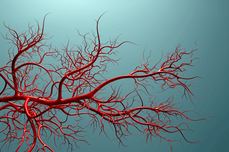
Back to the blood vessels we go!
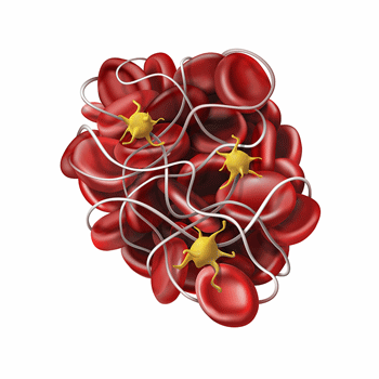
Now we’re looking at microclots, activated platelets, narrowed blood vessels, senescent blood vessels, thickened basement membranes, deformed red blood cells, issues with hemoglobin – the list of possible blood vessel problems just seems to go on and on.

There’s also the evidence that we have reduced brain and microvascular blood flows. Indeed, two of the big questions raised by David Systrom’s invasive exercise studies are why oxygen from the blood is not being extracted by the muscles, why the blood seems to be disappearing, and where the heck it’s going.
While most of the studies remain quite small – a big bugaboo in these fields – there seems to be a lot going on, and new researchers are joining the fray.
Two more blood vessel studies just popped up. One of them, not surprisingly, is from Germany, and the other is from Yale and Johns Hopkins in the U.S. This blog deals with the Yale/Johns Hopkins study.
The Gist
 It’s amazing how the blood vessel issue has grown in long COVID and ME/CFS. The list of possible blood vessel problems (microclots, activated platelets, narrowed blood vessels, senescent blood vessels, thickened basement membranes, deformed red blood cells, issues with hemoglobin) seems to go on and on.
- The “Vascular inflammation in neuropsychiatric long COVID” study started off on a nice note: “LC is a pressing public health issue that patients, providers, and researchers are eager to understand“. I don’t remember seeing such a blatant display of enthusiasm before, and it was good to see.
- This study examined markers of blood vessel inflammation in people with an acute COVID-19 infection, people who’d had long COVID for 1 or 3 years, and recovered COVID-19 patients.
- It found, as suspected, that acute coronavirus infection triggers a cytokine storm that pummels the endothelial cells lining blood vessels. A year later, the storm has died down, but a different kind of immune activation is present.
- The small blood vessels are still under considerable stress and are fighting back with repair mechanisms that are trying to limit the damage.
- It doesn’t entirely seem to be working. The study found clear correlations between markers of blood vessel inflammation and cognitive problems such as verbal learning, fluency, memory, depression, and anxiety.
- In the end, this was not a surprise. The authors noted that small blood vessel disease in the brain is strongly associated with, and predictive of, cognitive issues.
- They proposed that a “microvascular endotheliopathy,” which refers to an injury to the endothelial cells lining the body’s smallest blood vessels, was contributing to the cognitive problems and depression/anxiety.
- They believe their study provides “compelling evidence that… LC mental health symptoms likely have a neuroimmune and neuro-oxidative origin”.
- Three years later, an entirely different situation has developed. All signs of blood vessel inflammation, yet the long COVID persisted.
- The authors proposed that blood vessel healing may have taken place. It’s also possible that the small blood vessel problems may remain; i.e., the extracellular matrix and microvascular damage, and perhaps, most importantly, blood-brain barrier damage (from leaky endothelial cells) may all still be present, but the immune response died down over time.
- It’s also possible that the small blood vessels are in a senescent state that prevents them from operating normally and producing normal blood flows.
- It’s also possible that the brain – faced with blood flow problems – has rewired itself and that it’s now in a neuroinflammatory steady state. It’s perhaps notable that many of the brain regions associated with ME/CFS appear particularly vulnerable to inflammation in the blood vessels.
- More blood vessel studies have recently been published, and this team reported that they’re continuing to study their long-COVID patients – so we should continue to learn about the possibly vital blood vessel connection in long COVID and ME/CFS.
 It’s amazing how the blood vessel issue has grown in long COVID and ME/CFS. The list of possible blood vessel problems (microclots, activated platelets, narrowed blood vessels, senescent blood vessels, thickened basement membranes, deformed red blood cells, issues with hemoglobin) seems to go on and on.
Support Health Rising and Keep the Information Flowing!
Health Rising is not a 501 c (3) non-profit
The Study
The “Vascular inflammation in neuropsychiatric long COVID” study started off on a nice note: “LC is a pressing public health issue that patients, providers, and researchers are eager to understand“. I don’t remember seeing such a blatant display of enthusiasm before, and it was good to see. The study examined markers of blood vessel inflammation and added a nice twist: they assessed whether these markers affected cognition.
Two groups of long-COVID patients were included: 28 patients with COVID-19, 50 long-COVID patients who had been ill for about a year (Yale), and 114 long-COVID patients who had been ill for about 3 years (Johns Hopkins).
First, Though, a Rant about Criteria – Feel Free to Skip Forward

Six years later, still no standard, interim diagnostic criteria for long COVID? Really?
It’s amazing, disturbing, ridiculous – use what adjective you will, that 6 years later, we still do not have standardized criteria for diagnosing long COVID. For some who’ve been immersed in the ME/CFS field for decades, this is a stunning lapse.
The need to develop criteria to determine which patients could participate in an ME/CFS study was immediately recognized as a major problem, and considerable efforts went into addressing it. Over time, the Fukuda, Canadian Consensus, International Consensus Criteria, the IOM SEID Criteria, and others were put forth.
By 1994, the fledgling field came up with a faulty – but standardized – criteria (Fukuda criteria) which, honestly, largely worked. Post 1994, virtually every ME/CFS study used it. By 2003, the still very small ME/CFS field had picked itself up by its bootstraps, and – successfully challenging the Fukuda criteria – came up with a better criteria called the Canadian Consensus Criteria (CCC). By the mid 2000s, I would hazard to guess that most researchers (Bernard Wyller and the CDC excepted :)) were using the CCC.
The point is that the ME/CFS field recognized that developing good criteria was essential. Throughout this saga, it was lucky to have a crack epidemiological researcher in Lenny Jason, who kept pushing the issue.
Six years after long COVID showed up on the scene, though, the much larger and better-funded long-COVID field is still using nebulous and even unstated symptom criteria to assess a very heterogeneous disease.
Avindra Nath, Jonas Bergquist and others recently decried the diagnostic situation in Lyme Disease and other post-infectious illnesses. Jacqueline Becker, a neuropsychologist and coauthor on the paper, said, “If we want clinical trials that actually lead to treatments, we have to get the fundamentals right: we must confirm diagnoses, choose the right comparison groups, and treat patient populations as distinct rather than lumping everyone together. Patients deserve that rigor.”
They sure do. Getting the diagnostic rigor down is not sexy, but it sure is important. Ultimately, uncovering a biologically based diagnostic criteria is going to take time, but there’s no excuse for not having an international group of long-COVID experts meet to create interim criteria that researchers and doctors can use.
This blood vessel paper, for instance, said that the 50 individuals with neuropsychiatric Long COVID (LC) in the Yale part of the study were “carefully selected.”
They had to have at least one (one?) new, self-reported neuropsychiatric symptom (cognitive dysfunction, headache, etc.) for greater than 3 months, plus a confirmed COVID-19 test (nice touch), and had to take a “formal survey”. How little attention is given to this subject is evident in the fact that the name of the survey, if it has a name, is not even stated.
I’ll bet the researchers did, in fact, pluck out a good neuropsychiatric set of long-COVID patients, but the fact that the long-COVID field, after six years, still hasn’t taken the time and trouble (an international consortium could do it) to create a standardized definition of “neuropsychiatric long COVID”, or even long COVID in general, which guides patient selection across studies, just seems baffling.
At the very least, at the symptom level, let’s be able to compare apples to apples. While one study may have more Fuji apples, another more Granny Smith apples, and another more Honeycrisp apples, at least symptomatically, we know they’re all apples.
One would have assumed that with its huge sample sets, the RECOVER project would have been on this like bees on honey. In fact, it is making some progress, but, boy is it taking a long time.
The closest we’ve come is a 2023 RECOVER study, which listed the most common long-COVID symptoms. While the paper did highlight 12 “signature” symptoms (PEM, fatigue, brain fog, dizziness, GI symptoms, palpitations, sexual dysfunction, smell/taste loss, thirst, chronic cough, chest pain, abnormal movements), it did not provide a simple criterion clinicians could use.
Neither did a 2024 Update. It modified the signature symptoms a bit (post‑exertional malaise, fatigue, brain fog, dizziness, palpitations, change in smell or taste, thirst, chronic cough, chest pain, shortness of breath, and sleep apnea). Using its weighted symptom scores, it found that a remarkable 20% of COVID-19 patients met their criteria for long COVID (!).
The paper did produce 5 possible symptom clusters. Remarkably, 4 of the 5 clusters clearly denote ME/CFS-like subsets characterized by post-exertional malaise and fatigue.
The good news is that RECOVER’s next steps are to accumulate more data, including incorporating symptom trajectories (getting better, worse, or staying the same over time) and associating symptom types with immunologic signatures, viral persistence, autoantibodies, and autonomic and cardiopulmonary measures.
(Unfortunately, the observational side of the RECOVER project, which is driving the symptom typing, doesn’t appear to be gathering much, if any, multi-omic information).
In the end, it’s clear we will get better criteria, but, again, why not produce interim criteria for long COVID as a whole, and for the different subsets, to help guide researchers and clinicians?
This long rant was occasioned by the fact that this study involved two different cohorts, which produced very different results. That may well have been due to different durations, but since we don’t have good criteria, it could also have been due to different patient sets. Rant over! Back to a very interesting study.
Study Results
The results appeared to support a hypothesis that emerged in a previous study, which suggested that a cytokine storm during the initial infection hits the blood vessels really hard. After the infection was resolved (at least in the blood vessels), a different kind of inflammatory response set in. After some time, that died down as well, but the blood vessels may or may not have been left with long-term damage.
The Initial Big Blood Vessel Hit
Blood clots and inflammation gave the blood vessels a big hit during the initial infection.
The dramatically increased levels of acute-phase proteins AGP (p = 0.0005), CRP (p < 0.0001), and haptoglobin (p = 0.0003) in the COVID-19 group vs the long-COVID group indicated that a classic systemwide inflammatory response had occurred, which may have damaged the endothelial lining of blood vessels. This indicated that the endothelial cells lining the blood vessels have become activated and that clotting is occurring.
Struggling Blood Vessels A Year Later
A year later, things had changed. One group had recovered, and another had not. That big pro-inflammatory cytokine spike had largely disappeared in both groups. Now, the long-COVID group exhibited increases in different immune factors. They included elevated SAP (p=.0005), fetuin (p = 0.001), sp-selectin (p = 0.0001), sl-selectin (p=.03), and ADAMTS13 (p < 0.0001).
(Notice the huge probability factors (p = 0.0005, p < 0.0001) often found. They indicate there’s an extremely low probability (1 in 10,000 or 5,000) that the findings were due to random chance; i.e., these are really solid results.
Driving the Dysfunction?

Could activated platelets be driving the blood vessel inflammation?
The high p-value regarding the sp-selectin (p=0001) provides a very clear indication that while platelet/endothelial activation may have died down, it’s still present in long COVID. Several studies suggest that low-grade inflammation is keeping the platelets in long-COVID patients in an activated state.
Those activated platelets could be producing a lot of mischief. They could be sticking to the endothelium and releasing a host of pro-inflammatory factors that, in turn, activate the endothelial cells, increase blood vessel permeability (causing the blood loss/preload failure Systrom sees?), and cause microvascular damage.
One study suggested that an as-yet unidentified factor in the plasma is hyperactivating the platelets in long COVID. While microclots, NETs (see the last blog), autoantibodies, and damaged bits of the extracellular matrix weren’t assessed in this study, they could be activating the platelets.
A Short Treatment Interlude
Because platelet activation appears to be present and may be driving inflammation and vascular remodeling, platelet inhibitors seem a possibility.
Pretorius and colleagues found benefit from triple antiplatelet and anticoagulant therapy in long COVID, but dual or triple antiplatelet/anticoagulant therapy (except for aspirin) appears to be pretty serious business, and the field will likely need compelling evidence before it embarks on these trials.
On the supplement and diet side, omega‑3 fatty acids (fish oil, EPA/DHA), polyphenols/flavonoids (resveratrol, quercetin, grape seed extract, green tea catechins), garlic, ginger, ginkgo, turmeric, cinnamon, and Vitamin E could have modest effects.
The increased sL‑selectin (soluble L‑selectin) suggests that immune cells called leukocytes are being drawn to damage in the endothelium. Instead of producing a cytokine storm, however, they’re creating a kind of smoldering infection.
Fighting Back
A host of factors suggest that a year after the infection, the blood vessels are fighting back and trying to repair themselves.
Fetuin-A – The increased fetuin-A levels indicate the blood vessels are under stress and are attempting to stop calcium from being dumped into them. Calcium in our blood vessels is a no-no because it stiffens them, which disrupts the smooth flow of blood through them. That loss of elasticity and fine-tuning can, by itself, result in low oxygen conditions and reduced blood flow to the brain, muscles, and organs.
α‑2 macroglobulin – a protease‑inhibitor and extracellular matrix remodeler, α‑2 macroglobulin was the most elevated endothelial factor found in the long-COVID patients. A-2 macroglobulin tries to limit extracellular matrix damage – which was featured in a recent blog.
The elevation of α-2 macroglobulin suggests, as some studies have found, that extra collagen deposition may have occurred in the blood vessel lining. German studies have recently found increased collagen deposition in the basement membranes associated with ME/CFS patients’ endothelial cells. Increased collagen deposition may also be inhibiting blood from getting to the muscle tissues.
ADAMS13 – Because ADAMS13 supports microvascular blood vessel production and repair, increased ADAMS13 levels indicate the microvasculature is under stress.
An inflammatory environment, then, appears to be present in patients with long COVID. It’s much lower than in COVID-19 patients but much higher than in healthy controls. The demonstrates the stress the blood vessels are under a year after the acute infection has triggered several efforts to repair and maintain microvascular flows.
Importantly, once again, we have findings that jive with each other. If high SP levels are present, we might expect elevated fetuin, selectin, and ADAMTS13 levels. At times in ME/CFS, we’ve seen disjointed findings that were hard to know what to make of (and which were discarded), but there doesn’t appear to be any mystery here. The blood vessels have taken a hit.
Brain Fog, Depression, and Anxiety

The study suggested that inflammation in the blood was contributing to the brain fog, depression and anxiety found in long COVID.
The brain fog (reduced verbal learning, memory, fluency) and depression/anxiety that were associated with markers of inflammation in the blood vessels (sP-selectin, fetuin-A, AGP (α1‑acid glycoprotein) may have been the most important finding. Note that this study assessed inflammation in the blood, not the brain. The brain receives the same blood that circulates in the body, with one key exception: the blood-brain barrier filters out some substances.
Several of these substances, though, can weaken the blood-brain barrier, and a leaky BBB that allows sP-selectin and activated platelets to cross into brain tissue could produce or exacerbate neuroinflammation, and ultimately, brain fog.
Indeed, the authors noted that “chronic vascular inflammation, particularly in the context of cerebral small vessel disease, is strongly associated with and predictive of cognitive decline”. They proposed that a “microvascular endotheliopathy”, which refers to an injury to the endothelial cells lining the body’s smallest blood vessels, was at least contributing to the cognitive problems.
Given that, it was no surprise that the authors saw a clear biological foundation for both the cognitive problems and the depression/anxiety found in long COVID. They stated that the study evidence thus far provides “compelling evidence that … LC mental health symptoms likely have a neuroimmune and neuro-oxidative origin”.
They didn’t even get to the reduced blood flows to the brain! So we have some more ways to explain the brain fog present in long COVID and ME/CFS. Besides reduced blood flows to the brain and impaired metabolism, the inflammation associated with endothelial cell damage in the blood vessels could be weakening the blood brain barrier, allowing inflammatory substances to penetrate the brain and produce neuroinflammation. Combine that with the sticky, sludgy blood, and damaged small blood vessels in the brain, and you have a nice explanation for brain fog and a host of other problems.
Three Years Later: Healing, Chronic Damage, ????
Three years later, an entirely different situation has developed. All signs of platelet activation and endothelial disruption are gone (!), yet the long-COVID patients with their high rates of fatigue (95.5%), poor memory (90.2%), poor concentration (81.3%), headache (80.4%), post-exertional malaise (78.6%), and word finding difficulty (77.7%) remain.
Immune Exhaustion?
This pattern may fit with the idea – first elucidated by an ME/CFS Hornig/Lipkin paper – that these diseases change over time. Hornig/Lipkin observed a dramatic upregulation of the immune system followed by a dramatic reduction over time. At no time was the immune system in a normal state: it was either overly activated or under-activated.
The authors suggested that healing may have taken place, but it’s also possible that the blood vessel problems may remain; i.e., the extracellular matrix and microvascular damage, and perhaps, most importantly, blood-brain barrier damage (from leaky endothelial cells) may all still be present, but the immune response died down over time.
A Senescent Blood Vessel State?
Another possibility is that some blood vessels – perhaps existing in different parts of the body in different patients – have entered into senescent or SASP steady state (SASP). These blood vessels, perhaps located deeper in the body, are pumping out some cytokines, producing microclots, and having trouble opening and closing properly. This study would not have captured that state, but a recent study suggests it may be present.
SASP blood vessels tend to have thicker, stiffer walls (increased stiffness index, reduced pulse wave velocity), have damaged extracellular matrices, and may contain calcium deposits.
Because they’re not elastic enough, these blood vessels tend to pound blood into the microvasculature, potentially damaging the smaller blood vessels. This seems like it could nicely explain a PEM state that occurs after exercise. The small blood vessels get hammered, go hypoxic, and it takes some time for them to return to baseline.
The presence of SASP blood vessels could also help explain the neurovascular coupling problems observed in the brain. If they are present, the blood vessels may not be pliable enough to quickly deliver blood to different parts of the brain. Likewise, their inability to open properly may be impairing their ability to deliver blood to the muscles during exertion. SASP blood vessels have been described as “aged, inflamed, stiff, leaky pipes” (a nice portrayal of what it feels like to have ME/CFS!).
With its pulse wave velocity measurement, the Oura ring provides a coarse estimate of arterial stiffness, which it then translates into a “cardiovascular age”. My cardiovascular age according to Oura is usually 1-3 years younger than my real age, so it doesn’t appear that my larger blood vessels have stiffened. Because the Oura ring would not pick up stiffened blood vessels in the microvasculature, though, it’s possible that my small blood vessels have become stiffened.
Going Deeper?
It’s also possible that the problems have migrated deeper into the tissues, have rewired the brain, put the microglial cells on a knife’s edge, and destabilized the autonomic nervous system.
Senescent blood vessels can cause a leaky blood-brain barrier, which can let unwanted proteins, complement, and pro-inflammatory cytokines into the brain. The reduced capillary density and damaged capillaries that result could prevent the blood from smoothly and easily moving to different parts of the brain.
That introduces “noise” into the system, which can impair brain networking. It’s perhaps notable that the parts of the brain most at risk from immune factors coming from the body appear to have been most affected in ME/CFS.
In response to the altered blood flows, the brain begins to prune its synapses, resulting in a new hardwired state. At this point, you need to find a way to rewire the brain.
This situation – damaged capillaries, poor brain blood flows, hypoxia, etc., would put the immune cells of the brain – the microglia – on high alert. The inflammatory cytokines they produce would, then, exacerbate the situation.
The increased white matter hyperintensities found in ME/CFS (40 years ago!) and, more recently, in both ME/CFS and long COVID appear to fit this picture very well, as they’re now understood to be a signature of small-vessel disease and chronic microvascular injury.
Whatever the case, the good news is that researchers in general are digging deeper than ever into the blood vessels in both long COVID and ME/CFS, and this team reported that it continues to study its cohort of long-COVID patients. We can expect more out of them.
Finally, if this study is accurate, we now have two new subsets: early long COVID and later COVID that must be taken into account. The plot thickens!


Thanks, Cort, for this interesting post. Wouldn’t a statin (perhaps combined with antivirals) be a start for vascular repair until anticoagulant therapies can be developed? This would be for LC, not necessarily for ME-CFS.
The problem is not cholesterol, which is naturally produced in the liver and acts as a repair kit for the arteries. The inflammation in the arteries is the real problem. So, statins do not solve the problem. They restrict the supply of the repair kit. And that is not even mentioning the side effects. That is, in a nutshell. Blood thinners and anti-inflammatory medication for the “veins” could help. Since the mRNA vaccine causes your own body cells to continue producing spikes for up to 2 to 3 years, this triggers an additional inflammatory response. The coronavirus may also cause this problem briefly.Because your immune system eliminates this virus. And therefore, no more spikes keep being produced. I doubt whether this problem has anything to do with ME.
I think there is now widespread belief among cardiologists that statins work against inflammation and thus inhibit calcification. The cholesterol story was wrong. So I hear.
Have you seen that Iwasaki has reproduced Jacqueline Cliffs findings that HHV6 loads in saliva are connected to symptom severity in an LC cohort? And started two new studies into pathogens, especially herpes, with both an LC and an ME cohort!
🙂
I started hearing about statins about a year ago in connection with these diseases. From what I can tell they make sense! They Improved endothelial functioning – which several studies is disrupted in ME/CFS, FM and long COVID, I believe, by increasing nitric oxide availability, They also reduce leukocyte adhesion which was still going on in the 1 year long COVID patients and they enhance MICROVASCULAR blood flows. They also reduce oxidative stress, and reduce inflammation.
I don’t know what side effects they produce but at first glance they look to this laymen like a good idea:)
I think there is now widespread belief among cardiologists that statins work against inflammation and thus inhibit calcification. The cholesterol story was wrong. So I hear.
One of the best-known side effects is fatigue. And elevated liver enzymes and muscle breakdown. I know a number of people in my family and circle of friends who react very poorly to statins. They do not have long COVID or ME. Statins are not anti-inflammatory drugs. They do lower your cholesterol, that is certain! But with current scientific knowledge, as a layperson, this does not seem like a medicine for ME or even long COVID.
The Anti-Inflammatory Effects of Statins on Coronary Artery Disease: An Updated Review of the Literature
Statins have long been used for the protection against coronary artery disease (CAD). Their beneficial effect apart from cholesterol reduction lies in their pleiotropic properties. Emerging evidence from laboratory studies and clinical trials as well have pointed out the pivotal role of inflammation on the initiation and exacerbation of atherosclerosis; a major cause of CAD. Inflam-mation markers such as high sensitivity C-reactive protein and adhesion molecules are shown to in-crease in CAD patients and are used as prognostic tools.
It is well known that statins can actually re-duce the circulating levels of these agents slowing therefore the inflammatory process; interestingly not all types have the same outcome.
https://pmc.ncbi.nlm.nih.gov/articles/PMC5633715/
https://biologyinsights.com/are-statins-anti-inflammatory-a-scientific-look/
Interesting that it seems there is a form of anti-inflammatory response. But much research is still needed. Double-blind studies should be conducted to see if statins really work. Arterial calcification is a natural aging process. Cholesterol is an important substance. Determining what values are normal and which are not is also quite difficult to establish scientifically. In Europe, the normal values are often adjusted. This makes it seem as if many people have too high cholesterol, but in reality, that may not be a problem at all. I am not aware of studies that prove and have investigated which value leads to longer cardiovascular health. Connections have, however, been demonstrated. The problem with cardiovascular issues in ME, in my opinion, lies more in the regulation by the nervous system (or thickening of the blood) than in the arteries themselves being unhealthy. Therefore, I have more faith in blood thinners that also needs to be researched.
Turns out such studies are already underway:
https://clinicaltrials.gov/study/NCT06974084
This is so interesting. I am now on a statin after having high cholesterol for years and having heart disease in the family. I have noticed that in the last month I have felt better than I have in the previous few years, having more energy and a slightly better functioning brain.
I had been wondering if the statin was helping somehow but had doubted that.
pour ceux qui ne supporte pas les statines , il y a peut-être le pin maritime des landes. Le Pygnogénol (r).Le DR Younger en a fait une vidéo sur le sujet. Voici ce que que dit Grok;
Oui, il agit de manière similaire aux statines sur plusieurs points (sans être une statine, bien sûr) :Fonction endothéliale : Oui, bien démontré. Il améliore significativement la vasodilatation dépendante de l’endothélium (mesurée par FMD).
pubmed.ncbi.nlm.nih.gov
Disponibilité de l’oxyde nitrique (NO) : Oui. Il stimule l’enzyme eNOS → augmente la production de NO → meilleure vasodilatation et flux sanguin.
nutritionaloutlook.com
Réduction de l’adhésion des leucocytes : Oui (effet anti-inflammatoire via inhibition de NF-κB, réduction d’ICAM-1, etc.).
Microcirculation : Très bon effet (un de ses points forts, surtout aux niveaux des petits vaisseaux et capillaires).
Stress oxydatif et inflammation : Oui, puissant. Réduit les marqueurs oxydatifs (isoprostanes, radicaux libres) et l’inflammation systémique.
academic.oup.com
En résumé : Le Pycnogenol a des effets pléiotropes comparables à ceux des statines sur l’endothélium, le NO et l’inflammation, mais sans les effets sur le LDL-cholestérol aussi marqués.
I always thought it interesting that the core, fundamental symptom of ME/CFS – fatigue – is a constant, regardless of whether the immune system is overactive or under active. This has always strongly suggested to me that it isn’t the immune system that is the primary driver of symptoms. Rather, the initial immune assault damages something. And that ‘something’ it damages perpetuates the symptoms.
I think the brain / CNS is most likely, but it could also be other things, like blood vessels.
Matthias, I have learned over the years that you are much more well-versed in the science than I could ever be.
From a layman, though, what comes to mind with the blood vessel problems in this blog is “bailing a sinking boat” theory.
Is fatigue a constant? Sorta. But at times I have more capacity, which I then use (and then fatigue reasserts itself). This is the obvious push-crash cycle.
I was told very early on in my diagnosis that ME is not a “progressive” illness. The conclusion I come to is that at times–perhaps even imperceptible–I get better. My overall trendline is downward, but there are upward bumps. To me this suggests that though the condition is omnipresent, there are occasions (rare as they may be) when I can “bail the boat” faster than the water flooding in.
Again, what to me this suggests is that there is a tipping point. Thus, it might take a SMALL improvement to the blood vessel health/functionality to tip the balance and result in PERMANENT expansion of capacity.
If I was clear-headed I could have said all this much more simply. But maybe my idea here will resonate with some.
It is certainly looking more and more like that initial hit is very important. I don’t know what happened with me – I never had flu-like symptoms – but for those with an infectious onset, the initial hit appears to make a big difference.
Given the common overlap between joint hypermobility and ME-CFS I’m inclined to think vascular structure over brain, but just as likely ME-CFS is, as has been suggested previously, a group of heterogeneous conditions with a monolithic presentation.
Or potentially both? The neurovascular system is getting some research attention in the ME/CFS field.
Sure, as far as dysfunciton goes, but in terms of *tissue damage* I think, given overlap with various other conditions such as hypermobility, blood vessels are more likely than brain. Hopefully in the next few years we will know more.
I wish more PCPs had taken patients seriously back in 2021 when patients like me first began experiencing sudden bruising and petechiae, and had ordered appropriate hematology testing sooner.
It took me nearly a year to convince my PCP that I had inflamed blood vessels. Eventually, ELISA testing confirmed antiphospholipid syndrome (APS), which is associated with lupus, in addition to von Willebrand disease type 2 and Factor V Leiden.
When you get sick one of the ways your body responds is by producing histamine to widen your blood vessels to allow your immune system to travel more easily to where it needs to go. I know that Long Covid ,ME/CFS/POts are complex multi faceted illness however I have a Slow HNMT gene which means my body clears out histamine 30-50% slower from my brain and other organs than a normal person . This Slow HMNT gene roughly affects 15 to 20% of the population .. I wonder if this variant is one genetic variation that might make a person more prone to getting this type of chronic illness .
Hi Lisa.
Look at the results of the biggest ever gene investigation into ME by Edinburgh University, UK. The results of DecodeME were available last yearand they have found 8 genetic markers which are now being further investigated with the help of a £4 million bung from the UK government.
Interesting. Wirth and Scheibenbogen’s hypothesis proposes that efforts to get those blood vessels open by pounding in vasodilators is a key problem. They believe that upsurge in vasodilatory factors is a big deal. Interesting how histamine keeps popping up!
Super interesting. Thanks for the post.
There must be something that helps quell the vascular and neuroinflammation and the noise even if just a little and even if just a band aid.
ME is a distinct disease caused solely by continuous enterovirus infection of the nervous system. CFS is any of many other encephalitic-causing pathogens and toxins. ME has been known since 1908 and before that used to be called Superior Polio.
Thank you for explaining things so clearly and engagingly. I wish journal article authors could do a little better job of that. You are able to eat a lot of dry dust. My throat burns and I cough a lot.
There’s zero evidence for that assertion.
Too many valuable years and $$$ have been lost to futile viral theories around viral persistence driving ME/CFS. Cort even flogs the dead horse of ampligen again!
Long Covid research has repeated the error, although it has thankfully moved on to alternative theories sooner.
🙂 I understand the skepticism about viral persistence and I share it. A lot of smart people think it’s driving a subset of long COVID patients, though, and there’s actually good deal of work going on that’s examining it; i.e. I don’t; think the field has moved on – I think it’s dug deeper but at least we should be able to come to a conclusion about.
Ampligen is actually not an antiviral. It’s an immune modulator that turns down inflammation via the TLR3 pathway, and I know people who swear by it 🙂
A lot of people swear by a lot of things…..doesn’t mean they are effective treatments when allowing for placebo etc.
I’ve heard stories of people doing poorly on ampligen, too.
I’m curious to see where Polybio’s trial of antivirals lands. I am doubtful, but open minded enough to think that reactivated viruses *might* be a factor in long covid and potentially ME/CFS.
As I always say, I would indeed be delighted if my skepticism was unfounded and a drug is effective via an antiviral / immune mechanism.
“I’ve heard stories of people doing poorly on ampligen, too” but this is an issue? Isn’t this exactly what you would expect in a heterogeneous disease?
There’s actually quite a bit of evidence on Ampligen – they just didn’t have the two large RCT’s the FDA wanted. There’s a great deal of data from doctors using it. This is not anecdotal at all.
a subset of ME/CFS patients with ≥2 fold increased exercise response to rintatolimod. Substantial improvement in physical performance was seen for the majority (51.2%) of these severely debilitated patients who improved exercise duration by ≥25%. This magnitude of exercise improvement was associated with clinically significant enhancements in quality of life.
https://pubmed.ncbi.nlm.nih.gov/33119613/
Rintatolimod has achieved statistically significant improvements in primary endpoints in Phase II and Phase III double-blind, randomized, placebo-controlled clinical trials with a generally well tolerated safety profile and supported by open-label trials in the United States and Europe.
https://pubmed.ncbi.nlm.nih.gov/27045557/
The improvement observed represents approximately twice the minimum considered medically significant by regulatory agencies. The rintatolimod cohort vs. placebo also reduced dependence on drugs commonly used by patients in an attempt to alleviate the symptoms of CFS/ME (p = 0.048).
https://pubmed.ncbi.nlm.nih.gov/22431963/
In terms of drugs, I am most interested in bezisterim because of its supposed ability to cross the blood brain barrier.
Let’s hope! At least we have a big trial. 🙂
I believe fenbendozole also crosses the bbb
I wonder if the “Senescent blood vessels” that could cause a leaky blood-brain barrier, which can let unwanted proteins, and pro-inflammatory cytokines into the brain, are part of why many people with ME/CFS and Long Covid also have ‘Medication Intolerance’ aka ‘Drug Hypertensivity’ that induces an awful ‘Sickness Response’ that very different and unique to ‘Post Exertional Malaise’ but also can then trigger PEM afterwards. And it’s a real problem for any Talk of drugs helping cure or treat ME/CFS. Because half of us need to sort out the intolerance issue first.
Even the tiniest of doses can trigger the response. Symptoms often appear 2 to 6 hours after taking a new drug or molecule. So not immediate, which reduces the chances of it being MCAS (plus many of us don’t get those symptoms. We literally feel poisoned)
The good news is a group of us are trying to get to the bottom of this issue, because just under half of ME/CFS patients and many Long Covid patients have Drug Hypersensitivity, ranging from medicines to supplements. We’ve noticed the dose is too small to be a toxic reaction. And the delay in onset is too late to be IgE mediated. And too soon to be T Cell mediated. (although in biology there are exceptions to that rule)
So maybe inflammatory cytokines (IL-1, IL-6, TNF-α)?
Or could be ‘Pattern Recognition Receptors’ activating innate immune cells?
prostaglandins affecting the brain? Or possibly these molecules are getting past the blood brain barrier and triggering the Sickness Response directly with microglial cells?.
Anyway those of you with ideas on how to beat this problem are welcome to join the group which is on discord ‘Medicine Hypersensitivity in ME/CFS (LC welcome)’
https://t.co/FDgqhXa14I
So it’s more of a looking for answers or have good suggestions group.
for example I myself suspecting it is an immune based issue, tried taking prednisone while also taking a medication that I knew I couldn’t tolerate. Interestingly it totally blocked the intolerance issue (no symptoms). Which is quite remarkable considering how sick one normally gets. But the problem with prednisone is most of us can’t take it for very long (or at all) and it can worsen the condition. So we need to find a medication that has a similar way of blocking this sickness response without the wreaking ball effect of glucocorticoids.
Interesting! Daniel Clauw believes that the drug hypersensitivity is “just” another type of hypersensitivity aka pain, noise and sound hypersensitivity, but he didn’t mention the blood-brain barrier. It’s an intriguing idea and if its leaky from what I can tell the blood vessels would be very much involved in that. Jarred Younger has his immune cell invasion into the brain study going and a recent NIH grant is looking right at the BBB. 🙂
Message for Daniel Clauw…of all the illnesses I have had since 1985, MCAS is the one most likely to kill me or cause permanent damage. In 2016, an allergist used two small doses of antihistamines, Tagamet and Singular to confirm MCAS through a trial of medication. I woke up with blurry vision which got worse the next day. One or more of these tiny doses of medication had triggered narrow angle glaucoma which I will have the rest of my life and I can only hope to preserve my vision.
The second MCAS reaction occurred recently. The orthos were treating me with 1600 mg. of Advil a day for pain from a fractured wrist and bruised hip from a fall on concrete. I refused to take naracotics which they will readily give you. A month into this treatment, I noticed that the fingertips of my hand were peeling off. This is a sign of a very serious illness that can kill you. Now I can’t take any NSAIDS again and that includes aspirin.
Any idea on when Dr Younger’s research might be published? As you know, I am a long time advocate for the brain / CNS being the central aspect of this illness (following an *initial* immune assault). It was exciting to see Younger’s YouTube video last year we he talked about his preliminary findings apparently showing very strong evidence for neuroinflammation.
The Covid virus is not one thing. It is constantly mutating. When I got Covid (Omicron) in August of 2023, it affected my heart. High blood pressure and A-Fib started a month after I had tested negative for the virus. When this improved, we went to Maui to see our daughter in May 2024. I got another version of Covid which affected my ability to walk (they call it Covid-gait). I am still struggling with that. How can you do studies of a virus that is mutating multiple times a year with ever changing symptoms?
And why are no studies being done on the reason for the overwhelming increase in women with ME/CFS and Long Covid?
Interesting-sounding study being funded by Solve M.E. on a ‘mitochondrial stabiliser’
https://www.prnewswire.com/news-releases/new-research-funding-from-solve-me-advances-innovative-mecfs-and-long-covid-studies-toward-treatments-302769071.html
ME/CFS predominantly affects women (up to 75-85% of cases), yet research has historically suffered from underrepresentation of female subjects, inadequate sex-disaggregated data, and funding disparities. This gap contributes to a poor understanding of sex-specific phenotypes, biomarkers, and treatment pathways. NIH
Hi Cort,
Sharon here, New Zealand physician seeking advice.
I am wishing to come over and enrol in Dr. Nancy Klimas’s monoclonal antibody research trial for long COVID.
Essentially housebound after acquiring 2 acute COVID infections at work, 2022.
I will apply for a B2 visa to the states but wonder about an economical way to stay for the 6 months of the trial. Travelling back and forth adds jet lag which alters the analysis so not feasible.
Any suggestions appreciated
🙏
I hope you can make it. The only thing I can think is finding someone’s house to stay at. Our patient map is opening soon! Maybe someone will show up…????
Would HBOT be a consideration for remodeling endothelium? In my case I have severe lower extremity weakness and stocking glove neuropathy from Covid.This caused increasingly severe POTS and eventual severe ME/CFS. I’m convinced this can only be removed by high dose IVIG over two or more years. But the idea of HBOT despite the cost seems like the only other possible remedy.
Interesting idea! I’m just a laymen but I would sure think so. Adding in endothelial functioning and assessments of blood flows in the lower extremities to a HBOT trial would be great! Let’s hope it happens.
Regarding
‘ Pretorius and colleagues found benefit from triple antiplatelet and anticoagulant therapy in long COVID, but dual or triple antiplatelet/anticoagulant therapy (except for aspirin) appears to be pretty serious business’
it is important to note that this approach was initially invented by Dr. Beate Jaeger from Germany who combined H.E.L.P. Aphaeresis for COVID-19 patients and now with hypercaoagubility in Long-COVID patients as well, which is still being implemented with success. She had postulated already 06/2020 in Deutsches Ärzteblatt the use of Heparin and HELP.Aphaeresis. In 2021 she had already shared on BBC world news her observations of “macro” clotting in Long COVID patients.
Also, it is important to consider the role of a slow but direct injurious influence of spike protein on neurons (many published studies) and viral persistance that is driving the inflammatory response in the causation of neurological damage including the ‘Brain Fog’ in patients with Long-COVID.