


When Health Rising is off the air for a while I feel compelled to explain why. Sometimes when it rains it pours and its been pouring lately.
It started off with a catastrophic engine failure of the van (it needs a new engine), a 2 1/2 trip and back to get as cheap a rental car as possible. Then a fire to far to south produced such heavy, heavy smoke, that we (me and the dogs) were compelled to flee (leaving the trailer behind). The next day, Anni, one or our dogs got lost – resulting in some frantic searching, He ended up the next day a couple of hours away in Reno..
The Garnet fire shows no sign of abatting so for now we’re camping out in a little SUV and going into the Mammoth area intermittently to check on the trailer.
The good news is my health improved considerably recently after using a continuous glucose monitor which had me abandon sweets and most carbs. I will blog on that later. in the meantime, I’ve been itching to get at the Lipkin groups study which I found fascinating and now here it is This is a long and complex blog for a complex study that touches on more factors of ME/CFS than I can remember.
 When Health Rising is off the air for a while I feel compelled to explain why. Sometimes when it rains it pours and its been pouring lately.
This study suggested many systems were at their breaking point.
In our recent talk, Ian Lipkin noted how excited he was. The ME/CFS field, he thought, is on the cusp of something, and this preprint “Heightened innate immunity may trigger chronic inflammation, fatigue and post-exertional malaise in ME/CFS,” published in the Nature Journal “Metabolic Health and Disease” helps explain why.
Looking at how complex this study was (assessments before and after an exercise stressor, two cohorts with over 100 participants, proteomic, metabolomic, immune analyses, plus an infectious stressor – one could have guessed that it came out of Lipkin’s NIH ME/CFS research center. (It was funded by the Hutchins Family Foundation (Chronic Fatigue Initiative), the NIH Center grant, and the Ansell Family Foundation.
This large study, which I believe is a fundamentally important paper for this field (and for long COVID, if long COVID researchers will embrace it), demonstrates again how important it is that ME/CFS do well at the National Institutes of Health. Few other organizations have the resources to produce such large, complex, and comprehensive studies. An advocacy effort is underway to secure more funding for these vitally important studies.
The GIST

This study suggests that one thing leads to another and another and another
- My blog production has slowed considerably due to a catastrophic engine failure of my van, long drives to get a rental car, a fire that caused me and the dogs to flee the area, and finally, a lost dog who appeared the next day, safe and sound, about 2 hours away.
- I’ve been itching to get at this study which I feel is a fundamentally important one – not just because of what if found but because of how the findings hang together so well. What we really want are findings that tell a story and make sense with each other, and that’s what this study’s findings do.
- This study, out of the Lipkin group, used exercise and pathogen stressors to assess the immune system, the proteome and metabolome.
- So many cytokines were elevated at baseline that one analysis said this immune profile “screams baseline immune activation” with both the innate (early) and adaptive (later) immune pathways activated to the hilt. In other words, widespread inflammation appears to be present.
- Introducing pathogens to ME/CFS patients’ immune cells caused them to become hyperactive compared to healthy controls blood cells; i..e they were primed to react.
- Multiple findings indicated that the energy production systems in ME/CFS had gotten whacked – and in interesting ways. Several findings (citrate, phosphate) indicated that the muscles weren’t uploading the resources they needed to engage in exertion. Another finding (GDF15) suggested that the mitochondria in ME/CFS were under high stress.
- That pattern occurred again with the lipids / fatty acids which provide a crucial energy source for the mitochondria. The muscles were not pulling fats from the blood in order to provide energy. Plus, the boost in carnitine levels that usually occurs after exercise did not occur in ME/CFS.
- These findings suggest that people with ME/CFS may be stuck in a “carb-biased, oxidative-capacity–limited state; i.e., they demonstrate a metabolic inflexibility that prevents them from switching to the fuels they need to engage in things like exercise.
- The liver has been highlighted recently, and it was so in this study. High levels of glucuronic acid prior to exercise suggested high levels of metabolic stress were already present. Continued high levels after exercise suggest that the liver was under so much stress from toxins/free radicals that, instead of focusing on energy production, it prioritized detoxification and antioxidant production.
- This could indicate a different kind of “metabolic inflexibility“: the inability to switch from a focus on detoxification to energy production leaving the body locked into a “detox + defense” state.
- Toxicity reared its head again when the elevated ORN:CIT and ARG:CIT ratios before exercise suggested that nitrogen was being used in the urea cycle for detoxification purposes rather than being used to dilate the blood vessels or contribute to energy production.
- The urea cycle findings suggested that exercise had triggered arginine depletion, urea cycle stress, ammonia accumulation, and mitochondrial dysfunction in people with ME/CFS.
- Oxidative stress is always on the table with ME/CFS and, indeed, it’s possible that increased levels of oxidative stress found could help explain the kynurenine (see below), citrate, and fatty acid findings. This study was unusual in that it pinpointed a specific pathway involving copper that seemed under particular stress.
- A variety of signals indicated that a weakened extracellular matrix and microvascular blood vessels are not just prone to damage but that the repair mechanisms are not kicking in quickly, resulting in longer recovery times. Indeed one analysis suggested a scenario of “can’t recruit, can’t repair, breaks down instead” is occurring.
- Exercise turns on the complement system, potentially resulting in mast cell activation, connective tissue damage, fatigue, systemic inflammation, and cognitive impairment
- The deeper we go into the molecular foundations of ME/CFS, the more we find and, importantly, the more things map together. If ME/CFS were a fake disease, the deeper that molecular investigations go, the more they should flounder and fall apart. Things should get messier, not clearer, as it became clear that ME/CFS wasn’t a real disorder. The opposite is happening. The deeper researchers dig, the more they find, and the clearer the connections become.
- >A couple of major themes are present. Immune activation on a massive scale. Failures on multiple levels to provide the muscles with the necessary energy to exercise. Metabolic inflexibility that prevents the body from getting into “exercise mode”. A toxin and free radical-laden system that’s co-opting resources needed to deal with exertion. An extracellular matrix is under attack, but is unable to defend itself, and a variety of systems are so stressed that they fold when stressors show up.
- The really remarkable thing is that a coherent argument can be made that, like dominoes falling, virtually every one of the abnormalities cited above can be linked together.
- At the end of the paper, the authors proposed that innate immune activation in concert with high levels of oxidative and cellular stress was a good place to focus on.
- Regarding treatments: Innate immune system activation – metformin, the regulatory cytokine IL-37, and the mTOR inhibitor rapamycin. People with low baseline levels of 12,13 diHOME and high post-exercise levels of GDF15 – supplementation with 12,13-diHOME or treatment with GDF15 neutralizing antibody. Gut dysbiosis – prebiotics (inulin) and probiotics (F. prausnitzii). Metabolic disruption – Tryptophan metabolism – hydroxytryptophan or selective serotonin reuptake inhibitors. Low carnitine – supplement with carnitine,. Inflammatory response in women – estrogen supplements
Health Rising is not a 501 (c3) non-profit
Results
“Screaming Baseline Immune Activation”
Even “at rest,” it became clear that there was no rest for the immune cells in people with ME/CFS. With a remarkable range of cytokines (CXCL5, GM-CSF, IL-1β,IL-2, IL-6, IL-8, IL-23, IFN-γ, IL-13, IL-17, and TNF-α) elevated at baseline, their immune systems were churning away. One analysis stated this immune profile “screams baseline immune activation” with both the innate (early) and adaptive (later) immune pathways turned on to the hilt. In other words, a state of widespread inflammation appears to be present.
Hyperactive Immune State
In general, immune stimulation by a pathogen trigger resulted in higher levels of pro-inflammatory cytokines in ME/CFS, indicating their immune systems exist in a hyperactive state. After exercise, the PBMCs from females produced higher levels of many cytokines (GM-CSF, IFN-γ, IL-1β, IL-6, IL-10, and TNF-α in response to the HKCA (heat-killed preparation of Candida albicans) stressor than the males. This is presumably because women’s immune cells are more primed to act than men’s.
Energy Production Whacked
With many of the findings likely to impact or result from reduced energy production, energy production will play a significant role in this study. The increased plasma levels of GDF15 and increased levels of citrate in the ME/CFS patients after exercise seemed particularly telling.

The results post-exercise were often opposite to what was expected.
GDF15 is a general biomarker of cellular stress and metabolic strain that is particularly associated with mitochondrial stress. For its part, citrate plays a key role in the citric acid (Krebs cycle/TCA) cycle which provides the electrons the electron transport chain needs to produce ATP.
The reduced citrate levels in the healthy controls after exercise constituted a normal response. The increased demand for ATP during exercise required the cells to increase their uptake of citrate from the TCA cycle. Hence the drop in citrate levels in the healthy controls after exercise.
The citrate increase in ME/CFS after exercise is opposite to what should have happened and suggests that, instead of being used to produce energy, citrate was accumulating in their plasma. Metabolomic studies have found reduced citrate levels in ME/CFS patients at rest before, and needle biopsies by David Systrom found evidence of widespread citrate synthase deficiency.
The upshot is that in the ME/CFS patients, the TCA or citric (or Krebs- take your pick) cycle did not appear to be providing the fuel the electron transport chain needs to produce ATP. The predicted result – increased signs of mitochondrial stress – and high GDF15 levels in ME/CFS.
A decrease in phosphate levels in the healthy controls after exercise, but not in the patients, adds to this scenario. During exercise, phosphate should be pulled into the muscles from the plasma to refuel ATP, generate glycolysis, etc. While this happened in the healthy controls, it did not in ME/CFS patients. Once again, an important factor in energy production wasn’t being utilized.
The most likely explanation is mitochondrial issues, and indeed, this pattern is found in mitochondrial diseases, but can also be seen in metabolic inflexibility – a term we’re going to see frequently. (Metabolic inflexibility syndromes include type 2 diabetes and insulin resistance.) It is predicted to produce post-exertional fatigue or malaise.
All that was after exercise. The study also found evidence of mitochondrial dysfunction prior to exercise in subgroups of ME/CFS patients.
The Lipids Again
Always, it seems there are the lipids. These fat cells provide vital sources of energy and form the protective membranes that surround the cells. Plus, when oxidative stress is high, the lipid breakdown that results produces high levels of reactive oxygen species (free radicals).
Persistently high triglycerides and diglycerides both before and after exercise brought up a now familiar theme: the body’s inability to get into “exercise mode”. Again we see a pattern – the muscles failing to even try, it seems, to get the resources they need to function. During exercise, the muscles should be pulling fats from the blood and using them to provide energy, but the high tri and diglycerides levels after exercise in the ME/CFS patients indicated their muscles hadn’t done that.
The muscles should be drawing resources from the blood during exercise. They weren’t in ME/CFS.
This could be due to defects in lipid (fat) metabolism, and, indeed, past studies have suggested people with ME/CFS are having trouble getting fatty acids called carnitines into their mitochondria. The inability to use fats for energy would cause the body to rely more on glucose metabolism resulting in faster appearing fatigue and poor exercise tolerance.
Then came another strange abnormality. In healthy people, acyl-carnitines often rise after exercise, but they fall in the ME/CFS patients – suggesting that problems with fatty acid metabolism – a key component of ATP production – exist.
Lower levels of a lipokine (a lipid controlling hormone) called 12,13-diHOME, which stimulates fatty acid uptake by the muscles, apparently compounds the fatty acid problem by not signaling the muscles to use them.
Higher than normal linoleic acid and possibly high 12,13-diHOME levels after exercise suggest that the body is trying hard to increase fatty acid metabolism, but other findings (acyl-carnitine↓, citrate↑, GDF15↑, and weak phosphates) suggest the effort isn’t succeeding. Since the signal appears to be present, a fatty acid/mitochondrial production problem may be the key. Either the fatty acids are not getting into the mitochondria and/or the mitochondria are not utilizing them.
Altogether, this suggests that people with ME/CFS are stuck in a “carb-biased, oxidative-capacity–limited state; i.e., they demonstrate a metabolic inflexibility that prevents them from switching to the fuels they need to engage in things like exercise.
Again and again, we see the body failing to engage during exercise. We’ve seen this before in Dr. Hanson’s gene expression, lipid, metabolomic, and proteomic studies, all of which indicate that bodies of people with ME/CFS are unable, at the molecular level, to move into exercise mode.
A Metabolically Inflexible Liver?

Metabolic inflexibility – the inability to transition to the proper fuel source – may be present.
The liver has shown up more and more recently. Higher levels of glucuronic acid both before and after exercise in fasting patients could suggest an increased need for the liver to detoxify and clear toxins, hormones, or metabolites produced by oxidative stress.
High levels of glucuronic acid prior to exercise suggested high levels of metabolic stress were already present. Continued high levels after exercise suggest that the body was under so much stress from toxins/free radicals that, instead of focusing on energy production, it’s prioritizing detoxification and antioxidant production.
This could indicate a different kind of “metabolic inflexibility“: the inability to switch from a focus on detoxification to energy production, leaving the body locked into a “detox + defense” state and yet another compromised energy production system in ME/CFS.
The authors suggested that these findings, in combination with higher levels of bacteroidetes in the gut, could reflect dysbiosis (gut flora dysregulation).
Toxic Bodies = Urea Cycle Disruption?
The urea cycle is a core metabolic cycle that is responsible for detoxifying the nitrogen that results from amino acid breakdown. Since ME/CFS patients seem to preferentially use more amino acids than normal to power their cells, the urea cycle could be particularly important in this disease.
The elevated ORN:CIT and ARG:CIT ratios prior to exercise present another now familiar theme: nitrogen is being used for detoxification purposes rather than being used to dilate the blood vessels or contribute to energy production. The resulting endothelial dysfunction/ blood vessel stiffness/poor blood vessel dilation would fit with Wirth/Scheibenbogen’s hypothesis that blood vessels are constricted in ME/CFS.
The reduced ORN:CIT and ARG:CIT ratios after exercise suggest that exercise has caused the body to use nitrogen to support the blood vessels. While this could be a healthy response, it may also indicate that exercise has triggered arginine depletion, urea cycle stress, ammonia accumulation, and mitochondrial dysfunction. These kinds of problems appear to be especially relevant in fatigue-related conditions and diseases when exercise overwhelms the urea cycle. The authors suggest that a bottleneck in the urea cycle may exist at the argininosuccinate synthesis step. Problems with energy production could be a cause.
Chris Armstrong of the Open Medicine Foundation’s Australian collaboration is hot on the trail of ammonia ME/CFS.
Stanford ME/CFS researchers have proposed that an L-ornithine L-aspartate supplement (LOLA) could help reduce ammonia levels in ME/CFS.
Always….Oxidative Stress
The disturbance in antioxidant pathways is not new, but the specificity – the copper-dependent antioxidant pathways – was striking. Higher levels of retina-specific copper amine oxidase (AOC2) and copper homeostasis protein cutC homolog (CUTC) after exercise in the ME/CFS patients indicate that a copper-linked redox response to exertion has occurred. A match to “general fatigue” indicates that redox stress is having a substantial impact.
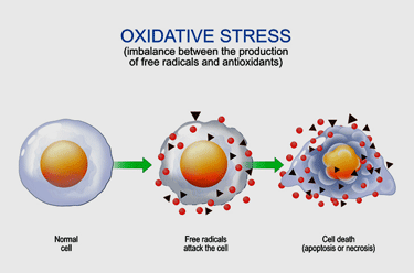
High levels of oxidative stress could be impacting many areas.
The AOC2 increase suggests exercise has increased levels of the free radical H202 (and ammonia). The CUTC upregulation reflects an attempt to mobilize copper to deal with an increase in reactive oxygen species (free radicals) triggered by the exercise. The increased levels of oxidative stress indicated could help explain the kynurenine, citrate, and fatty acid findings.
One doctor reported commonly seeing high copper levels in hair analyses of people with ME/CFS. Increased levels of ceruloplasmin – a copper transporter – have been found in fibromyalgia and have been associated with increased pain. Too much intracellular copper can damage the lipid membranes in our cells and damage the mitochondria. According to one website, estrogen, interestingly, may be able to elevate copper levels.
Neurotoxic Connection? The Tryptophan/Kynurenine Pathways
Lower KYN:TRP ratios before exercise and increased KYN:TRP ratios after exercise indicate that exercise in ME/CFS triggered a shift toward greater activation of the kynurenine pathway (ouch!). This has been proposed several times before.. The kynurenine pathways produce inflammatory and neurotoxic factors.
Higher kynurenic acid (KYNA) to KYN KAT activity ratios before exercise and lower after exercise reflect yet another pathway that’s failed to adapt properly to the stress of exercise. The diversion of kynurenine towards neurotoxic and neuroinflammatory pathways aligns with the metabolic inflexibility and high levels of mitochondrial/redox strain observed in other findings. The fact that elevated levels of 3-methoxyanthranilate correlated with general fatigue in ME/CFS suggests that activation of the kynurenine pathway is having real effects.
Indeed, the authors note that “energy depletion in the CNS…..(is) compounded by dysregulation in tryptophan-dependent pathways for the synthesis of KYN and serotonin”; i.e., it’s energy depletion meets neurotransmitter problems.
A similar pattern occurs in a series of chronic illnesses (chronic kidney disease, multiple sclerosis, cancer), and has been linked to impaired exercise capacity, fatigue, neuroinflammation, and poor recovery.
Weakened Extracellular Matrix (Connective tissues) and Blood Vessels
A variety of signals ( integrin cell surface interactions, VEGF signaling, leukocyte adhesion and diapedesis, analytes in the extracellular matrix protein (ECM) degradation pathway, lower CD93 and cartilage oligomeric matrix protein (COMP) high34 high-temperature requirement serine protease A1 (HTRA1), lower tetranectin (CLEC3B) levels) have left a weakened extracellular matrix and microvascular blood vessels prone to damage. When stressed, both fold, leaking blood or gut contents. Reduced levels of L-selectin (SELL) and adhesion G-protein coupled receptor (GPCR) D1 (ADGRD1) suggest that repair processes are inadequate and are late to the game – prolonging postexertional malaise. It’s yet another system that cannot stand the stress.
Can’t Recruit, Can’t Repair Either: Breaks Down Instead…
One analysis of the following findings (no change in plasma DAP levels after exercise, reduced leucate levels before and after exercise, baseline levels of citrulline (CIT) trended toward reduction, and reduced baseline levels of TFF1 characterized the result as“can’t recruit, can’t repair, breaks down instead”.
These findings suggest that weakened connective tissues, which just can’t “hang together” properly, weakened blood vessels, especially the smallest blood vessels (which may become leaky and unable to provide nutrients to the muscles), leaky gut, which past studies have shown accompanies intense exercise in ME/CFS, and, of course, reduced repair processes are present. Possible results after exercise – gut problems, tendon and muscle problems, blood pooling, reduced blood flows to the brain, all of which require longer than normal recovery times…(It just goes on and on.)
Stress Calcium Handling
A series of factors (S100 analytes, Class B/2 pathway markers, calcitonin receptor (CALCR), modifying protein 3 (RAMP3)) found before and/or after exercise suggest that exercise produces disturbed or stressed calcium handling in tissues, possibly affecting energy production, and producing inflammation and nervous system issues in the post-exertional period. Wirth/Scheibenbogen have proposed that calcium handling issues play a key role in energy production problems in ME/CFS.
A Mast Cell Match? Complement Activation
Increased levels of C1R and complement factor H-related protein 4 (CFHR4) in ME/CFS after exercise suggest exercise turns on the complement system, potentially resulting in mast cell activation, connective tissue damage, fatigue, systemic inflammation, and cognitive impairment. Interesting, one of the first ME/CFS exercise studies – by the CDC, no less – found the same kind of complement activation (c4) and proposed that the complement system might be responsible for the “inflammation-mediated postexertional malaise”.
A recent review overseen by Akiko Iwasaki proposed that complement activation plays a role in the coagulation, inflammation, and vascular injury in long COVID.
More Systems Under Stress

Multiple interconnected systems appear to be under stress in ME/CFS
Higher pre-exercise levels of eukaryotic translation initiation factor 1 (EIF1) and ubiquitin-conjugating enzyme E2 D3 (UBE2D3) indicate that even at rest, the cells are turning proteins over faster than normal. (Remember the high metabolic stress at rest…)
That happens when oxidative/mitochondrial or ER stress and/or low-grade inflammation is present and aligns with other findings suggesting that an already strained network is primed to be overwhelmed by exertion.
Reduced levels of contactin 4 (CNTN4) and CNTN4 and EPHA4 prior to exercise suggest another system – the nervous system – is not ready for the stress exertion will impose on it. Increased levels of ELAVL2 suggest that a fragile nervous system tries to cope, but decreased levels of NRXN1 after exercise suggest that the exercise ends up impairing signaling in the central nervous system – potentially producing brain fog/fatigue.
Ditto with the reduced pre- and post-exercise tetranectin (CLEC3B) levels. Because tetranection repairs damage to the extracellular matrix, i.e., the connective tissue, and connective tissue damage is synonymous with exercise, tetranection or CLEC3B levels should rise with exercise. The fact that they didn’t suggests that repair mechanisms that should kick in after exercise didn’t result in a slower recovery period after exercise, which is what we see in ME/CFS. It’s yet another system that appears to be folding under the stress of exercise.
The Symptom Tests
Being able to show that these abnormalities are associated with symptoms is a major test. Several of them were. They included
- f S100A8 and C1r – Elevated post-exercise levels o in ME/CFS correlated with MFI scores of reduced activity/motivation
- 12,13-diHOME – baseline scores correlated with Multidimensional Fatigue Scores of physical fatigue.
- GDF15 – post-exercise levels were positively correlated with MFI scores of general fatigue, physical fatigue, and reduced activity
- CNTN4 – reduced levels correlated with higher general fatigue scores
- Tetranectin (CLEC3B) – Reduced pre- and post-exercise levels correlated with higher MFI scores of physical fatigue and reduced activity/motivation
The authors proposed prioritizing treatments that regulate the metabolism and reduce inflammation.
The Deeper We Go – the More We Find

The deeper researchers dig into ME/CFS the clearer the connections become
The deeper we go into the molecular foundations of ME/CFS, the more we find and, importantly, the more things map together. If ME/CFS were a fake disease, the deeper that molecular investigations go, the more they should flounder and fall apart. Things should get messier, not clearer, as it became clear that ME/CFS wasn’t a real disorder.
The opposite is happening. The deeper researchers dig, the more they find, and the clearer the connections become. That’s crucial because in a multisystemic disease, what we need more than anything else is to be able to explain how so many different systems can be affected. This study does that in spades.
Major Themes
This study suggests that one thing leads to another and another and another
A couple of major themes are present. Immune activation on a massive scale. Failures on multiple levels to provide the muscles with the necessary energy to exercise. Metabolic inflexibility that prevents the body from getting into “exercise mode”. A toxin and free radical-laden system that’s co-opting resources needed to deal with exertion. An extracellular matrix is under attack, but is unable to defend itself, and a variety of systems are so stressed that they fold when stressors show up.
The really remarkable thing is that a coherent argument can be made that, like dominoes falling, virtually every one of the abnormalities cited above can be linked together. They may be the logical conclusion of an energy-depleted, toxin-overrun system. The authors noted how interconnected all this appears to be:
“The persistent cellular stress, characterized by impairments in urea cycle, xenobiotic metabolism, and protein metabolism, may initiate response pathways that perpetuate mitochondrial dysfunction and immune dysregulation.”
In their press release, the authors reported that these “interconnected pathological processes” do not appear to be new but are often “observed in chronic inflammatory conditions”. While we are probably going to find unique things in ME/CFS, what we really want to find are “pathological processes” that are present, being worked on, and treated in other diseases.
Thousands of drugs and treatments, only a small fraction of which are currently available to ME/CFS, are present. Showing that the same pathological processes are present in ME/CFS should open the door to more treatments.
At the end of the paper, the authors proposed that innate immune activation in concert with high levels of oxidative and cellular stress was a good place to focus on. The end product of all these pathological processes, the authors believe, is “systemic inflammation,” which is what we might expect in a disease that affects so many systems.
Treatment Implications
The authors suggested
- Innate immune system activation – metformin, the regulatory cytokine IL-37, and the mTOR inhibitor rapamycin
- Gut dysbiosis – prebiotics (inulin) and probiotics (F. prausnitzii)
- Metabolic disruption – People with low baseline levels of 12,13 diHOME and high post-exercise levels of GDF15 –
supplementation with 12,13-diHOME or treatment with GDF15 neutralizing antibody - Tryptophan metabolism – hydroxytryptophan or selective serotonin reuptake inhibitors
- Low carnitine – supplement with carnitine
- Inflammatory response in women – estrogen supplements
No Smoking Gun Yet
They indicate that multiple, interconnected systems have become destabilized. While Lipkin noted that we stil don’t know how ME/CFS got started, studies like this are revealing at a deep molecular level why people with ME/CFS can get so sick. What we need are biomarkers that lead to “targeted interventions” and by elucidating multiple problems at the molecular level this study is pushing us along this path.
“While what gives rise to ME/CFS remains obscure, understanding the ways it disrupts the body’s various biological processes on the molecular level is revealing biomarkers for specific ME/CFS subtypes that may inform clinical research and lead to targeted interventions,” senior author W. Ian Lipkin, MD.
The next steps appear to be validating biomarkers – this study has many potential biomarkers – that can be used to assess treatments. That’s what brings pharma in. Long COVID has the VIPER biomarker project – adding ME/CFS to it would be a huge boost for this disease – and given the unique findings ME/CFS researchers are uncovering, possibly for long COVID.
Check out a recent talk with Ian Lipkin
Like this Big Blog?
Support Health Rising and Keep the Information Flowing
Health Rising is not a 501 (c3) non-profit


Phew
In your previous article, I dissed this study. After reading it properly, that was unfair. It’s a good study.
My problem is that we still seem to be going in circles in terms of finding the smoking gun.
Perhaps there is no smoking gun. Perhaps ME/CFS is a complex, systemic illness, and we need to resign ourselves to that. There won’t be one magic treatment. Hopefully there will be options to work on the different systemic issues and improve quality of life.
In terms of ‘immune activation’ – they found that in younger people but not older people. This seems to back up previous studies that show immune activation earlier in the illness, but not later.
Yet the older people still have ME/CFS.
Which suggests to me, as it has for a long time, that an immune insult triggers the illness, but does not perpetuate it.
It seems undeniable now that there is chronic inflammation and metabolic disturbances. Again, why? And then, how to address these?
It’s good that the study authors touch on some treatment ideas. Including rapamycin. I wonder how PolyBio’s study is going.
What this study showed for me was how many weakened systems are present and how they can virtually all be connected together! One thing just leads to another. Instead of immune cells being the cause of inflammation the authors said that all these dysfunctions may be producing the inflammation they found. Something very organized is happening in ME/CFS patients as a whole.
So many things are in the pits that its hard to say where it starts but because they are all potentially connected I think we will have an answer at some point. It may take AI to puzzle this out.
In the meantime – validate the biomarkers (please) – and start looking for treatments. It was encouraging to see the authors say that these processes are happening on other disesases.
The findings in many ways reinforce the findings of the recent, major Edinburgh study.
A number of general, systemic issues. Nothing apparently specific to ME/CFS.
So yes, I guess that is a blessing in a way. In that, a number of other illnesses are characterised by many of the same sorts of inflammatory/ metabolic issues, and hopefully therefore increases the chances of meaningful treatments being confirmed – within the next couple of years, please!
I still believe the brain is a big part of ME/CFS. Are the brain issues a flow on from these wider systemic issues? Or does it fundamentally control and influence the wider systemic issues? I had been thinking the latter, but perhaps it really is one big, multi-directional systemic thing.
There’s an excellent lead article in the latest New Scientist about inflammation and the brain. Quotes Andrew Miller several times
12,13-diHOME doesn’t seem to exist as a supplement?