


Geoff’s Narration
The GIST
Warning: This is a LONG blog. (It just kept growing and growing – which is a good sign)
The Molecular Antidote to the RECOVER Project: the IMPACC Study
NIAID’s IMPACC study is the molecular antidote to the RECOVER Initiative’s study.
With 60 co-authors spread across over 25 US universities who followed over 1,000 hospitalized COVID-19 patients, the latest paper from the NIAID-funded IMPACC network, “A multiomics recovery factor predicts long COVID in the IMPACC study“, is a serious effort to understand long COVID. No fewer than 24 NIH grants powered this paper. It’s probably the largest longitudinal (followed patients over time) long-COVID study yet, and it’s going to get a lot of attention.
Simply put, the IMPACC, or “IMMuno Phenotyping Assessment in a COVID-19 Cohort IMPACC Network”, is the molecular antidote to the NIH’s much larger (and stodgier) RECOVER long-COVID Initiative. Instead of assessing 30,000 people using more or less standard lab tests, as RECOVER has, IMPACC is going deep into “omics” studies (genomics, proteomics, metabolomics, transcriptomics) and deep immune work (immunophenotyping, antibody profiling) in an attempt to understand what went wrong on the molecular level in long COVID.
While IMPACC doesn’t have nearly the funding of the $1.65 billion RECOVER project, the 50-$100 million it’s received is nothing to sneeze at. One has to congratulate the NIAID for so rapidly funding (May 2020!) and continuing to fund this large project.

The fly in the ointment is the total focus on hospitalized COVID-19 patients. We’ll see, though, that many of its findings mirror those showing up not just in non-hospitalized patients but in ME/CFS patients as well.
THE GIST
-

Does it all come down to blood that just isn’t reaching the tissues?
With 60 co-authors spread across over 25 US universities who followed over 500 hospitalized COVID-19 patients, the latest paper from the NIAID-funded IMPACC network, “A multiomics recovery factor predicts long COVID in the IMPACC study” is a serious effort to understand long COVID. It’s probably the largest longitudinal (following patients over time) long COVID study yet, and it’s going to get a lot of attention.
- Immune functioning was a strong focus in a study that also included metabolomics and proteomics. A major goal was to develop a “multiomics recovery factor; i.e., a molecular blueprint of recovery (and non-recovery).
- The study was able to reliably identify which patients were likely to come down with long COVID as early as three days following infection. People with the biology described below were more likely to develop long COVID.
- It wasn’t surprising to see high levels of inflammation early (and later) in people with long COVID, but it was exciting to see that problems with blood vessel inflammation, in particular, were prominent.
- Male sex hormones were another prominent factor. The more male sex hormones a COVID-19 patient had, the better off they were, and the lower the levels of male sex hormones, the more likely they were to come down with long COVID.
- Interestingly, male sex hormones protect the blood vessels, and low testosterone levels have been associated with blood vessel inflammation.
- Nancy Klimas’s modeling efforts suggest that testosterone is protective in both men and women for ME/CFS, and the first stage in her ME/CFS drug trial includes boosting male sex hormones.
- Male sex hormones also affect other factors that potentially impact ME/CFS, including muscle mass and protein synthesis, energy availability / metabolic tone, red blood cell production, bone maintenance (high rates of osteoporosis in fibromyalgia), and sexual/reproductive functions.
- All in all, the reduced male sex hormones could contribute to symptoms like fatigue, reduced exercise tolerance, muscle weakness, and lower motivation/libido.
- Heme metabolism was the last major factor associated with long COVID. Heme metabolism refers to the breakdown of heme – the part of hemoglobin which contains the iron ring which oxygen attaches to.=
- When red blood cells break open heme must be quickly recycled before it damages the blood vessel walls.
- Enter a recent ME/CFS study from the Open Medicine Foundations, Alain Moreau, which found reduced haptoglobin levels in people with ME/CFS both before and after exertion.
- Because haptoglobin neutralizes free heme, the low haptoglobin levels suggested that exertion was destroying red blood cells were in such large amounts that the haptoglobin levels were not keeping up. It also suggested the blood vessel walls were being damaged.
- Haptoglobin lab tests are available to the public. Check out the blog if you want to get your haptoglobin levels assessed.
- These findings appear to fit perfectly with the red blood cell deformability finding which suggests that stiffened, fragile red blood cells are having trouble deforming enough to get into the small capillaries that feed the tissues.
- Recent European ME/CFS findings back up that finding and add to it. They suggest that oxygen is being held on the red blood cells too tightly, that deformed red blood cells are blocking the capillaries, that massive collagen deposition is preventing the capillaries from reaching the tissues, and that this is all result in damage to the endothelial cells lining the blood vessels.
- Moving over to long COVID, several studies suggest that red blood cells are being destroyed, that the microvasculature (capillaries) is in ruins.
- An Australian study which found high levels of dead endothelial cells in the microvasculature suggests the red blood cell problems may start there. Endothelial cells line the blood vessels. When they die their membranes blister, rupture, and detach, leaving jagged and sticky blood vessel walls behind. Red blood cells trying to pass through these narrowed, roughened blood vessels are likely to be torn apart.
- To make matters worse, the dying endothelial cells activate the complement system, which attacks the red blood cells, punching holes in them. The low oxygen conditions that ensue stiffen the red blood cell membranes, making them more prone to rupture as they try to pass through the jagged blood vessels.
- The microclots, found in these diseases consist of amyloid-like aggregates, inflammatory proteins, and red blood cell debris that result from the dying endothelial cells and red blood cells.
- Because all of this is happening in the microvasculature, it’s difficult for test results to pick it up. Ironically, the deformed red blood cells, first found in ME/CFS in 1986 by Les Simpson, may have been the canary in the coal mine for these diseases.
- Since each red blood cell passes from the arteries into the microvasculature and then into the veins hundreds of times a day, they may be getting damaged every pass through the circulatory system.
- The Australian study suggests there’s nothing inherently wrong with the red blood cells – they’re simply getting whacked every time they enter a capillary.
- In total, the studies suggest that the blood vessels are not functioning properly (endothelial dysfunction); narrowed, jagged, debris ridden capillaries are preventing oxygenated red blood cells from getting to the tissues; fragile, stiffened, deformed, ATP deficient, oxygen grabby red blood cells (see a recent Polish study) with low haptoglobin levels are getting broken up, and leaking toxic materials into the capillaries, and the autonomic nervous system responsible for moving the blood around has gone wonky.
- It may all begin with the immune system attacking the endothelial cells lining the blood vessels perhaps because parts of viruses have become embedded in them. (This is Bruce Patterson’s hypothesis from five years ago. Autoimmune processes or problems with mitochondrial production could also be at cause.
- Note, though, that much of these hypotheses rely on a few studies, some of which have not been published yet.
- Getting back to the IMPACC study, the immune findings mirrored those in ME/CFS which suggest that the innate immune system (the main inflammation producer) is in overdrive trying to compensate for an underperforming adaptive immune system.
- Drugs that tamp down the innate immune system might be helpful (and are being tested in long COVID). Other drugs that improve blood vessel or red blood cell functioning might be helpful as well.
- All told, the inflammation, male sex hormone, and heme/blood vessel findings appear to be full of potential. In the end, what many of us suspected – that the blood is just not getting through – may be the key after all.

Support Health Rising and Keep the Information Flowing!
Health Rising is not a 501 c (3) non-profit
The Study
Is the blood just not getting through?
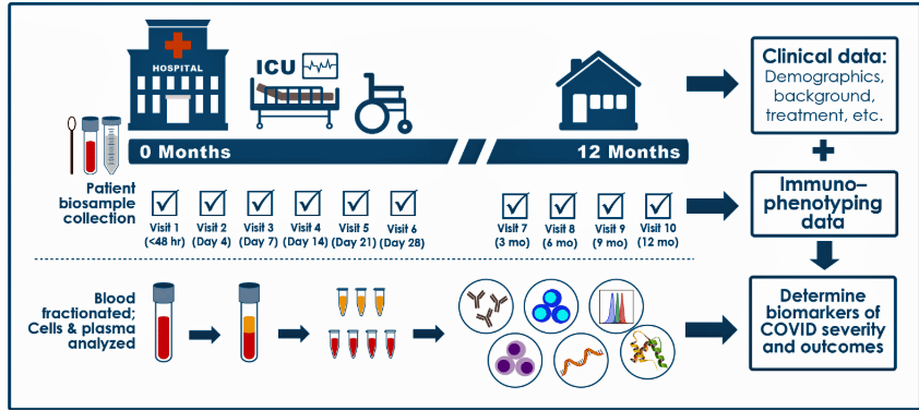
This study used machine learning techniques to analyze data from over 500 patients, collected in the 12 months following their hospitalization for COVID-19. The immune system was given a strong focus. The data included immune cell (PBMCs), gene expression (transcriptomics), serum O-link and plasma proteomics, plasma metabolomics, and blood mass cytometry(CyTOF) protein levels. A physical functioning score was used to determine which patients recovered and which did not. A major goal was to develop a “multiomics” recovery factor; i.e., a molecular blueprint of recovery (and non-recovery).
This study was unusual in its size (>500 patients), its depth (many omics assessments), and its span (1 year).
Blood Vessel Problems
Inflammation – in particular, inflammation affecting the blood vessels – was highlighted.
Rather remarkably, the study was able to reliably identify which patients were likely to come down with long COVID as early as three days following infection. People with the biology described below were more likely to develop long COVID.
It wasn’t surprising to see high levels of inflammation early (and later) in people with long COVID, but it was exciting to see that problems with endothelial/vascular inflammation, in particular, highlighted the blood vessels. Even the gut got into the act when two gut-derived amino acids (phenylacetylglutamate, phenylacetylglutamine) associated with inflammation in the blood vessels popped up.
- Score one for the blood vessels – long an area of interest in long COVID and ME/CFS.
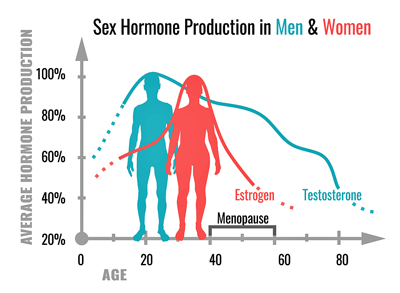
Male Sex Hormones (androgenic steroids pathway)
Seeing the “androgenic steroids pathway” involved in steroid hormone metabolism pop up was somewhat, but not entirely, surprising. It would seem bizarre if the sex hormones weren’t involved in some way in these female-dominated diseases. This study shines a light on a potentially key factor – the male sex hormones – that has shown up big time in Dr. Klimas’s ME/CFS studies but perhaps hasn’t received the attention it should. (While these hormones are termed male sex hormones and are present in higher levels in males, they play an important role in females as well.)
The more male sex hormones the COVID-19 patient had, the better off they were, and the lower the levels of male sex hormones, the more likely they were to come down with long COVID. The pregnenolone pathway, a potent inhibitor of inflammation, was particularly affected.
Problems with the male sex hormone pathway make sense in several ways in long COVID/ME/CFS/FM. While the authors focused on its impact on inflammation, this pathway also affects many areas highlighted in these diseases, including muscle mass and protein synthesis, energy availability / metabolic tone, red blood cell production, bone maintenance (high rates of osteoporosis in fibromyalgia), and sexual/reproductive functions. Reduced male sex hormones, then, could contribute to symptoms like fatigue, reduced exercise tolerance, muscle weakness, and lower motivation/libido.
Plus, they cohere so nicely with the blood vessel findings. We want pathways to show up and interact together to deliver a big punch, and it turns out that reduced male sex hormones and blood-vessel inflammation can go hand in hand. This is the first time I can remember that male sex hormones have been linked to blood vessels in these diseases.
With her model showing that testosterone is protective in men and often deficient, Dr. Klimas added testosterone to her ME/CFS clinical trial.
Lower testosterone levels have been associated with blood vessel inflammation and increased oxidative stress, and in long COVID, they’ve been associated with increased symptoms. Testosterone helps keep endothelial cells that line the blood vessels healthy, and low testosterone and blood vessel inflammation can form a vicious circle because they reinforce each other.
Noting that similar androgen steroid findings showed up in a 2020 ME/CFS study, the authors suggested low levels of male sex hormones were helping to produce the fatigue, post-exertional malaise, and sleep disturbances found in both ME/CFS and LC.
Reduced testosterone levels have been found several times in both ME/CFS and fibromyalgia. Nancy Klimas’s models suggest testosterone is protective in ME/CFS, and some doctors have prescribed testosterone for fibromyalgia. Klimas’s supercomputer treatment model proposes normalizing the sex hormones first, then stopping brain inflammation with etanercept (Enbrel), and then resetting the HPA axis and reducing inflammation using mifepristone, is the way to go in ME/CFS.
In a small but intense study, Jarred Younger measured testosterone, progesterone, estradiol levels, and cortisol levels in 8 women with FM for 25 days straight while having them record their pain levels. He found that both progesterone and testosterone were inversely correlated with pain levels; that is, the higher the FM patients’ progesterone and testosterone levels were, the lower their pain was.
One doctor has been using testosterone gel to treat fibromyalgia.
Impaired Heme Metabolism
Blood vessel inflammation and altered metabolism of heme – a part of hemoglobin (pictured) – will open the door to a wide range of possible injuries. (Image from RFZYNSPY_2021_Wikimedia_Commons)
The heme metabolism finding is going to introduce a very rich vein indeed as it gets extended to recent blood vessel and red blood cell findings.
Heme metabolism refers to the cycle of building and breaking down heme – the iron-containing molecule in hemoglobin that carries oxygen in our red blood cells. The heme metabolism/blood vessel/red blood cell themes will occupy much of the rest of this blog.
Each hemoglobin molecule contains 4 heme groups, each of which contains an iron atom that oxygen binds to. If the red blood cells break open, though, they will spill heme into the plasma, and there the trouble starts. Free heme is highly oxidative and will damage the blood vessel walls.
Stress-Induced Damage
“Our findings suggest that hemolysis-like processes (red blood cell destruction) … may underlie symptom exacerbation in ME and could be modulated by inherited factors.” Moreau et al.
Haptoglobin comes to the rescue by capturing and neutralizing free heme. Not only did Open Medicine Foundation researcher Alain Moreau find lower haptoglobin levels in ME/CFS at baseline and after exercise (using the cuff) in two ME/CFS cohorts but he also found that they were associated with increased post-exertional malaise and cognitive problems. (Exertion did not reduce haptoglobin levels at all in healthy controls.) He proposed that reduced haptoglobin levels could be a biomarker of PEM in ME/CFS.
Reduced haptoglobin levels suggest massive red blood cell destruction may be occurring in ME/CFS.
Further analysis suggested a genetic component was present: people with certain HP gene variants were more likely to have low haptoglobin levels, more symptoms and more problems with PEM.
This suggests that exertion is causing fragile red blood cells in ME/CFS to break open and spill their guts, throwing free hemoglobin and heme into the bloodstream. With so much free heme floating around, supplies of its neutralizer – haptoglobin – run out, leaving the highly toxic free heme to attack the blood vessel walls. (Other studies suggest damaged blood vessel walls may play a key role in long COVID.) ME/CFS patients who have a genetic predisposition to “stronger” haptoglobin that is more effective at neutralizing heme do better while people with “weaker” haptloglobin have more PEM.
In this scenario, the inflammation in the blood vessels is not the result of an innate immune response run amok, but is the result of exertion, in particular, causing a massive destruction of red blood cells.
Testing Your Haptoglobin
This finding can be tested using standard laboratory tests. Because haptoglobin levels will likely be normal at baseline given the huge “normal” range (30-200 mg/dl), it would be best to measure both at baseline and after a physical or cognitive stressor. (Most doctors are not aware of these tests or how to assess them in ME/CFS.)
The key finding would be a significant drop in haptoglobin levels after exertion. The study found a 50% drop in haptoglobin levels 90 minutes after the cuff stressor was applied but a reduction of 20% or more in your personal baseline would be a strong indicator that your red blood cells were breaking open and haptoglobin levels were being depleted.
If you’d like to get tested, here are the laboratory codes for the:
Haptoglobin (The “Chaperone” Level) – This is the primary test mentioned in the 2026 study. It measures how much “protective” protein you have available to soak up toxic heme.
- Labcorp Code: 001628 (CPT 83010)
- Quest Code: 502 (CPT 83010)
- Alternative (Sonora Quest): 9239 or HAPT
Haptoglobin Phenotype/Genotype – This identifies your genetic “type” (Hp1-1, 2-1, or 2-2). The study found that those with Hp 2-1 had more severe PEM and cognitive symptoms.
- Labcorp Code: 504350 (or sometimes listed as 001636 for Phenotyping)
- Quest Code: 91361 (Haptoglobin Phenotype)
- Note: This is usually a one-time genetic test to see your baseline susceptibility.
Ferritin – checks your iron stores. In ME/CFS, this may be high (indicating inflammation) even if you feel like you have low iron symptoms.
- Labcorp Code: 004598
- Quest Code: 457
- Sonora Quest: 9210
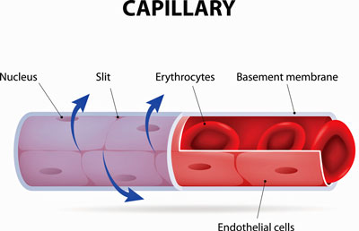
Red Blood Cell Deformability
In turn, the haptoglobin finding appears to feed right into the 2019 red blood cell deformability finding in ME/CFS.
The large blood vessels look great – they are full of oxygen – but the oxygen can’t make it to the tissues because stiffened red blood cells are shattering as increased blood pressure pushes them into the small blood vessels/capillaries that feed the muscles. Some research suggests that capillaries may be abnormally narrowed in these diseases, putting even more pressure on fragile red blood cells.
There’s good evidence that the blood is either not getting through to the tissues or, at the very least, is not getting its oxygen through to the mitochondria. Systrom’s invasive exercise work has shown that oxygen is not getting used up in about half of ME/CFS patients. Plus, at least three small studies have shown that blood flows through microvasculature are inhibited in a large subset of ME/CFS patients.
Not getting enough blood to the tissues and brain could, in turn, trigger sympathetic nervous system activation and produce racing heart rates in ME/CFS and/or hyperadrenergic POTS.
Things seem to be adding up, but we’re not done, though, not by a long shot. We haven’t taken into account what the Germans and Dutch recently found in ME/CFS.
The Europeans Step In
Research into red blood cells and small blood vessels is booming in Europe. At the 2025 Charite Conference, Puta reported that the stiffened red blood cells in ME/CFS and long COVID were holding onto their oxygen too tightly. He also proposed that these deformed cells were physically blocking capillaries thus preventing oxygen from reaching mitochondria.
His work validated his compatriot Wilhelm Bloch’s 2022 finding that deformed red blood cells in long COVID were retaining oxygen. In what’s getting to be a pretty common comment, Bloch said he’d never seen anything like that.
Two of the three prize-winning posters at the 2025 Charite Conference involved the blood vessels. Anouk Slaghekke’s (Vrije Universiteit Amsterdam) exciting finding (“Microvascular Dysfunction and Basal Membrane Thickening in Skeletal Muscle in ME/CFS and Post-COVID: from Pathology to Diagnosis”) suggested that massive collagen deposition is making it hard for blood to reach the muscles.
Timon Kuchler’s (Technical University of Munich) “Prolonged Endothelial Dysfunction in Post-COVID Syndrome Patients Compared to COVID-19 Recovered and SARS-CoV-2 Naive Individuals” likewise charted long-standing blood vessel problems.
Put these together, and we potentially have problems at every juncture: oxygen gets stuck on deformed red blood cells, capillaries aren’t forming, and many of those present aren’t working, to boot. We should start seeing papers from these researchers come out.
Long COVID
Red blood cells are starting to get a long look in long COVID as well. A recent study linked significant reductions in RBC deformability and increased aggregation (clots) to microvascular issues. A 2022 study found massive red blood cell damage and clots that the authors thought were clogging the microvasculature (capillaries) and reducing blood supply to the tissues.
Several studies suggest ME/CFS and long COVID are microvascular diseases.
A remarkable study of formerly hospitalized COVID-19 patients found that even after their lung failure had been successfully treated, their microvascular system remained in ruins.
Severely reduced capillary oxygen saturation (low oxygen levels in the microvasculature) highlighted a now common theme: the oxygen isn’t getting through.
Greatly increased rates of oxygen extraction suggested that oxygen-starved tissues were pulling as much oxygen as possible from the red blood cells present. Finally, reduced “functional” capillary density (i.e., a reduced number of capillaries that are actually carrying red blood cells) was found, echoing Slaghekke’s ME/CFS finding.
All in all, it looked like multiple hits to the small blood vessels could be occurring: not only were their oxygen levels low, but they were either damaged or weren’t getting much blood flow, or both.
The authors proposed something long suggested in ME/CFS: that systemic tissue hypoxia (low oxygen levels) are present. Low oxygen levels in the small blood vessels across the body sounds like an almost ideal way of decreasing functionality.
Australia Goes Deep
Researchers keep digging deeper. An Australian Nature study “Ischaemic endothelial necroptosis induces haemolysis and COVID-19 angiopathy” may have gotten near the heart of what’s happening.
This 2025 study suggested that the problem does not begin in the red blood cells but in the small blood vessels themselves. The study found high levels of dead endothelial cells. These very active cells that line the blood vessels regulate blood flows, produce pro-inflammatory cytokines, prevent blood clots from forming, and allow immune cells to reach the tissues when needed. They’re so involved in the immune response that some consider them to be innate immune cells.
These are also the cells where atherosclerotic plaques that block blood flow in the arteries form. Transfer that general idea (blocked blood vessels) to the microvasculature, and you just might have explained ME/CFS and long COVID.
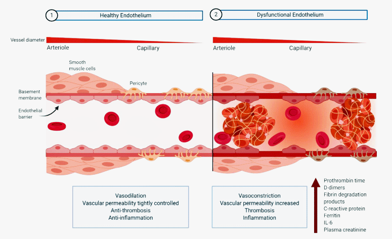
Endothelial damage caused by the SARS-CoV-2 coronavirus. (Image from -Bernard_Limonta_-Mahal_-Hobman_Endothelium-Infection_Dysregulation_Coronavirus_2020_ MDPI_Wikimedia_Commons)
The endothelial cells appeared to be undergoing programmed cell death. As this happens, their membranes blister, rupture, and detach, leaving jagged and sticky blood vessel walls behind. Red blood cells trying to pass through these narrowed, roughened blood vessels are likely to be torn apart.
To make matters worse, the dying endothelial cells activate the complement system, which attacks the red blood cells, punching holes in them. The low oxygen conditions that ensue stiffen the red blood cell membranes, making them more prone to rupture as they try to pass through the jagged blood vessels.
Something apparently novel then happens in long COVID. The red blood cell fragments seal over the damaged endothelium. That prevents microvascular bleeding but blocks the small “lumen” or space through which red blood cells pass on their way to the tissues. The authors called this process a “a previously unrecognized haemostatic mechanism preventing microvascular bleeding. (Is it any surprise that something heretofore “unrecognized” is found in ME/CFS and long COVID?)
Enter the microclots that Pretorius, Kell, and Nunes have found in ME/CFS and long COVID. Classical clots are produced by a kind of fibrin-platelet cascade which does not appear to be happening in these diseases. They are often associated with bleeding. Microclots, on the other hand, consist of amyloid-like aggregates, inflammatory proteins, and red blood cell debris. They’re not associated with bleeding. Because they’re missing the fibrin component that anti-coagulants attack, anti-coagulants often don’t work.
In any case, they further block blood flows. The authors also proposed that small blood vessel hypoxia (low oxygen levels) are present bodywide in people with long COVID.
Severity Doesn’t Matter
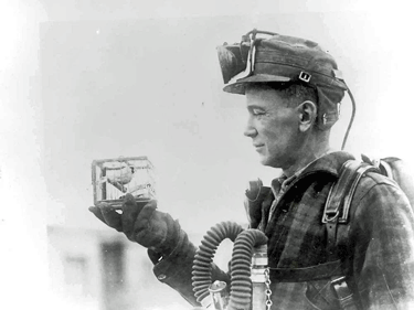
Were Les Simpson’s 1986 red blood cell findings the canary in the coal mine, indicating that the microvasculature was profoundly disturbed in ME/CFS? (Image from McCaa, Bureau of Mines, Wikimedia Commons)
Note that the Nature study focused on severely ill, hospitalized COVID-19 patients, but their microcirculation ended up looking like that found in long COVID and ME/CFS! The same process appears to be occurring in them, but happens more quickly and severely in them. Indeed, early studies suggested that the microvasculature was primarily affected in COVID-19.
Because all of this is happening in the microvasculature, it’s difficult for test results to pick it up. Ironically, the deformed red blood cells, first found in ME/CFS in 1986 by Leslie Simpson, may have been the canary in the coal mine for these diseases.
Simpson and others have found deformed red blood cells in the major blood vessels of ME/CFS patients, not in the microvasculature. If a toxic microvasculature is deforming the red blood cells, how are they showing up in the larger blood vessels?
The answer is easy – each red blood cell passes from the arteries into the microvasculature and then into the veins hundreds of times a day. During each pass, they may be getting damaged, which shows up in blood taken from a vein. The Australian study suggests there’s nothing inherently wrong with the red blood cells – they’re simply getting whacked every time they enter a capillary.
Putting It All Together
This scenario can explain so much. These findings suggest that even before people with ME/CFS or long COVID begin to exert themselves, they’re already behind the eight ball.
Their blood vessels are not functioning properly (endothelial dysfunction); narrowed, jagged, debris ridden capillaries are preventing oxygenated red blood cells from getting to the tissues; fragile, stiffened, deformed, ATP-deficient oxygen, grabby red blood cells (see a recent Polish study) with low haptoglobin levels are getting broken up, and leaking toxic materials into the capillaries, and the autonomic nervous system responsible for moving the blood around has gone wonky.
Is it any wonder that the system goes bananas when it’s asked to exert itself?
The Necroptopic Disease?
If it all begins with an endothelial cell breakdown (necroptosis) in the small blood vessels, what causes that? We’re back to inflammation again – but a particular type of inflammation – inflammation driven by TNF-a, interferons, and perhaps most importantly, activation of the complement system, particularly C5b‑9.
But why would the immune system attack the endothelium? Perhaps because it detects bits of viruses, such as the coronavirus or herpesvirus, in these cells and attacks them. Indeed, SARS‑CoV‑2 RNA or proteins have been found in the endothelial cells. An inability to fully eradicate a pathogen could be responsible.
Bruce Patterson proposed this scenario was happening five years ago. Patterson was criticized for promoting his hypothesis on social media, but he’s put his money where his mouth is and a large clinical trial testing his hypothesis is underway. His goal is to stop the immune system from attacking the endothelial cells.
Other possibilities are present. Autoantibodies could also be weakening the endothelial cells by attacking receptors (B2 adrenergic, M3/M4 muscarinic, AT1R, and ETAR (angiotensin and endothelin receptors, GPCRs)) on them. Impaired mitochondrial production could also cause the endothelial cells – when put under stress – to initiate necroptosis (cell death).
Nice Ideas…
There seems to be no dearth of ways to explain ME/CFS and long COVID, and we’re in a rather familiar place with the blood vessel and red blood cell findings. It seems impossible that they don’t play a role, but note that these findings hinge on a few, mostly small studies. Even the ME/CFS finding of red blood cell deformability hangs on a single 2019 study. The evidence is growing (three studies have found atypical red blood cell shapes in long COVID) and this area – particularly in ME/CFS – is getting active research. Time will tell us more.
Several approaches exist that can test the necroptosis hypothesis. Biopsies, plasma tests, etc., could determine if it’s present and is fragmenting the red blood cells. Assessing microclots and endothelial and red blood cell health before and after exercise seems like a no-brainer at this point.
Endothelial cells (ECs) from long COVID/ME/CFS and healthy controls could also be exposed in the lab to low-oxygen conditions and/or inflammatory factors to assess whether long COVID/ME/CFS ECs are more fragile. Endothelial cells could also be exposed to ME/CFS/long-COVID serum to see if something in it (autoantibodies?) is breaking them up. (An early long-COVID study (2022) found that LC plasma was altering the shape of the red blood cells. Red blood cell deformability could be assessed before and after exertion to see if exertion increases it as the hypothesis proposes.
This is clearly a rich arena of interest that should grow over time.
Immune Findings
Returning to the IMPAAC study, its overall immune findings were also consistent with those in ME/CFS. Elevated levels of innate immune cells (neutrophils/monocytes) and reduced levels of B-cells suggest the innate immune system is trying to compensate for problems in the adaptive immune system.
The type of inflammation in LC and ME/CFS may be similar. Interestingly, several studies suggest that the same kind of monocyte CD14+CD16− found in this study may play a significant role in ME/CFS. Indeed, one ME/CFS study suggested that monocytes may play the key role in ME/CFS.
A recent long COVID study highlighted an interferon/monocyte connection, and another suggested that chronic IFN-y production may be a biomarker in long COVID. Note that while subsets undoubtedly exist in long COVID, the blood vessel, sex hormone, and immune findings may be core factors that pervade the entire long COVID cohort.
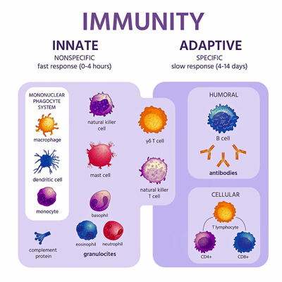
Once again, researchers propose that the innate immune system is attempting to compensate for a deficient adaptive immune response.
As noted earlier, all the significant factors – the heme metabolism issues, the reduced male hormone levels, the increased inflammation, and innate immune cell activity – were present as early as 72 hours after infection. Put together, people with these factors were more likely to come down with long COVID.
Interestingly, reduced iron and hemoglobin levels 2 weeks to 1 month after a SARS-CoV-2 infection also put people at risk of coming down with long COVID. The authors believe the reduced iron levels impaired immune cell functioning. The adaptive immune system’s inability to clear the virus allowed it to persist longer. The innate immune system jumped in to help, but the amped-up response caused the inflammation that whacked the blood vessels.
Treatment Implications
The authors didn’t get into treatment implications. The study, though, clearly suggests immunomodulators that knock down the innate immune system, such as JAK Inhibitors (3 long-COVID trials underway), may be helpful. Drugs that improve red blood cell health such as Pirfenidone are being assessed to see if they stop the “smoldering” inflammation found in long-COVID patients’ lungs and blood vessels. Drugs that improve endothelial cell health and functioning (statins, pentoxyphylline, ACE inhibitors, calcium channel blockers, nitrates, iron supplementation) might be helpful as well.
Conclusion
In the end, it was exciting to see an important long-COVID effort, whose findings, at first, looked a bit out of the mainstream, feed right into emerging findings in ME/CFS and long COVID. Given these diseases’ exercise limitations, it’s always seemed likely that the blood just wasn’t getting through and that the blood vessels must be involved in some way.
This study’s focus on three aspects – male sex hormones, heme metabolism, and inflammation – which reinforce each other ended up, once other ME/CFS and long COVID research findings were included, implicating blood vessels and the red blood cells. These researchers appear to have struck a rich vein (:)). Hopefully, that vein will get the “dig” it deserves.
Meanwhile, ME/CFS researchers, with their focus on red blood cells and the microvasculature, once again appear to be ahead of the curve.


Myoglobin, (sort of hemoglobin’s cousin in muscle cells) also binds to haptoglobin. Myoglobin does not bind as strongly to haptoglobin as hemoglobin does, but recent studies have shown extensive damage to muscles after exercise (was it in FM or ME/CFS?).
https://pubmed.ncbi.nlm.nih.gov/3756181/
Interesting.
My daughter had a real boost, for a while, after taking an iron supplement (she was at low end of normal in terms of iron levels). It was quite amazing. She went from sleeping 16-17 hours a day to 11-12 hours.
Unfortunately the benefits subsided. But I thought it was interesting and perhaps pointed to some sort of mechanistic factor in ME/CFS.
Its really unfortunate ieron is not pursued more particularly in POTS https://www.healthrising.org/blog/2016/11/19/iron-man-pots-chronic-fatigue-syndrome-recovery-ferritin/
Cool. So what’s the ‘So What’?
Especially for treatment?
Pregnonolone?
Testosterone?
On the Big T – where is Klimas’s study at?
Lots of options and probably many we have not thought of – JAK/STAT inhibitors, various ways to protect the blood vessels and red blood cells. Since not a lot of work has been done it will, yes, take time – but the idea that the blood is just not getting through has always made sense to me. A nice vein (lol) to dig.
Well, the way I read the study is that the problems start with sex hormones. They are low, that results in more inflammation, and that impacts the blood. It’s probably more circular than that, but that seems to be the main causal chain they describe.
That’s why I asked about steroids and Klimas’s study.
It’s hard to know where it starts! If these findings are correct there appear be a combination of things that allow this process to start.
One is low testosterone which weakens the blood vessels. if a pathogen or part of a pathogen is lodged in the endothelial cells, low testosterone might play a role in allowing that to happen (?) or weakening them further. A pathogen lodged in the endothelial cells could trigger the inflammation which ultimately results in the small blood vessels failing and dragging the red blood cells down with them. Note that Patterson proposed the pathogen idea years ago and his approach seeks to stop the immune system from attacking these cells.
Or autoantibodies could be attacking receptors on the endothelial cells. That possibility is what made the adrenergic and muscarinic receptor idea so intriguing….it provides a way to whack the blood vessels. Or mitochondrial problems could be getting in the way of everything.
The innate immune activation/bollixed adative immune response fits well here because it suggests a way for the pathogen and inflammation to get entrenched.
Every time I see the same ideas pop up in different papers and even different illnesses I get excited.
In the end the key idea is that all this results in the microvasculature collapsing and preventing blood from getting the last mile to the tissues. I’ve kind of just intuitively thought this is likely happening in ME/CFS for decades – so I’m happy to see this line of reasoning show up.
As to treatments – time will tell. First is identifying what’s happening. I fervently hope this line of research gets a lot more work.
That’s why, conceptually at least, Klimas’s trial makes a lot of sense – working on multiple systems that could be implicated. Do we not know what progress on the trial is? Sorry if I sound impatient, but I want to see more ‘walk’ and less ‘talk’.
I don’t know the status. If I remember correctly, they were gathering the data (???)for the trial that was “always about to begin”. I was surprised to see, though, that the trial was changed to improve testosterone levels first. She’s involved in a lot of stuff including a huge long COVID study. Hopefully, we’ll see the results sooner rather than later.
Thanks.
Any chance of a blog on her work? And in particular the trial.
The research on disease mechanisms is interesting and great but I am sure like most people I am most interested in treatments, of actually getting better!. Successful treatments (even symptomatic rather than curative) will of course advance our understanding of disease mechanisms.
And makes sense that LC in the states impacts more menopausal women than men of the same age.
I have mentioned several times that I have received some benefit from high quality, highly bioavailable curcumin supplements, especially in terms of PEM. Interestingly, curcumin is a JAK-STAT inhibitor
Which did you take? @mathias
I was first diagnosed with Chronic Encephalopathy and Immune Deficiency in 1985 by two immunologists, one a professor of immunology at our local university. ME/CFS didn’t even have a name then. They immediately put me on a form of heparin obtained on a orphan drug status from Germany. Some of my earliest symptoms were horrible headaches which could be knocked out with a shot of heparin. I also was reactive to almost everything: food, chemicals, chlorine in water, etc. The orphan drug helped with that. What did my doctors know way back then that we “are just discovering now”?
I have often wondered if this intravascular inflammation in me/cfs replicates that of sepsis, once referred to as ‘malignant intravascular inflammation’. We know metabolically, me/cfs looks identical to SIRS (viral sepsis), so it’s quite possible it’s the same albeit a lower grade but chronic inflammation.
Interesting. The difference appears to be that, if I have it right, malignant intravascular inflammation usually involves thrombin generation and clotting, while a different kind of clotting occurs in ME’/CFS and long COVID. The microclots, though, found in these diseases should be able to help produce their own form of “malignant intravascular inflammation”. Interesting!
Less deformable red blood cells sounds like a very interesting finding for the symptoms, but it is a pity that it was discovered so long ago and that so little further research has been done. I know that my blood is a bit stiffer than normal blood from healthy people. The doctors did nothing about it and found it rather strange. I believe that all ME patients have this phenomenon. Much more research should be done on the vascular system and red blood cells. There is certainly a problem there. The fight-or-flight response would then be a compensatory effect, with consequences as well.
This is just a very subjective observation, but when I am in bad pem, the only way that I can describe one of the symptoms is that my blood feels “scratchy”. This blog explains why!
Great Cort – thanks:
Notably, chronic inflammation and stress-related depression also play an important role.
Endothelial cells (ECs) in the brain are strongly implicated in depression because dysfunction in these cells can disrupt or compromise the blood–brain barrier (BBB). Such impairment may allow peripheral inflammatory signals to influence neural circuits involved in mood regulation.
In some cases, selective serotonin reuptake inhibitors (SSRIs) may help reverse certain neurobiological mechanisms of depression, although their effectiveness depends on receptor dynamics and individual neurobiological factors.
Glad you highlighted Dr. Les Simpson’s pioneering work with red blood cells and ME/CFS, Cort. One thing I’ll add about this is that he found those who showed evidence of deformed red blood cells often responded positively to therapy with evening primrose oil (e.g. Efamol or other quality source). It’s one of the supplements that unquestionably helped me and I continue to use, after trying a lot of things over the years.
Interesting about evening primrose oil. It’s known to slow blood clotting but obviously if it helps red blood cells it has other properties as well. I love that mainstream still thinks it has no uses. It’s safe to use so I’ll give it a try. Thanks for posting this!
You’re welcome, TAllen! When Dr. Simpson analyzed my blood back in 1996, his notes provided the following recommendation for dosing with Efamol:
* Under 50 years old – 8 x 500 mg capsules daily
* Over 50 years old – 9 x 500 mg capsules daily
I’ve been managing on 2000 mg daily so you might want to follow the Golden Rule with ME/CFS and start “low and slow”, to be gentle on your system and avoid taking more than you need (it’s not cheap stuff!)
Thank you for this information, Scot. I’m wondering, given the recommended high dose, is there a reason why 500 mg capsules are suggested instead of 1000 mg?
Because Efamol originally only came in 500 mg capsules, Lise. I take the 1000 mg capsules 🙂
Thank you again, Scot. One additional question – it appears that you started taking the Efamol in / around 1996. And you also mentioned that you’ve been “managing” on 2000 mg daily. Did you begin at the recommended high dose? If so, at what point / what signaled to you that you were able to calculate reducing the dose to the 2000 mg you currently use?
Hi Lise, I started at 8 x 500 mg capsules for two reasons. One was that was what was recommended by Dr. Simpson, and two was that I wasn’t yet familiar with the “go low and slow” rule we ME/CFS people typically do best with when starting new medications or supplements. After several years of supplementing with EPO (Efamol), I decided to see if I could reduce the dose without any negative effects – so I started dropping the dose in 500 mg increments, and found I did fine on 2000 mg/day. If I was starting that treatment again, I’d just start at a low dose and work up toward the recommended dose. It’s a bit of an optimization exercise. Like I said, it’s costly stuff, so I was just trying to find the point at which I derived maximum benefit for $ spent. Hope it helps you!
Thank you again. Apologies, another question: I see that Efamol is made in the UK but is sold by various sellers on Amazon. I also noticed that reliable brands like Metagenics and Pure Encapsulations make their own version of EPO. Do you have recommendations for a trustworthy source?
No problem, Lise. I have tried EPO from other manufacturers and also other sources e.g. Borage Oil, and was disappointed with all of them; they just didn’t have the equivalent positive effect. Right now I get mine from a town just 25 minutes up the highway (Naked Naturals, in Parksville, Vancouver Island, Canada) that offers a great price, so I don’t have to order online. You could experiment with alternative suppliers/sources, or you can just go with Efamol from the start. If you research Efamol, you will notice their refining process does not involve heat, so it’s possible that their product is indeed different from others.
I’ll mention that most of the research on Efamol and ME/CFS was done using “Efamol Marine”, which is a blend of Efamol EPO and fish oil. I take an equivalent amount of fish oil daily, to emulate the effect. That might be what allows me to use less than what Dr. Simpson recommended – not sure!
Thank you for your generosity of time and information, Scot. Yes, I already take fish oil, so that part is good to know. I trust in your history with this supplement so I’ll continue to navigate what trustworthy source I can tap into.
Wishing you well.
Great work as usual, thanks Cort!
When Buhner said it’s all about protecting endothelial cells, in Lyme & co-infections, he wasn’t wrong! Japanese Knotweed or resveratrol made of the same can help protect, but I reckon it’s a long game. But then, that’s what this disease is all about, isn’t it? Long. Tho’ not so much a game….
Thank you for your generosity of time and information, Scot. Yes, I already take fish oil, so that part is good to know. I trust in your history with this supplement so I’ll continue to navigate what trustworthy source I can tap into.
Wishing you well.
Thanks Lise, wishing you success with EPO supplementation!
Well, this is so interesting, and so helpful to my own decades long history of ME! I struggle with having enough androgenic hormones, and have been told by doctors, now that I am 61, that I’ve probably had that problem all my life. So the testosterone theory makes sense. Also, I’m intrigued by the heme iron issue. I have hemochromatosis, and I’m wondering lately how much that has been involved in my ME since childhood. Thank you so much for writing this! I’m giving this to two docs of mine. This gives me hope that research might actually help me in the long run.
Interesting. Together with Matthias’ daughters experience of first improving on iron supplement and then benefits disapearing it seems iron status ‘tries to adapt’ to minimize long term damage.
In the case of many ME/CFS patients with low haptoglobin and possibly in Matthias’ daughter, the body may attempt and succeed in reducing the danger higher levels of iron imposes. More haptoglobin means more iron recycling and higher risk associated with increased iron load up to hemochromatosis:
https://ashpublications.org/blood/article/105/8/3353/20590/Haptoglobin-modifies-the-hemochromatosis-phenotype
So when the body ‘estimates’ that higher or even normal levels of iron pose a risk, it may try and reduce iron levels by reducing haptoglobin. In that sense, low haptoglobin could be the bodies way to tell that it is reluctant to increase iron levels.
Why would that be? Iron has many properties to boost energy production, from carrying oxygen in hemoglobin and myoglobin to be part of energy production in the mitochondria. But increased energy production has a darker side effect: whatever increases energy production can also amplify ROS and immune response including excessive inflammation and auto-immune issues. That sounds like Naviaux’s “Dauer” or inhibition to shelter from an unknown danger.
I think we know that inflammation is a huge part of this illness. Everything links back to it. Including blood and brain issues (inflammation’s impact on neurotransmitters) the key question remains – what perpetuates it? Hormones, viral reactivation, other things – or all of them together?
My estimate is that it is a form of ‘hysteresis’. That is a term used in technology for things that don’t turn off at the same point as they turn on. For example a water boiler may turn on only if temperature drops below 50 °C and heat the water all the way up to 65 °C. After that, it’ll rest and do nothing until temperature drops again below 50°C.
A situation more related to danger could be someone smoking under a fire alarm triggering an evacuation of the entire building and people are only allowed if every single alarm and room in the building has been checked to be sure there is no fire anywhere. Just extinguishing the sigaret and the alarm going off isn’t enough to let people back in.
In case of ME/CFS: everyone has a number of pre-existing weaknesses ranging from genetic weaknesses, previous infections, poor sleep, food intolerances… and so on. Yet the person remains healthy enough to function well to even very well in daily life. Then something like another infection or a long peroid of too much exhaustion tips the alarm point. The alarm gets triggered, the body goes into resting and recovery mode / inhibition and ‘refuses’ to go to the old way of functioning until the number of problems is reduced to a very low point because ‘it got freaked out by crashing down by the straw that brokes the camels back’.
If so, we need to reduce risks and things that are averse to our health to a ridiculous low level to regain our former functionality. We need to live five to ten times as healthy compared to before in order for our body having enough trust it can return to normal.
This totally resonates with me, Dejurgen. From all the recovery stories I’ve reviewed, all of the approaches/therapies/treatments all have one thing in common – they helped to contribute to a real, felt sense of safety IN THE BODY. To this end, I’m learning to use somatics and finding some success. I think my body is paying attention and that it could find that level of trust again – just not clear when!
Back in the early 90’s a hematologist/researcher from either Australia or New Zealand stopped in RI to lecture on red blood cells in CFS, and also to collect blood samples. From his years of research he had some interesting findings.
– Red blood cells (RBC’s) come in a variety of shapes (plates, cups, etc) and are frequently changing shapes. Healthy populations around the world do have a very similar distribution of each shape. He found that patients with CFS, downs syndrome, and another group had ‘signature” distributions of shapes that was recognizable from the general population. This was true all over the planet.
– Since RBC’s are a larger diameter than capillaries, they have to squish to get through. Many of us already have existing Reynauds (small peripheral cappillaries), which makes for backups in circulation in extremities, like fingers and toes.
– With CFS that backup becomes more profound. For me that led to achy lower legs, when standing still for even 3 minutes.
– One of his findings was that for chronic CFS our circulation could be improved by taking large doses of Oil of Evening Primrose (OEP). Something in it helps make the RBC cell membrane more flexible during formation.
– Per his instructions I started taking 3900mg/day. For the last 20 years or so, I only take 1300mg.
– He also mentioned that it improved circulation for both pre and post surgeries, and also at higher altitudes. zero adverse side effects for me.
Thanks for sharing that Gidget. I had not head EPO being used in ME/CFS before this post.
I am a New Zealander, and came down with CFS in 1990. I remember there was quite a lot of profile of this research and evening primrose oil at the time. I remember taking it but much lower dose than this. I think it helped a little bit, perhaps the dose was not high enough. It could help in a few ways – red blood cells, inflammation.
What symptoms have you found have been relieved by it?
I think it may have been Dr. Simpson himself, who I referenced above.
It takes the body 4 – 8 weeks to replace all of our red blood cells, so it would take that long to have a totally new crop of more flexible RBC’s, and before seeing any effects from taking Oil of Evening Primrose. The change was subtle. Before, even standing at the sink for 5 minutes to wash a few dishes, my lower legs would ache. I would have to alternate raising up and down on my toes, and bending one knee at a time to raise the foot back and up. A few months later I could get through the dishes. 35 years later I still have Reynauds and still suffer from CFS but my fingers stay somewhat warmer and I can walk a mile. Standing still, like at a coctail party, is still exhausting, but I can do it with only a mild crash.
Dr. Simpson also mentioned that evening primrose oil’s effect on RBC’s is helpful for recovering from physical trauma, like surgery. That I can also attest, after going through two total knee replacements and two total hip replacements. I healed well for being in my 70’s.
It’s so interesting to see others mention taking Efamol since 1990s. It has been part of my ME journey too. ( I remember reading about the deformed stiff red blood cells back then too).
In 1990s Efamol was the first treatment that I tried that actually made any difference. For me, it took away the horribly painful inflammation at the base of my skull, and was a general anti-inflammatory.
I took the Efamol marine study to my GP and persuaded him to prescribe it for me: 3000mg per day with fish oil (800mg I think).
Unfortunately when the NHS removed Efamol from the prescription list, I couldn’t find any other brand that worked the same. It seems that Efamol had gone out of production after this. I believe Efamol was made in Scotland by someone who created it to help his wife’s disorder – I may have that wrong. I did tracked it down again eventually, but being produced by a different source. I don’t feel it has the same benefits that I used to get (but I no longer have inflammation at the base of my skull) and stopped taking it.
A change of topic: what thoughts do you have Cort on nattokinase/ serraptase/ lumbrokinase to deal with the microclotting/ blood disorders? I certainly find they makes a difference to cognitive processes.
Dr Simpson was from New Zealand. I was tested by him too. Unfortunately EPO did not improve my symptoms.
Brain fog this evening so I’m not sure how to express this. Could the stuff you describe going on in the small vessels, ripping up the red blood cells a bit each time they go through etc, help explain the delayed crash we experience with PEM? And our “recovery” from crash the replacement with healthy RBC?
Yes, all that damage produces a low oxygen environment. When the exercise stops oxygenated blood rushes back in producing what’s called a “reperfusion” injury. Oxygenated blood hitting a low oxygen environment produces, oddly enough, huge amounts of oxidative stress – free radicals – and that means a big spike in inflammation and damage. The PEM as I understand it, is caused by that spike in post-exercise damage, and the time it takes to clean the blood vessels up. Plus because the tissues haven’t been receiving oxygenated blood they’ve been relying on anaerobic energy production which produces its own toxins that need to get flushed out.
This is a very exciting hypothesis to me. Time will tell!
This makes a lot more sense than the hypothesis I’ve been given before, that adrenaline is the reason for the delay. That wasn’t jiving with my lived experience. I’m really looking forward to the “here’s what we do to fix it” phase!
The sticky-blood hypothesis again? That’s a well-worn L.P.
Personally, I’ve too much testosterone for my own good, but it seems to evaporate when I’m very ill (‘man’ flu 😉
I reckon that could just be a normal part of ‘sickness behaviour’ designed to prevent men having heart attacks when they ought to be in bed – sports-car engines are notoriously mechanically unreliable. A symptom rather than a trigger?
It only got two or three small studies way back when – so it was never really investigated until recently. The endothelial wall breakdowns causing complement to attack the red blood cells and deform them, plus the microclot and microvasculature findings suggesting that capillaries aren’t getting to the tissue push this well beyond the “sticky blood” findings. This time a process has been identified that could be causing multiple problems in the blood vessels and red blood cells.
Interesting blog. And just a few personal anecdotes. Not once, during LongCovid/LongVax has my CRP or ESR been elevated. Nor, Interestingly enough, has ferritin EXCEPT when I received an much needed iron infusion last summer (Guessing, by the way that I’m feeling, that I’ll need another very shortly). I have been polycythemic since long before C/V and have had, paradoxically, borderline or low iron status also that probably should have been treated long before it was . The infusion I received DID help with symptoms.
I wouldn’t be that euphoric about that Long Covid study. All persons were hospitalized, 66% of LC patients were obese and average age was 57, so I don’t think that’s a good sample representing the general LC patients.
Furthermore their ‘Recovery Factor’ only is able to predict if someone has LC or not with 69% accuracy, which is far from impressive.
I, like you, was under the assumption that studying hospitalized COVID-19 was fruitless for understanding ME/CFS-like long COVID but what really intrigued me was the fact that so many of the findings – which I pointed out and was really the reason I wrote the blog – were similar to those found in long COVID or ME/CFS, and, in fact, ME/CFS findings were mentioned several times in the paper. It may be that it doesn’t matter if you were hospitalized or not. Note, if I remember correctly, that these patients’ lung functioning had returned to normal.
It’s going to be fascinating to see over time how much hospitalized LC patients differ from non-hospitalized patients. RECOVER should be able to tell us a lot about that.
If you look at the early studies of ME outbreaks they found that different severities and symptoms were found in the beginning but as the time went on the patients looked more and more like each other. It’s possible the same thing is happening here.
Thanks for the note on the recover factor – I never found what its predictive capability was.
Some valid points. However I think many of those hospitalised had mild-moderate covid? And the recovery factors were just as predictive for them as the patients with severe symptoms?
Fair point on the 69% matter, although statistically that is pretty strong.
Also, I don’t think the authors are saying the recovery factors – like lower androgen levels – are definitive in terms of causation.
I think what the study is clearly showing is that long COVID is an inflammatory illness. Lower levels of androgens, amongst other things, could contribute to that inflammatory status. And that could be a factor that helps to ‘cause’ the condition, as well as perpetuating it (ie. making it chronic).
If you have lower levels of androgens, then increasing them *might* help the inflammation and the condition overall. I know a couple of people who have benefited significantly from testosterone.
Stress reduces testosterone. So given that many with ME/CFS, including myself, had high stress leading up to the illness, it all quite makes sense.
The endothelial/vascular inflammation setting the whole thing off seems to line up quite well with the Wirth + Scheibenbogen unifying model. Am I right in that assesment?
They certainly track with regard to the importance of the blood vessels. A central component of their model is that autoantibodies are attacking receptors on the blood vessels – which the blog noted was one way to account for the blood vessel problems. They proposed that the blood vessels were so scrunched down that they were being flooded with vasodilators like bradykinin that produced inflammation – and inflammation is core part of both models. Bradykinin also sensitives nerve endings – so it’s nasty stuff in large amounts.
They also believe poor mitochondrial functioning plays a key role – which could also explain why the red blood cells and endothelial cells fail. Wirth has proposed that the microvasculature is implicated and has even proposed that microvascular issues extend to diseases like endometriosis.
There is a drug called Sulodexide that supposedly rebuilds the epithelium and was researched for Long COVID. Have you heard about it?
Could you do a post about it?
https://pmc.ncbi.nlm.nih.gov/articles/PMC8710949/
Also this hypothesis would explain why some people report improvement from Nattokimase/Serrapeptase?
A number of viruses can cause blood coagulation problems.
Coagulation disorders in coronavirus infected patients: COVID-19, SARS-CoV-1, MERS-CoV and lessons from the past
https://www.sciencedirect.com/science/article/pii/S1386653220301049
(Not much is ever said on this forum about MER-COV which killed 35% of those infected.)
Human Herpesvirus 6 (specifically HHV-6A and 6B) is also associated with coagulation problems and vascular issues, particularly through inducing a prothrombotic state, endothelial dysfunction, and triggering thrombotic microangiopathy (TMA). Reactivation can cause platelet reductions (thrombocytopenia), especially in immunocompromised patients.
I have long believed that HHV-6 A is the cause of ME/CFS.
Hold up. Testosterone and DHEA regulate nitric oxide signaling in endothelial cells. If inflammation suppresses steroidogenesis, could THAT impair microvascular oxygen delivery?
Kinda connects low androgen signaling, endothelial dysfunction, and impaired oxygen extraction during exertion (PEM)—yes?
So many potential connections!. I wonder if testosterone levels don’t have to be abnormally low in the context of these diseases to cause problems; i.e. normal testosterone levels may not be so normal.
I’ve been a fan of Dr. Naviaux’s research for years, which stated that the O2 was not being used at the mitochondrial level. For energy, the body was forced to provide day to day energy through adrenaline, which it was designed for. The 3rd level would use an inefficient amino acid method. So, if O2 delivery failed, adrenaline would be tapped out for day-to-day energy, and amino acid use wasn’t efficient enough, it would send the fight or flight cortisol loop acting like a toilet bowl flushing us slowly into another crash. The fact that the areas we were sickest with (brain, cardio-pulminary, digestive organs) were also the systems needing the most oxygenated blood, just couldn’t be a coincidence. Imagine that if ANY cell without O2 was unable to find the energy needed, we’d be negatively affected. I am likely oversimplifying a complex process, but all I can say is that everything that happens to me could now be explained. For the past decade, I’ve been able to analyze every crash, and research projects would always fit into my model. No O2 at the cellular level would also seem to be a theme of this research, though far more complicated than my model. I did try to explain the theory but was told to leave it to the researchers but they’ve have had 35 years of my life to do so. Most research starts with the symptoms and work backwards to find a cure, but with so many symptoms shared by other illnesses, ME/CFS specific research would get washed out and help would never be forthcoming. We need to find the beginning of the problem for ME/CFS to be solved. Perhaps studies like this one will get us there. I’m fully aware that there are far smarter patients on this website, but we’ve all needed to be our own clinicians.
EDIT -which adrenaline was NOT designed for.\
(…) ”which stated that the O2 was not being used at the mitochondrial level. For energy, the body was forced to provide day to day energy through adrenaline ”(…)
This is how i felt for a long time…… makes sense to me
“Blood Vessel Problems” Are Common but Still Underdiagnosed
Athens – Cardiovascular risk factors are common in people with antiphospholipid syndrome (APS) but are often insufficiently monitored and controlled. This is reported by an international research group in the journal The Lancet Rheumatology.
“Cardiovascular risk factor control in antiphospholipid syndrome, and differences between primary and systemic lupus erythematosus-related antiphospholipid syndrome (SURF-SLE and APS project): a cross-sectional study of 1003 individuals from 11 countries”
https://www.thelancet.com/journals/lanrhe/article/PIIS2665-9913(25)00257-7/abstract
This is not about the red blood cells and blood vessels per se but I have low aldosterone which means I can’t hold onto liquid/water how I should. I use sea salt water to help with that but it does seem to lead to low blood volume overall and if I don’t use enough sea salt it feels like the blood is not getting to my brain until I lie down for a while.
For more than 2 years life has been hell for me. Im positive i have long covid which affected my microvascular. I know this because somehow it broke loose and everything is being pushed in to my legs. I can feel it everywhere, literally everywhere and it has been killing me. But doctors say, impossible go see a shrink. This has been crazy! Luckily the more goes into my legs the better i start feeling. What a nightmare.