




Geoff’s Narration
The GIST
 Health Update – Fire Weather … This was supposed to be a hot, fire-ridden summer out in the Western U.S., and it has been.
My pattern of running into issues continues. This is the 6th strenuous episode since I crashed 7 weeks ago.
With three near symptom-free days behind me, I was ordered to move because of the Mt. Ward fire near Ely, Nevada (which was not tending in my direction at all (darn!). (It was a spectacular sight, though :))

Breaking camp is a bear and it set me back. A week later, and following some very hot weather, I’m better but not back to baseline.
The good news is that while I’m still pretty symptomatic, my resting heart rate (which had tanked) finally appears to be back to normal, and my HRV – which zoomed up (not actually a good sign) was normal this morning. Plus, the weather has cooled considerably, so hopefully better times are ahead.
Onto a most interesting blog!
One of the great questions has been where the muscle fatigue comes from – the brain or the muscles (or both)?
One of the great questions in these diseases is where the muscle fatigue comes from. Is the brain somehow clamping down on the muscles, or are the muscles simply pooping out, or are both happening?
The question goes at least as far back as 2004, when Chaudhuri and Behan published their tome, “Fatigue in Neurological Disorders“, in the Lancet. In truth, there is evidence for both in ME/CFS and long COVID.

Could neuroinflammation be behind all this?
THE GIST
-
- Health Update – a nearby fire forced me to break camp, which inevitably interrupted my recovery. Things are looking up now, though.
- The study, “Central origin of fatigability in Myalgic encephalomyelitis/chronic fatigue syndrome revealed by multimodal neuroimaging“, is a follow-up of Nath’s 2024 intramural Deep Phenotyping ME/CFS study and used some of the same participants.
- Using a handgrip test in which participants attempted to maintain force as their muscles became fatigued, the researchers measured electrical activity and oxygenation/blood flow in the brain, as well as electrical activity in the muscles.
- The ME/CFS patients were able to produce normal amounts of force at the beginning of the study but then declined quickly.
- The brains of the healthy controls showed a continued increase in brain electrical activity as their brains sent signals to recruit more and more muscle units as their muscles became fatigued.
- The brains of the ME/CFS patients, though, showed a quick uptick in electrical activity and then a quick decline. For whatever reason, their brains did not engage in the muscle recruitment process – resulting in a quick reduction in force and increased fatigue.
- The same pattern repeated itself with the blood flow/oxygenation imaging. While the healthy controls recruited a wide swath of brain regions as they worked to keep their handgrip force up, the ME/CFS patients did not. It was as if their brains were sitting the exercise out.
- A test assessing how well the brains were communicating with the muscles and vice versa suggested that the brains of both groups were communicating OK with the muscles, but the muscles of the ME/CFS group were not communicating well with the brain.
- The strangest finding came, though, when they found that electrically, at least, the muscles of the ME/CFS patients never seemed to be in a fatigued state. When muscles become fatigued and ATP production drops, their electrical frequencies drop.
- This happened in the healthy controls, but despite the fact that the ME/CFS patients’ muscles quickly stopped producing normal amounts of force, this never happened with them. That led the authors to the odd conclusion that their muscles did not enter a state of normal fatigue.
- How to explain that? While the authors emphasized that the process was not conscious or deliberate, they fell back on some old canards: fear, pain, and issues with perceived effort could have all stopped the brain from engaging properly during the exercise and failing to recruit new muscles.
- While they noted that their study could not confirm this, they suggested that fatigue in ME/CFS is caused by the brain, and that centrally acting drugs and/or magnetic stimulation might help the brain engage in exercise more.
- That was the end of the paper, but that’s not the end of the story.
- The muscles send signals to the brain as well, and it’s possible that damaged muscle could be telling the brain to shut down and stop recruiting them. The authors discarded this possibility because their (rather primitive) muscle biopsies did not reveal any problems.
- The biopsies done, though, were examined for gross structural defects and did not assess muscle physiology, which could have been impaired without the muscles showing dramatic structural problems.
- Rob Wust has dug much deeper into long-COVID patients’ muscles before and after exercise, and he has discovered exactly the type of physiological problems in them that the NIH team didn’t look for.
- Plus, Wust’s lab work found that muscles that had been separated from the brain were still unable to generate energy. That suggests that an inherent energy problem exists in the muscles.
- His newer work has found significant muscle damage in both ME/CFS and long-COVID patients. If Wust is correct, damaged muscles could be telling the brain to shut down and stop trying to recruit them.
- Another less likely possibility is that the brain’s inability to recruit enough muscles causes the ones that have been recruited to become overloaded and then damaged. They would then send messages to the brain to shut down.
- The most interesting possibility, though, concerns a paper Health Rising reported on several years ago. Adding infectious factors to the brains of laboratory animals produced neuroinflammation and reduced mitochondrial activity in the muscles (but did not disrupt the muscle fibers).
- This hypothesis neatly ties together infectious onset, neuroinflammation (Younger), and a kind of muscle fatigue that would not show up in the tests the Bedard group did.
- Interestingly, preliminary results from the Open Medicine Foundation muscle biopsy study suggest that the mitochondria are indeed affected in ME/CFS.
- It’s clearly time for comprehensive studies that assess brain activity, muscle metabolism and activity, blood flows, and immunology to see if we can get at the driver of the mysterious fatigue found in these diseases.
Support Health Rising and Keep the Information Flowing!
Health Rising is not a 501 c (3) non-profit
The Study
This small study (15 ME/CFS; 19 healthy controls), “Central origin of fatigability in Myalgic encephalomyelitis/chronic fatigue syndrome revealed by multimodal neuroimaging“, came down firmly on the brain side. It’s essentially a follow-up of Nath’s 2024 intramural Deep Phenotyping ME/CFS study and used some of the same participants.
The exertion stressor used – a 30-second handgrip exertion/rest test done 16 times – was pretty simple. The participants were asked to maintain a certain level of handgrip pressure over the 30-second blocks. Because it became harder and harder to do that over 30 seconds, the researchers were able to assess how the brain and the muscles coped with the stress of maintaining the handgrip pressure.
During the handgrip test, they measured:
- Grip force – the maximal force the participants were able to produce over a short period.
- Muscle electrical activity (EMG) – assessed the electrical activity in the muscle to see how the muscles responded.
- Cortical electrical activity with EEG – as the test proceeded, the motor cortex should have increased its activity; i.e., it should have recruited more and more muscle fibers in order to keep the handgrip strong.
- Regional brain activity with BOLD fMRI – since BOLD MRI measures blood flow, blood-oxygen-extraction, etc., the authors wanted to know which parts of the brain became activated as the participants strained to keep the handgrip pressure up.
- Directional corticomuscular coherence (CMC) between the brain and muscle and muscle and brain – asks how tightly the brain and muscles, and the muscles and brain, are communicating.
If their hypothesis that the brain plays a key role in muscle fatigue was correct, then abnormalities should show up in each of these tests – and, in fact, they did.
Results
“ME/CFS did not change their muscular and brain activity” the authors
Initial Grip Strength
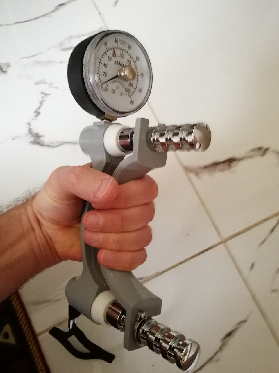
Handheld dynamometer (Image by Ashayou_Wikimedia_Commons_Creative_Common_CCO1.0)
Both the ME/CFS patients and the healthy controls were able to produce about the same handgrip force/strength at the beginning. This will be the sole completely normal finding in the study – and it will turn out to be an important one.
The rest of the tests indicated that while the brains and muscles of the healthy controls became activated, neither the brains nor the muscles of ME/CFS patients did. From what we could tell from these tests, it was as if, except during the very beginning of the handgrip test, they never got the message that exertion was occurring. (It’s going to get a lot more complicated than that…)
Not Just Reduced Blood Flows (the strange BOLD MRI findings)
The handgrip test in the healthy controls resulted in an increased BOLD MRI signal (blood flows, blood oxygenation, oxygen extraction) in a wide swath of the brain (sensorimotor, thalamus, cerebellum, temporal parietal junction, middle temporal gyrus, mid-cingulate, precuneus/cuneus).
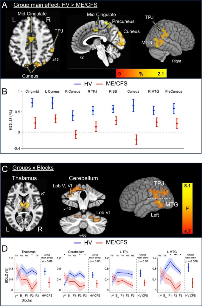
The top section shows the places in the brain where activity as measured by the BOLD MRI was greater in the healthy controls during the handgrip tests. (No part of the brain was more activated in the people with ME/CFS.) The C and D sections show what happens as the handgrip test proceeds. Note in D how, at the beginning of the experiment, the brain activation is actually a bit higher in the ME/CFS (red). As the test proceeds, though, the brain activation in the healthy controls (blue) climbs, while the activation in the ME/CFS patients’ (red) brains actually drops.
The regions that were activated made sense as they assess sensory input, force production, motor control, movement, effort, pain, attention, and the state of the body.
The fact that so many areas of the brain became activated as the healthy controls strained to maintain their handgrip force indicates that maintaining strength requires multiple areas of the brain to work together.
The BOLD findings in the ME/CFS patients started off normally but instead of increasing, actually declined as the test proceeded; that is, the parts of the brain needed to maintain physical exertion actually became a bit deactivated.
The authors did not give an explanation why this strange pattern occurred but asserted the early normal/later decline pattern likely meant that reduced brain blood flows, i.e., neurovascular coupling, were not solely responsible.
Indeed, the declines seen in the electrical activity in the ME/CFS patients’ motor cortex (see below), which does not depend on blood flows, suggest that something more than reduced brain blood flows is going to play a role.
A number of things could explain this:
- Reduced neuron recruitment – The motor cortex needed to become more and more activated and recruit more and more neurons as the fatigue increased. If it failed to do this, the BOLD readings could remain flat.
- Neurovascular coupling – the ability to send blood to the parts of the brain that need it – might be OK at rest but poop out when the brain is put under strain. If this is true, then reduced brain blood flows could play a major role.
- Reduced cerebral blood flows – blood flows to the brain as opposed to solely within the brain – might not have been sufficient as the test proceeded.
- Signals from the muscles – due to metabolic strain/energy stress, and pain – could be telling the motor cortex to shut down.
Electrical Glitch
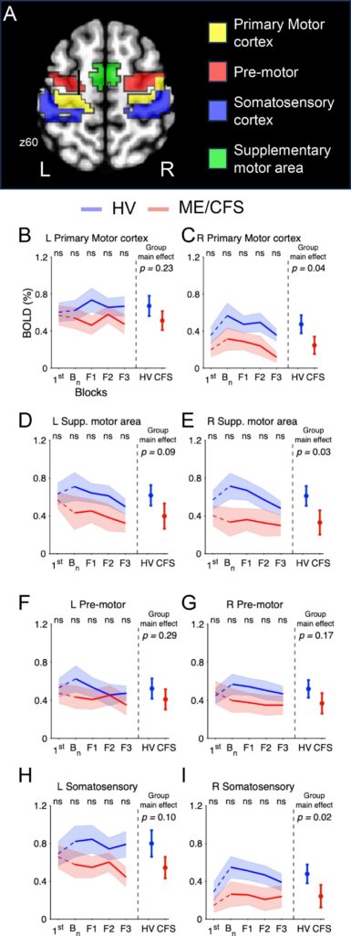
The fMRI assessed the electrical signals in the bilateral primary motor cortex, pre-motor, supplementary motor area, and somatosensory cortex (Top image). Note that activation in these areas initially rose in the healthy controls but immediately dropped, or rose less, in the ME/CFS patients. Note also that the primary motor cortex was not the big problem. Other, more secondary parts of the brain that are involved in exertion were. The finding does not suggest that the
As the muscle gets fatigued, the motor cortex and its supporting areas should light up and recruit additional muscle motor units; increase the firing rates of the muscle, and more. That’s how the muscle is able to maintain strength even as it’s becoming fatigued. Over time, the brain will reach its limit, and the activity will decline.
The EEG findings indicated that electrical activity in brain areas that produce movement and force increased dramatically in the healthy controls, but in most cases, not at all in people with ME/CFS. Note that the supplemental motor areas were most affected in the ME/CFS patients.
The low motor cortex/supplemental motor/premotor/somatosensory activity in the people with ME/CFS probably prevented signals for muscle activation from reaching the muscles.
Brain-Muscle: Muscle-Brain Communication
The corticomuscular coherence test assessed how well the brain communicates with the muscles and how well the muscles communicate with the brain.
Interestingly, the brain-muscle connection was relatively normal in ME/CFS, but the reduced muscle-brain connection in ME/CFS suggested that the muscles were not sending normal levels of signals to the brain. This finding may gain in significance later.
No Muscle Fatigue !?

The really strange reading. At least electrically, the ME/CFS muscles did not show the expected signs of fatigue.
Finally, and quite significantly, there was the Dimitrov Index (DI). The DI measures how the frequency of the electrical signal changes as the muscle works.
As muscles fatigue, the intracellular ATP-dependent Na⁺/K⁺ pump activity loses steam, which causes muscle fiber conduction velocity to decrease, the fast-twitch muscle fibers to drop out, and the slow-twitch muscle fibers to ramp up.
This results in lower electrical frequencies in the muscles and a higher Dimitrov index. This is a normal muscle response to muscle fatigue.
Except it didn’t happen in the ME/CFS patients. Instead of the DI increasing, it remained about the same and even dropped a bit. It was as if their muscles never entered the normal fatiguing process.
Interestingly, the ME/CFS muscle electrical activity was not diminished – it simply didn’t shift as it should have during exercise. Despite being highly stressed, electrically at least, they didn’t look like fatigued muscles. They were kind of inert!
The DI results led the authors to a most interesting conclusion – they couldn’t technically say the ME/CFS muscles were fatigued (!).
“Put together, these results show that the ME/CFS had limited engagement of their neuromuscular system that was substantially less than what was observed in HV. This resulted in an early decline in performance, and thus, an early onset of physical fatigue; although, we should be careful before labeling this as fatigue since their neuromuscular system showed much reduced engagement.”
In the paper’s conclusion, they again noted:
“In healthy individuals, fatigability is the objective decline of performance over time. Fatigability in ME/CFS should be interpreted differently. While our results suggested that ME/CFS fatigability has a central origin, we would argue that… the fatigability process in ME/CFS did not fully take place.”
Not Engaging – Brainwise or Muscularly
So, now they had to explain how both the brain and the muscles of the ME/CFS patients simply did not engage. Why did they not pick up? That immediately brings up the effort question; i.e. were the ME/CFS patients trying hard enough? (Here we go again!).
This study did not answer that question, but they noted that reduced engagement was not conscious: “Importantly, this lack of engagement was not conscious or deliberate.”
Still, they went back to some old canards: the fear, conscious or unconscious, of triggering post-exertional malaise, musculoskeletal/joint pain, or elevated perceived exertion could be responsible.
After noting that central-acting drugs have not proved helpful in ME/CFS, they nevertheless suggested that drugs (or brain stimulation) could help the brain engage more when presented with exercise.
They reported that the most likely explanation for their results was that the fatigue in ME/CFS is caused by the brain, but that the study could not prove that. The explanation wasn’t surprising, though. It was the one they reached in the intramural study.
Taking a broader view in the conclusion of the paper, though, they shifted and stated:
“that a cascade of events affecting the autonomic, neuroendocrine, immunologic, bioenergetic, and physiologic systems… are most likely the source for the lack of engagement of the neuromuscular system.”
Other Explanations
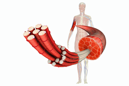
Citing the rather primitive muscle assessments done in the deep phenotyping study, the authors discarded the possibility that damaged muscles might be playing a role.
The authors agreed that damage to the muscles could have prevented them from responding, but they didn’t think this was likely because the muscle biopsies in Nath’s “Deep Phenotyping” study did not show “major” problems with muscle-fiber composition.
The muscle assessments done in that study were quite primitive, however. The one biopsy per patient simply assessed muscle type and relative muscle fiber size. A gene expression analysis of the muscles was unilluminating, but the sample size was very small.
Muscle physiology was ignored. The ability of the muscles to generate ATP, ATP or phosphocreatine availability during or after exercise, oxygen extraction, microvascular blood flows to the muscles, the muscles’ metabolic response to exercise, lactate, pH or other metabolite levels, fatty oxidation and calcium handling, and many more things – many of which have been raised in past ME/CFS studies – were not done.
The muscles could have looked just fine, structurally, but still had considerable abnormalities.
In the end, the muscle biopsy assessment may have been the weakest part of Nath’s intramural study. The evidence wasn’t much to hang a hypothesis on.
Other Explanations
Several other possibilities exist.
Damaged Muscles?
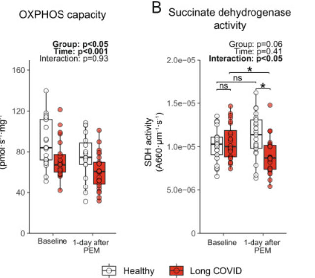
Wust found significant reductions in aerobic energy capacity and enzyme activity in long COVID.
The 2024 Wust group’s long-COVID paper, “Muscle abnormalities worsen after post-exertional malaise in long COVID“, did dig very deeply. In fact, that paper’s findings make it hard to believe that muscle issues do not play a role in the fatigue and reduced strength that accompany exertion.
Wust’s study, while not large (n=46), was still considerably larger than the Deep Phenotyping study. Unlike Nath’s study, Wust did find a higher proportion of more fatigable type II glycolytic fibers at baseline. He also found reduced aerobic energy production capacity (oxidative phosphorylation), reduced reliance on aerobic energy production, and reduced power output per skeletal muscle.
One day after a strenuous exercise bout, things considerably worsened. Wust found reduced SDH activity, worsening metabolic abnormalities; increased muscle-fiber necrosis; increased muscle atrophy and immune cell infiltration in the muscles (a big no-no).
As noted earlier, the Bedard paper authors banked their conclusions in part on the inability of the ME/CFS patients’ muscles to respond electrophysiologically to exertion. The fact that they never reached the kind of fatigue state expected led them to believe that the motor cortex must be failing to activate them.
If the muscles in the ME/CFS patients were in as bad shape as Wust’s long-COVID patients, though, it’s possible that they couldn’t respond correctly.
If that’s true, then the equation potentially flips – the brain asks the muscles to activate (note that they did react normally at first), but as the test went on, they quickly became depleted/damaged and sent a message up the spinal cord to the brain – “Stop that! You’re hurting me”. The brain says “OK – Sorry! (:)) and stopped muscle recruitment (motor cortex) and activating the brain regions (BOLD MRI) needed to keep the force up.

Were damaged muscles telling the brain to stop activating them?
The muscle-brain connection is solidly based in science. The muscles produce over 50 proteins that affect the brain, and affect so many processes in the body that they’ve been called a second endocrine organ.
One result from the Bedard handgrip study – the normal coherence between the signals coming from brain to the muscles and the reduced coherence between the signals the muscles are sending to the brain – suggests that this is what happened.
One of the Wust group’s study results produced quite a challenge to the top-down brain hypothesis. By testing the muscle biopsies in the lab, Wust completely separated them from the brain.
His finding that long-COVID patients’ muscles generated less power per unit of muscle than the healthy controls in the lab indicated that the muscles themselves lacked power. Neither reduced effort, conscious or otherwise, nor a balky motor cortex could explain that result.
His latest ME/CFS/long-COVID study, “Skeletal muscle properties in long COVID and ME/CFS differ from those induced by bed rest“, suggested that skeletal muscle issues such as reduced capillary supply and mitochondrial impairments characterized both ME/CFS and long-COVID patients. Neither of those factors, of course, were assessed in the Bedard or Nath studies.
Wust’s findings don’t mean that the brain isn’t playing a role – the handgrip study suggests that it is – but that it’s probably more complex than the brain simply shutting down muscle recruitment.
It’s still possible that the brain is driving the whole thing – just not in the way that Bedard proposed.
The Brain-Muscle Whack Possibility
Is it possible that the reduced brain activity shown in this study could have damaged the muscles by not supporting them when they tried to work? As the fatigue proceeds, the brain should be recruiting new muscle units. What if that doesn’t happen and the same block of muscles gets “tortured” again and again?
Presumably some damage could result. The fly in the ointment in this idea is that the overworked muscles should still have entered a fatigue state characterized by a high DI. In fact, the ME/CFS patients’ muscles – now really struggling under the load – should have had screamingly high DI ratios. Instead, they had a low DI index.
Because DI only reflects the electrical properties of the muscles, though, it would miss problems with ATP production, metabolic stress, microvascular problems, etc.
In this conception, the ME/CFS muscles become stressed and send signals to the brain asking it to recruit more muscle units, but it fails to do so, placing more stress on the muscles (which the DI is unable to pick up) and leading to the exertional damage that Wust found.
Infection/Neuroinflammation – the One Hypothesis to Rule Them All?

Perhaps the most comprehensive explanation (but also the newest) is that infection-induced neuroinflammation damages the energy processes in the muscles, leaving the muscle fibers largely intact, but preventing them from producing normal amounts of energy.
The last possibility may make the most sense of all. A 2024 study prompted by the coronavirus pandemic produced neuroinflammation in the brain by introducing a variety of infectious factors into laboratory animals.
Oddly enough, it found those factors reduced muscle mitochondria activity while preserving the muscle fibers that the Nath intramural/deep phenotyping study looked for but failed to find.
The senior author of the study stated:
“This is more than a lack of motivation to move because we don’t feel well. These processes reduce energy levels in skeletal muscle, decreasing the capacity to move and function normally,” Dr. Aaron Johnson
He proposed that neuroinflammation may shut down the muscles on purpose in order to save energy. The study found that even after an infection is cleared, the process can become chronic.
Johnson’s results are based on animal studies, but he pointed out that the process is evolutionarily conserved – suggesting that the same process is probably seen in humans.
This scenario potentially ties together the Bedard brain and Wust muscles findings, Jarred Younger’s neuroinflammation findings, and Systrom’s preliminary muscle findings (see below) into the mix to boot (!).
The authors proposed JAK/STAT inhibiting drugs – which are being assessed in long COVID – and IL-6 inhibitors be trialed.
Conclusion
While the authors’ conclusion that fatigue in ME/CFS is “central” (i.e., caused by the brain) is supported by their study, other long COVID and ME/CFS studies suggest that muscle abnormalities will play a major role. The big question now is which one is driving the muscle fatigue, and ultimately, PEM.
With Wust continuing his work and the Open Medicine Foundation digging deep into ME/CFS, we will learn much more about the role muscles play. The Open Medicine Foundation’s muscle biopsy study, which uses exertional stress, has been underway for a couple of years and should be nearing completion.
With the study about 2/3 done a year ago, Dr. Systrom provided preliminary results from the 50-ME/CFS person study. Thus far, it appears that people with ME/CFS have an “acquired” (not genetic) mitochondrial problem that results in a reduced number of mitochondria and impairs the muscles’ ability to produce energy.
Some people also have reduced activity in the last step in producing ATP – the electron transport chain (:() – while others, apparently trying to compensate for the reduced mitochondrial levels – have increased ETC activity.
Note that reduced muscle mitochondrial activity is what Johnson found occurred after he produced neuroinflammation by introducing infectious factors into laboratory animals.
In the end, this study opens up many intriguing avenues. It’s clearly time to merge brain imaging studies with muscle metabolism, brain and muscle blood flow, and immunological studies. Using exercise stressors would be critical to understanding how PEM develops. This field is ripe for exploration.
Support Health Rising and Keep the Information Flowing!
Health Rising is not a 501 c (3) non-profit





My experience as a patient is that in ME/CFS there are different types of fatigue and exhaustion. The easiest to discern are an exhaustion that is just like the one that accompanies infections and inflammatory processes, which made me think from the beginning that I had an infectious disease. After episodes there is clearly regenerative fatigue. Also, there is mental fatigue following the physical fatigue. And then there is deep mental fatigue from living with the overwhelming situation of ME/CFS that can again feel like a deep physical exhaustion sometimes, and I am sure others have even more. It’d be important to qualitatively map them and think about possible causes and develop proper concepts before investigating them biomedically.
I mentioned that study by Johnson more than a year back.
The brain is a big part of this disease, probably the key.
Dr Andrew Lloyd was saying that at the back end of the Dubbo study nearly 20 years ago.
Some patients went mental with him – couldn’t contemplate that a virus / immune system may not be the primary issue perpetuating the illness.
Sad.
What specific data base this claim? Relying on the hypotheses of Nath and Pointing—which push a re-psychiatrization of the illness concept based on weak signals from single, non-replicated studies—overlooks where the empirical weight of the field actually lies. Over the past twenty years, the evidence for an immunological origin centered on latent viral persistence has built substantially, alongside plausible, mechanistic models showing how primary immune dysregulation subsequently impacts the central nervous system.
Chris Ponting (University of Edinburgh) has recently shifted toward a neurological or psychiatric framing of ME/CFS, despite a lack of substantial empirical evidence to support such a broad claim.
https://www.deutschlandfunk.de/me-cfs-heilen-2-2-forschung-nimmt-fahrt-auf-100.html
Neurological does not equate to “psychiatric”; the latter is a subset of the former, with a higher proportion of enviro influence…
Fifty years of neuropsychiatric research haven’t brought a single real breakthrough in our understanding of psychiatric conditions. Quite the opposite: we are finally seeing growing awareness regarding untenable claims—like the idea that depression is a simple chemical imbalance cured by drugs, the uncritical acceptance of schizophrenia’s heredity, or the structural flaws in concepts like ADHD, autism, and neurodivergence.
The air is quickly coming out of these hyped topics, and it will soon be clear that billions in taxpayer money went toward dead-end research.
Do we as ME/CFS patients really want to jump on a bandwagon that is falling apart, driving deeper into a desert of ignorance regarding the true causes of these conditions?
If this field has yielded no genuine understanding at the very center of its focus, why believe that the same people who have mispsychiatrized ME/CFS for decades—and actively hindered progress—will suddenly help advance our understanding now?
https://www.sickkids.ca/en/research/research-programs/neurosciences-mental-health/
*I stopped reading after your first sentence, which is a inaccurate and emotional negative generalization. For example, Canada’s premier children’s hospital/research center summaries some of their breakthroughs:
https://www.sickkids.ca/en/research/research-programs/neurosciences-mental-health/
As a clinical psychologist, I am glad we are moving beyond an overreliance on broad spectrum psychotropic medications for mental health disorders, to better targeted interventions. Psycho-social interventions will/should also remain a 1st or 2nd order intervention for mental health difficulties — that is the origin of most mental disorders.
CFS/ME is not a mental health disorder, but might benefit from some advances in that area, as Cort has shown.
Reply
Mehmet Kozak
Mehmet Kozak on August 13, 2026 at 2:20 am
Dismissing a structural critique as “emotional” is a standard ad hominem deflection to avoid engaging with factual outcomes.
Pointing to a hospital’s promotional page does not refute the stark reality admitted by the field’s own leading neuroscientists—such as former NIMH directors Thomas Insel and Steven Hyman—who publicly acknowledged that decades of massive research investment failed to yield a single objective biological biomarker, mechanistic understanding, or novel therapeutic target for major psychiatric conditions.
A hospital’s routine clinical programming does not erase the total lack of biological breakthroughs in psychiatric neuroscience. Expecting a discipline that has failed to decode the basic pathomechanisms of its own core conditions to advance our understanding of a severe physical neuro-immune disease like ME/CFS remains entirely illogical.
It’s far more than Nath.
Jarred Younger is building a solid evidence base for the brain in ME/CFS.
Nancy Klimas – a long term advocate for a central role for the immune system in ME/CFS – proclaimed after Nath et al’s study that ‘It’s all in the brain’. that idnicates she was convinced by the evidence.
And here’s a recent Australian study:
https://www.sciencealert.com/scientists-discover-a-potential-driver-of-chronic-fatigue-syndrome-hiding-deep-in-the-brain
there’s plenty more
BTW – suggesting the issue lies in the brain does not in anyway suggest the issue is psychiatric.
I don’t see Jarred Younger as part of that movement. For years, he has been working to show that active inflammatory processes are indeed occurring in the brain. I’ve never heard him suggest that the cause is solely neurological; he has consistently acknowledged that it could well be immunological or infectious.
Unfortunately, you are mistaken about Avindra Nath’s work. His studies are actually at the forefront of the flawed ideas about the brain that have emerged in psychiatric research over the past two decades.
I’m looking forward to a well-researched book coming out this fall that critically examines this shift toward ‘neuropsychiatry’—specifically its underlying assumptions, methodology, and aims: https://marianne-apostolides.com/books/go-no-go/
Even after twenty years of empty scientific hype, too many people still treat ‘neuro’ as a mark of rigor and high quality. In reality, the opposite is true: whenever ‘neuro’ is attached to a field, it’s often a clear red flag for sloppy thinking, poor study design, and a flood of low-value research that isn’t even worth replicating.
I stopped reading after your first sentence, which is a inaccurate and emotional negative generalization. For example, Canada’s premier children’s hospital/research center summaries some of their breakthroughs:
https://www.sickkids.ca/en/research/research-programs/neurosciences-mental-health/
As a clinical psychologist, I am glad we are moving beyond an overreliance on broad spectrum psychotropic medications for mental health disorders, to better targeted interventions. Psycho-social interventions will/should also remain a 1st or 2nd order intervention for mental health difficulties — that is the origin of most mental disorders.
CFS/ME is not a mental health disorder, but might benefit from some advances in that area, as Cort has shown.
Dismissing a structural critique as “emotional” is a standard ad hominem deflection to avoid engaging with factual outcomes.
Pointing to a hospital’s promotional page does not refute the stark reality admitted by the field’s own leading neuroscientists—such as former NIMH directors Thomas Insel and Steven Hyman—who publicly acknowledged that decades of massive research investment failed to yield a single objective biological biomarker, mechanistic understanding, or novel therapeutic target for major psychiatric conditions.
A hospital’s routine clinical programming does not erase the total lack of biological breakthroughs in psychiatric neuroscience. Expecting a discipline that has failed to decode the basic pathomechanisms of its own core conditions to advance our understanding of a severe physical neuro-immune disease like ME/CFS remains entirely illogical.
Lina – Whatever opinions work for you; go for it.
Unfortunately, you are showing the phrase that “a little knowledge is a dangerous thing”.
End of interaction.
You make a fair point: I don’t claim to have a detailed technical understanding of neuropsychiatric methods. As a trained historian, however, I rely on rigorous source criticism and look to the consensus of the most respected authorities in the field—like former NIMH directors Thomas Insel and Steven Hyman. Evaluating high-level evidence and track records is precisely how one avoids getting sucked into fields of research that remain speculative and fail to deliver scientific progress.
Really interesting findings. I wonder if the muscle-to-brain signaling could actually be a key piece of the puzzle here. Could the brain’s reduced activation be a response to problems originating in the muscles rather than the other way around?
Theoretically, anything is possible.
“In healthy individuals, fatigability is the objective decline of performance over time. Fatigability in ME/CFS should be interpreted differently. While our results suggested that ME/CFS fatigability has a central origin, we would argue that… the fatigability process in ME/CFS did not fully take place.”
Seriously? That is the reason these researchers say our muscles don’t objectively fatigue while they do in HC???
Look at *their* own plot in figure 2A. This plots the force normalized to MVC (maximum voluntary contraction) over time.
What it shows: in every single measure one can see (initial force at start of a new grip period; decline during the grip period…) the force ME/CFS patients can produce is lower compared to HC as the exertion effort continues. Especially the drop during each grip period is a lot bigger in ME/CFS.
=> According to that very same definition of fatigue they use:
ME/CFS ability to maintain force over time as exertion continues is *a lot* worse and therefore the (total system of brain plus nerves plus blood and energy supply) SHOWS A LOT MORE FATIGUE in ME/CFS.
SO according to their own rules: the fatigability process in HC did not fully take place.
Sighhhh
Sometimes I can feel The polling/ pinging of my muscles by my brain especially The arms and hands. Just this systematic pinging. I have been rescuing Kittens and it has been really Stressful and my fatigue is off the chart. I actually Think after 10 years, I have once again reactivated The EBV That resulted when I got the lumbar epidurals. That was What led to ME/CFS.
Really sorry about The west. So many Fires. We, in The Central and Eastern US are getting The crazy rain and tornadoes. Look out on Drought.gov.
https://www.drought.gov/current-conditions#:~:text=As%20of%20September%2023%2C%202025,to%20the%20U.S.%20Drought%20Monitor.
On the Fourth map down you can see a link to ” Wildfire”. you’ll be stunned. Geoengineering. 🤨
Thanks!
Here is the simple answer— Mitochondrial Prof at Columbia University says that Brain did not evolve to think they evolved to manage energy.. so all effort framing is nonsense.. at molecular level Acetylcholine is affected first in case of low atp as cholinergic neurons use 40-45% of atp for Acetylcholine synthesis.. as soon as brain senses atp deficiency it shuts down peripheral atp use for Acetylcholine synthesis and this affects neuromuscularjunctionsand some other select functions.. we have borderline Acetylcholine based on symptoms and confirmed by leading researchers.. the Acetylcholine drop is not uniform across all Acetylcholine functions but selective to ensure survival.. as soon as we give mestinon the strength returns.. neuromuscular junctions are functional and transmit signal.. pls request Dr Nath to look at patients who do not tolerate any antihistamine and respond well to nicotine to find his answer..
“If that’s true, then the equation potentially flips – the brain asks the muscles to activate (note that they did react normally at first), but as the test went on, they quickly became depleted/damaged and sent a message up the spinal cord to the brain – “Stop that! You’re hurting me”. The brain says “OK – Sorry! (:)) and stopped muscle recruitment (motor cortex) and activating the brain regions (BOLD MRI) needed to keep the force up.”
This resonates with my own experience, trying to exercise with weights and heavy labour after an initial partial recovery from ME/CFS. While I did become stronger/bulkier at first, the harder I exercised, the weaker/skinnier I got. It was almost like the experience of the post-Polio weight lifters described in Dr. Richard Bruno’s book “The Polio Paradox”.
Our bodies are sophisticated and designed with contingencies and backup systems so we survive for as long as possible. For example, we have many more neurons than we actually need to function, which explains why those who are hit with Polio can regain function (as long as they don’t over exert themselves and kill the over-sprouted and somewhat weakened neurons that grew to mitigate the polio damage – like the weight lifters did).
It makes total sense that we have “instrumentation” monitoring our systems/structures and when a situation is detected that could cause serious harm, our bodies partially shut down as necessary to conserve cellular life. Sort of like a circuit breaker.
On the flip side, if the instrumentation is faulty, and flags problems that don’t really exist, then that could, say, support a more central origin for the problem (i.e. something that brain retraining by itself can fix). However, from my personal experience, it’s somewhat easier for me to believe the former. That’s not to say that there isn’t a resulting central component that develops after the body has endured a certain amount of pathological trauma – our bodies don’t distinguish between the different types of trauma.
I cannot understand why more people aren’t jumping up and down about trying microdoses of Mounjaro. It has been well documented now by many and varied medical specialists about the beneficial effects of reducing inflammation in the whole body, including the brain. At very low doses, titrating up to around 0.75mg, the potential benefits surely far outweigh any side effects or risks.
I wish it did more for me, but after 6 months all I noticed was worse sleep.
It can make one lose muscle.
Hi, from Australia, thanks for your blogs Cort. You may be interested in this study by an Australian University that indicates that ME/CFS patients have an impaired glymphatic system (not saying it is the only issue). https://news.griffith.edu.au/2026/07/03/brains-waste-clearing-ability-impaired-in-me-cfs-patients/
Glad they’re looking into it – not surprised at all they found problems. Thanks!
Fellow Aussie here. Griffith University is doing some quite interesting research with their minimal budget.
https://www.griffith.edu.au/research/health/national-centre-neuroimmunology-emerging-diseases/themes
Cort, this has been a tough time for you. I know that very soon you will find that place of rest and rejuvenation you so well deserve.
Thank you for once again putting aside your own needs to research and write this very interesting article.
Thanks, Jeanie. We’ve emailed about sleep and boy has my sleep taken a hit. It’s slowly getting better 🙂
Hi Cort,
Reading your analogy takes me back to my earlier hypothesis.
The key point for me is what happens when the neuromuscular system is already in a fatigued or damaged state:
Muscle fatigue and energy availability. As muscle becomes fatigued, ATP production and energy availability may become insufficient to sustain normal contraction and electrical activity. This could contribute to declining force and increasing fatigue.
A protective shutdown mechanism? Is it possible that damaged or metabolically stressed muscle sends feedback to the central nervous system that reduces further motor-unit recruitment—a protective mechanism intended to prevent additional damage?
Loss of motor neurons. Another important issue is whether the author adequately considered motor neurons that may have been permanently destroyed. If the available motor-neuron pool is already substantially reduced, the nervous system has fewer motor units available to recruit, regardless of the condition of the remaining muscle fibers.
Overloading of surviving motor units. This seems particularly relevant to post-polio syndrome (PPS). During the original poliovirus infection, many motor neurons were permanently destroyed. Surviving motor neurons compensated through collateral sprouting, with new axonal branches reinnervating denervated muscle fibers. This produced enlarged or “giant” motor units, sometimes several times their normal size.
Those surviving motor neurons and their associated muscle fibers are therefore being asked to do considerably more work. Over many years, this increased metabolic and functional demand could contribute to progressive dysfunction of the enlarged motor units, muscle-fiber stress, and possibly mitochondrial abnormalities.
This raises an important question: Is the problem necessarily a lack of ATP production, or could ATP be available but the neuromuscular system be unable to effectively translate that energy into sustained muscle force?
In other words, reduced motor-neuron availability, impaired recruitment, neuromuscular transmission, mitochondrial dysfunction, altered ATP production/utilization, and immune or inflammatory activation may all contribute to the fatigue and loss of force seen in PPS.
That would also help explain why simply increasing energy availability might not solve the problem: if the limiting factor is the number or functional capacity of the surviving motor units, the bottleneck may lie upstream of—or alongside—ATP production.
Although I raised this question with both Wüst and Akiko Iwasaki, neither appeared to consider the complex mechanisms underlying muscle weakness in post-polio syndrome (PPS), or whether those mechanisms might provide a useful comparison for the muscle weakness and fatigue being discussed here.
Details can be found: https://swaresearch.blogspot.com/2024/12/post-polio-syndrome-pps-summary-and-key.html
This was Dr. Richard Bruno’s hypothesis for ME/CFS as explained in his book “The Polio Paradox” chapter 17 (Fatigue by Another Name).
Thank you Scot.
I’ll order Dr. Richard Bruno’s book.
I’m currently living in Germany, and over the past two years, poliovirus has been detected in the sewage systems of several German cities.
https://www.ecdc.europa.eu/en/news-events/detection-wild-poliovirus-wastewater-germany-risk-and-recommendations
You’re welcome, Sieglinde. If I recall correctly, Bruno suggests that the substantial eradication of the polio-virus set the stage for other enteroviruses to become prevalent; some of which can damage the body in similar ways that polio does. I can send you a pdf of the chapter if you’d like (email me at srm3555@telus.net)
This company can test your DNA for the best meds to use for depression, anxiety, ADHD, etc. The VA is paying for my brother-in-law to be tested. He is 100% PSTD. It would be great if they would expand to medications used for ME/CFS and Long Covid.
https://clarityxdna.com/
Cort, I really appreciate your sharing of your own personal health challenges. I wish you well!
I also appreciate your own spin on studies you’ve read, and I only hope that all the researchers out there subscribe to and read all your articles. Without doing so, they run the risk of tunnel vision and selective perception in order to prove their theories.
It might seem that they’re obviously reading everyone else’s studies but what I’ve heard from doctor friends is that they are often too busy to read the vast number of studies out there. Your blog does a great job of pulling it all together in one place.
I just want to give a shout out for how much Cort does to give us the current news on whats happening with me/cfs in spite of all the ups and downs he is going through. I really want you to know you are appreciated and to take this opportunity to say thank you and please continue to take care of yourself as well.
Thanks LAC! I do hear that some researchers do – and yes, I have also heard that some researchers don’t have time to explore all aspects of these fields. 🙂
Well put LAC and Shira. Reading and considering all information provided is so important. Thank you Cort for using your “energy” for all to benefit, it is very much appreciated.
Hope you can remain in place and recover. Best of luck.
Ditto LAC, Shira and Mandy.
Thank you Cort for devoting so much of your precious energy to make this website such a valuable resource. The heat this summer must have been really tough for you – it’s been bad enough here in the UK. Hope your health picks up again soon.
Please – your thoughts on cancer in relation to ME and Long Covid.
My partners sister was recently diagnosed with cancer. There has been an increase in cancer since Covid. I have quite a few ME friends who have died of cancer pre Covid. So young and of course no one wrote ME on their death certificates. Like myself they had longterm severe ME and couldn’t tolerate any meds. So I did a bit of research online. The following is a PDF from a cancer magazine.
https://cancerworld.net/cancer-related-fatigue-might-research-into-long-covid-help-find-causes-and-cures/
The article was written in 2021 – I was amazed that the cancer researchers were saying 25% of those who recover from cancer are left with ME. It was interesting that they are also pinning their hopes on Long Covid research to understand cancer fatigue.
I also googled cancer and Long Covid. It suggested they are seeing a connection between the 2 illnesses. Presumably anecdotal – has there been any actual research looking at the relationship between Long Covid and cancer?
Thanks! Very little research has been done on ME/CFS and cancer. The only thing that might relate was a study that looked at mortality and ME/CFS and didn’t, as I remember, find increased mortality from cancer. It was not a big study and we need more comprehensive ones. Hopefully, it’s accurate!
PS The above link doesn’t open as a PDF – I just tried it.
Cort I know you have problems with insomnia. I’ve had ME for decades with severe insomnia. An ME friend suggested I try magnesium. I’m currently experimenting with a magnesium spray. You spray it on your skin and rub it in. Early days but it does seem to be helping – the first thing ever that’s helped my insomnia. My diet is good – it’s taken 20 years to gradually build up my intake of food. My magnesium levels in a regular blood test are normal. Maybe the levels in my muscles are low? Can’t tolerate vitamins or minerals orally – so wondered if a spray could help.
What if one reason for the different problems is bottlenecks in nutrients?
For example, can muscles be activated properly if the body is deficient in choline (and therefore also acetylcholine)? Might that be why some of us respond to pyridostigmine?
With the electron transport chain, might the difference between those who had more and less activity be that some are missing vitamins and minerals (such as zinc) that are required for that process?
If different patients have different nutrient bottlenecks, perhaps, that could account both for the inability to keep producing ATP through aerobic respiration and for the differences in which exact chemical pathways are lagging and which are trying to compensate.
Also, I wonder whether it would explain why we sometimes feel better for a while with a new supplement and then crash again–might it be that we’ve fixed one bottleneck only to uncover a new one later down in the process (or else a new one in a different process that we depend on more as we become more active)?
Thanks for this excellent coverage, Cort!
We are all better for your research, too-tier composition and freely sharing.
Wishing you safe travels and more symptom free days. Thanks again.