




Geoff’s Translation
The GIST
The brain has been showing up a lot recently. (vintage image by Oberholster Venita from Pixabay)
Back to the brain! Years ago, Andrew Lloyd of the Dubbo studies noted that the symptoms in ME/CFS were all symptoms produced by the brain – and we are overflowing with brain studies right now.
We just looked at the Bedard group’s study, which proposed the brain is the major player in fatigue. A new paper on brain norepinephrine was recently published, and Griffith’s group in Australia has published two brain studies – one on the glymphatic system, and one on water movement through the brain – which the current blog focuses on.
The Study
Brain imaging studies are usually small, and this one by Singh, while not bad for a brain imaging study, is of that ilk (37 ME/CFS, 19 Long COVID, 27 healthy controls). The Griffith group has been digging into the brain in ME/CFS for years. Their latest study, “Microstructural alterations in brain tissue of ME/CFS and long COVID using diffusion tensor imaging and diffusion kurtosis imaging“, expands on an earlier one and brings something new to the field.

It adds something called diffusion kurtosis imaging (DKI) to the diffusion tensor imaging (DTI) that’s usually done. Instead of blood, DTI assesses how easily water moves through brain tissue, and in which directions. Because healthy axons, which the nerves use to communicate with each other, affect water movement differently than unhealthy axons, DTI is often used to assess the microstructural integrity of white matter that the nerve axons are made of.
The GIST
The brain has been showing up a lot recently (Vintage image-by-Oberholster-Venita-from-Pixabay)
- Brain studies are all the rage right now. This is the second of a series of three blogs on new findings in ME/CFS and/or long COVID.
- This Australian team from Griffith University has been leading the charge on brain imaging studies in ME/CFS for years. This time they added a new factor as they assessed how water was diffusing down the axons (the long connecting part of the neuron) in the brain.
- This water diffusion trick tells them something about axon health and how well axons communicate between different regions of the brain. It turns out that many problems in the brain are caused by poor connectivity or communication between its different regions.
- This turns out to be so in ME/CFS. Two abnormalities in nearly the same place in the axons in the left cingulum suggested that this part of the brain – largely involved in cognition – may have gotten hit hard.
- Because this long neural pathway links the cingulate, frontal, parietal, and temporal regions, problems with attention, executive control, memory, emotion, and internally directed cognition could result.
- A bell went off when I saw that multiple problems were found in the supplementary motor area as well. The supplementary motor area was among the most underactivated regions of the brain identified in the Bedard study the last blog focused on.
- The SMA is a most intriguing part of the brain because it’s involved in “motor planning,” i.e., movement, including initiating and organizing voluntary movement. It’s where the brain transitions from the intention “I want to move” to the actual production of movement.
- In my experience, it’s that transition from intention to movement that often seems so tricky. I might want to move; I might think or feel I can move, but once I start moving, the problems begin. Problems here would translate into more effortful movement, reduced motor drive (reduced muscle recruitment), poor coordination, and increased fatigue.
- Lastly, problems were found in a long, dense, thick wire called the corpus callosum that connects the two hemispheres of the brain. Problems with sustaining attention, having difficulty switching tasks, mental fatigue, movement, and sensory stimuli could result.
- All in all, the regions identified make sense with what we know about ME/CFS. While other brain regions that clearly play a role in ME/CFS deal with “inputs” (assessing sensory data, energy production, agitation, exhaustion), these neural pathways deal more with “outputs” – like movement.
- Pairing all the brain regions affected in ME/CFS together seems to lead to an inevitable conclusion: movement or exertion will be very effortful.
- These types of brain scans cannot tell us why there appear to be blockages or weaknesses that impair connections between different regions of the brain. Nerve damage could be present. Neuroinflammation, metabolic issues or blood flows could also be causing.
- One possibility is that something in the extracellular spaces outside the axons could be disrupting water flow. That’s an intriguing possibility given that Rob Wust in the Netherlands has found that enlarged basement membranes in the capillaries are affecting blood flows in the muscles. Could a similar problem also be present in the brain?
- Could interruptions in fluid flows(water, blood, lymph, cerebrospinal fluid – brain; blood-muscles) across the body be behind the systemic symptoms in ME/CFS? Time will tell.
Support Health Rising and Keep the Information Flowing!
Health Rising is not a 501 c (3) non-profit
Because DTI studies are focused on the axons, they’re concerned with the brain’s “wiring” or how the different regions of the brain communicate with each other.
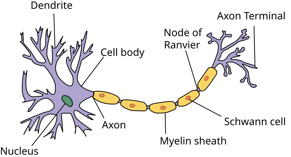
DTI/DKI assess the movement of water along the long axons that connect one nerve cell to another. (Image from Anatomy and Physiology US National Cancer Institute’s SEER Program, DHP 1080_ Wikimedia_Commons)
DTIs can tell us, for instance, if the prefrontal cortex is not communicating well with, say the limbic part of the brain. It cannot tell us why, however. For instance, it cannot differentiate between demyelinated axons, damaged axons, or processes like neuroinflammation that could impair nerve transmission as well.
Diffusion kurtosis imaging (DKI) uses the same (but longer) MRI and adds a more sophisticated modeling and statistical analysis. It can also assess water flow through the surrounding tissues and detect pathologies that DTI would miss. It’s particularly useful in assessing neurodegeneration, ischemia, and aging.
Various measurements are done using DTI. They include:
- FA: We want free water movement in the brain along nerve axons. Increased FA could result from axon issues, edema/neuroinflammation, myelin loss, etc.
- MD: average overall diffusivity. Increased MD levels would indicate increased water content.
- AD/RD: diffusion parallel/perpendicular to the fitted primary direction. They are sometimes interpreted as axon- and myelin-related proxies, but neither is a direct measure of axonal injury or myelin content.
Results
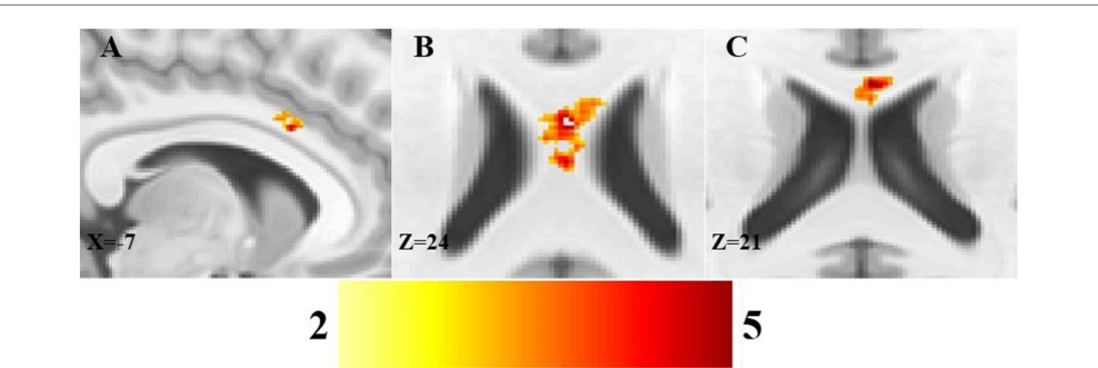
The ME/CFS results.
Cognition Gets a Double Hit
The increased FA on conventional DTI and increased KFA on diffusion kurtosis imaging in the left cingulum were a strong reading because abnormalities showed up on both DTI/DKI in almost exactly the same region of the brain. That is strong, suggesting the Australian group is onto something there.
The increased FA suggests that something is off with the axons, which is interfering with water movement. This could be due to problems with nerve fibers, cellular swelling, or alterations in the extracellular spaces surrounding the nerves.
The cellular swelling that interferes with the free flow of water in the brain is intriguing because it could result from an infection and/or microglial or astrocyte activation (neuroinflammation).
The increased DFI suggests that something is altering the direction of the water flows. It could be something to do with the axons, the myelin coating them, the glia cells that hold them in place, or again – the extracellular space surrounding them.
If you experience brain fog, an inability of the cingulum pathway to relay signals normally may be one reason why. A long nerve pathway linking cingulate, frontal, parietal and temporal regions, the cingulum is involved in attention, executive control, memory, emotion and internally directed cognition.
The Supplementary Motor Area (Again)
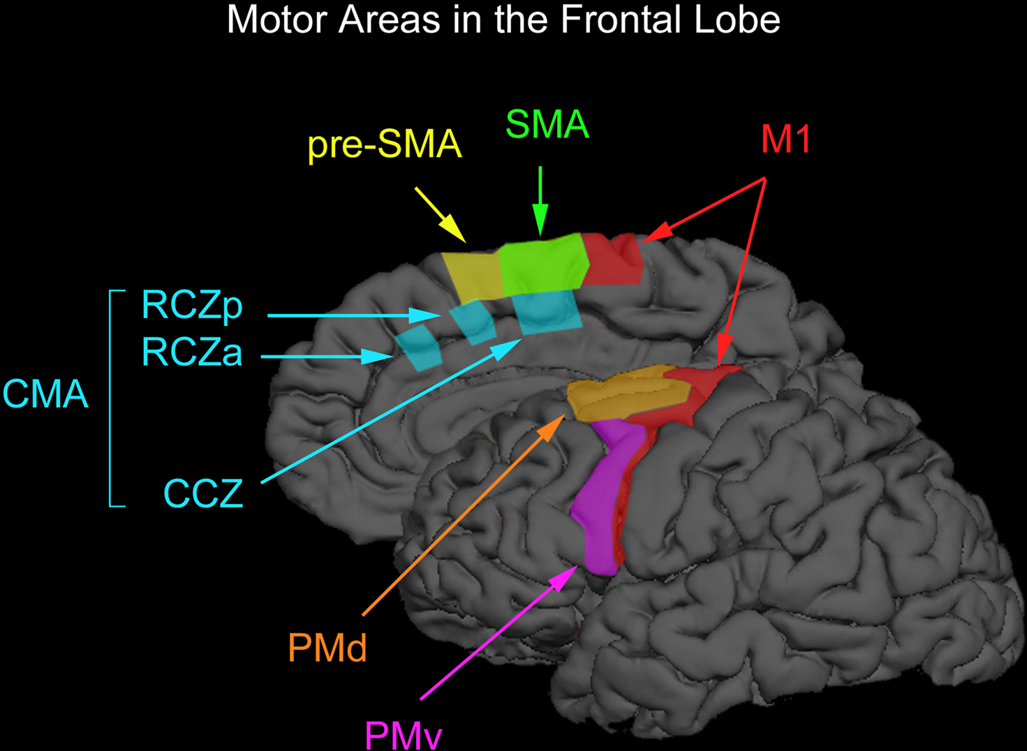
The motor areas found in the frontal lobe. The SMA is in green at top. (Image from Chouinard PA and Paus T (2010) Front. Hum. Neurosci. 4173. doi10.3389fnhum.2010.00173)
The significantly lower values across all three DTI measures (MD, AD, and RD) strongly indicated that water movement is obstructed in all directions within the supplementary motor area (SMA).
The SMA is a most intriguing part of the brain because it’s involved in “motor planning,” i.e., movement, including initiating and organizing voluntary movement.
The name “supplementary” motor area kind of downplays how important this part of the brain is. It’s an intriguing area for ME/CFS because it’s in the SMA that the brain transitions from the intention “I want to move” to the actual production of movement.
In my experience, it’s that transition from intention to movement that often seems so tricky. I might want to move, I might think or feel like I can move, but once I start to move, the problems start. Problems here would translate into more effortful movement, reduced motor drive (reduced muscle recruitment), poor coordination, and increased fatigue.
This is also, notably, one of the same brain areas where the Bedard paper found reduced activation. In fact, the supplementary motor area was one of the most underactivated parts of the brain.
These two findings suggest that the movement problem in ME/CFS may be primarily in the deeper brain pathways involved in planning, rather than in the motor cortex. They also align with the idea that the brain is a major source of fatigue in ME/CFS.
The findings might suggest there’s less actual axon damage than there are problems in the microenvironment around the axons. That’s probably a good thing as the less nerve damage, the better! That could point us toward neuroinflammation or connective tissue problems that are impairing the free flow of fluids. More on that later.
A Big Wire Gets Hit
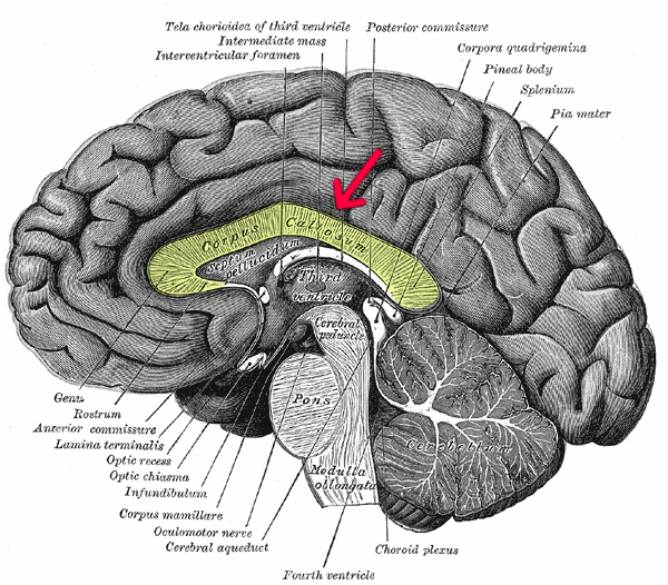
A big wire gets hit! Damage to this connector could affect sustained attention, difficulty switching tasks, mental fatigue, movement, and sensory stimuli. (Image from Gray’s Anatomy 1918)
The authors proposed that the decreased axial kurtosis in the genu/body of the corpus callosum “probably means there is axonal injury or loss of integrity”. The corpus callosum finding makes so much sense given what we know of these diseases. It is a large nerve fiber that connects the two hemispheres of the brain and affects a triad: cognition, movement, and sensory function.
Problems with sustained attention, difficulty switching tasks, mental fatigue, movement, and sensory stimuli could result.
The decreased axial kurtosis suggests that the microstructure of the axons in the corpus callosum itself has been affected. It does not mean that the axons are damaged per se – they may just be weaker and not working well. They could be thinner than usual, the myelin protective coating may be affected, or extracellular space problems could be kicking in.
Impaired Diffusion
We don’t know what’s causing these issues, but note that this study measured “diffusion,” i.e., the free flow of water along neurons. Problems with the extracellular spaces were particularly prominent in two of them.
The authors didn’t focus on extracellular spaces, but they’re interesting because a changed or remodeled extracellular matrix could easily inhibit the flows of solutes around the neurons.
That brings us back to, who else, Rob Wust and the extracellular matrix remodeling his group found in the microvasculature of muscles. It looked like thickened basement membranes were blocking blood flow to the muscles. They believed these changes were reducing oxygen/nutrient delivery to the muscles and toxic metabolite removal from them.
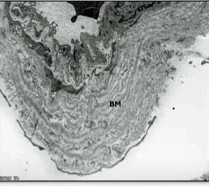
Check out the massive basement membrane (BM) found in an ME/CFS/long-COVID patient. Could something similar be happening in the brain?
If extracellular-matrix/basement-membrane were also “remodeled” in the brain, they could impair the movement of water/solutes from the tiny interstitial spaces surrounding the neurons to the perivascular spaces surrounding the blood vessel.
Infection-triggered chronic inflammation/neuroinflammation could do the trick. So could, interestingly, problems producing energy. If ATP-dependent ion pumps fail, water could flow into the cells, causing them to swell, which could impair fluid flows outside the cells. Impaired cerebral blood flows could be responsible as well.
Note that while axon damage could be occurring, it’s not required to produce any of this.
In this scenario, the glymphatic system and cerebrospinal fluid flow would likely be affected. A recent Griffith paper and a recent Stanford paper suggest a clogged-up glymphatic system could play a major role in ME/CFS.
That potentially fits an intriguing theme in these diseases – problems with the free flow of liquids, whether they are in the brain (blood, water, lymph, cerebrospinal fluid), or the muscles (microvascular blood flows).
Connections
It doesn’t seem like damage to a single brain region is going to be it for ME/CFS or long COVID – and that’s probably a good thing. Instead, the problems will probably lie in damaged connections between brain regions.

The deeper researchers dig, the more connections they find – a very good thing. The light bulb, or driver, at the heart of all this is still unclear, but could be inflammation, metabolic problems, and/or disruptions to fluid/blood flows.
Quite a few brain networks are impacted in ME/CFS. Some of the major ones include the salience network, default mode network, the prefrontal cortex and its network, and autonomic‑brainstem circuit. These networks can produce symptoms such as sensory overload, a wired-but-tired feeling, brain fog, reduced attention spans, and difficulty standing and exercising.
The brain regions that popped up in the last couple of studies – motor planning / motor cortex / SMA / cingulate cortex / corpus callosum / brainstem – fit quite nicely into the picture. While the other regions deal more with inputs, these regions deal more with the output side of the equation; i.e., they come into play when you decide that you want to walk across the room.
Here’s the problem for them. They have to take into account the signals already present in the brain (low energy, high pain/sensory stimuli, autonomic issues) and then try to add movement on top of them. The inevitable outcome is a high degree of effort.
They’re showing a kind of brain that’s designed to knock you down and keep you down. No wonder functionality is so poor.
It’s a pretty devastating picture, but the nice thing is that it all makes sense. We’re not seeing weird brain regions that no one can make head or tail out of popping up in these studies. Even with their small sample size, the brain imaging studies are producing a coherent picture, and that’s crucial for this field.
Every study opens up new possibilities. By adding DKI to the equation, the Griffith researchers showed that it’s not just the axons but also the regions outside them that are affected in these diseases.
A follow-up study could include brain imaging to assess neuroinflammation and myelin damage, and would evaluate brain vascular basement membrane or extracellular matrix markers, as well as perivascular spaces, to determine whether anything is impeding the free flow of water and solutes in the brain.
That would tell us whether the nerves themselves are damaged, whether inflammation could be a culprit, and whether the passageways through the brain are being squeezed.
The systemic nature of the findings in ME/CFS and long COVID suggests that a fundamental, body-wide factor is responsible. Something that inhibits the free flow of fluids (blood, water, lymph, and/or cerebrospinal fluid) would surely fit the bill. Time will tell!




“In my experience, it’s that transition from intention to movement that often seems so tricky. I might want to move, I might think or feel like I can move, but once I start to move, the problems start.”
That I know well. Especially when I was at my worst, it was very common of wanting to stand up from a chair, doing ‘in the brain, the intend’ everything I usually do and nothing happened. Not a single bit of movement. Not even feeling a slight tension in my leg muscles. I then had to focus manyfold (emergency peak adrenaline strength!) as strong as normal for my legs to move. A similar stronger need to focus was nescessary for near any action ranging from thinking to swallowing food. And as many of you who experienced this too will be able too tell, that amount of focus is very exhausting and rapidely coming with a great payback cost.
It’s exactly the opposite of the ‘PACE study’ conclusion: patients with ME/CFS do not do less effort to get anything done, but massively more! Telling they are afraid trying to do anything and will only get better if they try harder isn’t going to help them if this study and what it means holds true!
Yikes; that is not a comment, that is a diatribe — a lot of energy.
The first two paragraphs are nothing but a description of symptoms; the first paragraph by Cort and the second by me.
That leads to the third paragraph: contrasting the result of the above research and both our experiences to the PACE study, patient reports and the ideas in above study. I wrote this for several reasons.
1) The outcome of the PACE study, providing CBT + GET, is still allowed to be promoted (and effectively promoted) as a scientificaly proven safe and helpfull treatment in many places. That science is under attack from many big names and organisations including multiple medicine Noble Price winners and the biggest statistical organisation in the world, to name but a few of the very long list of reputable sources of well argumented criticism on the papers’ methodology, results and conclusions.
2) Thousands upon thousands of patients reported a strong to very strong and often permanent decline in remaining health, abilities and quality of life coming with their PACE style CBT + GET treatment. That statement goes beyond it being a biased view of the situation. See https://www.pnas.org/doi/10.1073/pnas.2426874122, with title “Patient-reported treatment outcomes in ME/CFS and long COVID”.
See Fig2: 70% of patients who took the theraphy reported they got at least moderately worse, with 50% much worse against roughly 5% got at least moderately better and 1% much better.
One can argue there will be some reporting bias, but these numbers are truelly massive. If self reporting would invallidate such very clear results, then they would even more invalidate the very weak / none results the PACE study yielded after external evaluation of the date obtained after a lengthy legal battle. After all the bulk of the results the PACE study relies on are symptom severity self assessments before and after the study.
3) Yes, I am one of those people who got much much worse during and after such CBT + GET theraphy. I admit that creates an emotion. It is however not an emotion of hate, but a very strong drive to find actual answers to this dissease and try and devote what few energy and ability I have left to try and help our deeply suffering community. You can debate the quality of my public contributions so far. Rather then hating the study and those who are responsible for it, I chose to learn and embrace the study. Yes, embrace it! One of the strongest challenges patient lead research has is that it is very easy to disqualify the knowledge that non-medical schooled patients possibly can gather. It is easy to disregard all of their ideas if they miss out on some basic concepts. That is where PACE comes to the rescue. Discrediting all of patients’ work by one or a few wrong ideas plus their lack of medical schooling is easy. But PACE is, according to world reknown scientists and leading scientific organizations, full of holes and problems. And as a mathematically schooled person I can say the statistics used in the paper are horrible. I can be disqualified for not having a proper medical education, but the authors of that paper demonstrated a worrying lack of knowledge (or effort to correctly apply it) of statistics. Both medical knowledge and statistical knowledge are equally important in a non-blind patient questionnary based study. I learned to turn my emotion in strength, not hate nore diatribe.
4) The brain study, discussed in this blog, with quite likely much stronger methodology and much more objective outcomes gives a good explanation why the entire hypothese upon which the PACE trial was build, the idea that patients get deconditioned due to fear of movement and exertion and that that is a main contributor to their illness, would be simply wrong. As I said, if it holds true so upon further analysis and after external confirmation of these results. Thoses are things I strongly encourage.
5) The end result of 4) could mean a final dismissal of the PACE study as valid and correct science. With it, the horrible outcomes of the resulting CBT + GET theraphy on patients (which has often been directly or indirectly forced on patients) as demonstrated in the PNAS paper I linked could finally come to an end. There is no need nor desire to put any more large numbers of (before treatment) half functional people into a wheelchair or make them bedbound. Yes I agree. I say this with emotion. Not with an emotion of hate and angre I could hold as one who was put in a wheelchair by such theraphy, but by the possitive and energizing emotion of hope that drives me to try and be of help to current and future patients.
I am giving constructive feedback. If you want your opinions to be read (by more people), you would keep them much more concise and coherent — i.e., edit them.
“Yikes; that is not a comment, that is a diatribe — a lot of energy.”
What was constructive about this? You played on the messenger, giving zero arguments as to why this was diatribe or was full of energy.
Now again you hide behind the facade of being objective and commenting without charged emotion. You try and take the higher ground by it, yet you go flaming against an author building a reasonable argument why this brain research could become a key piece in finally puting the PACE study and its results where it should be.
Maybe that’s what caused your original passive yet obvious aggressive reaction: because it could put the final stake through this study that should not have been published and certainly no longer should be upheld after all those deep flaws that are exposed in it?
People who actually try and help patients do not have the same luxury that the defenders of PACE have. They can be concise as there are no more arguments left to defend PACE and its current day practice.
Hi Harold. I’m sorry you have such a hard time reading.
I know it’s exhausting.
I’ve had a lot of success in using AI to simplify things.
I put Dejurgen’s info above into ChatGpt in blocked mode, so it’s not saved, and asked for 2-3 sentence summary of each point.
That way you can read it slowly and come back and read it
it several times as you have energy.
See below.
1) The PACE study’s CBT + GET treatment is still promoted in many places as a scientifically proven and safe treatment, despite substantial criticism of its methods, results, and conclusions. This criticism comes from many respected scientists and organizations, including Nobel Prize winners and major statistical experts.
2) Thousands of patients have reported becoming significantly, sometimes permanently, worse after PACE-style CBT + GET. In a large PNAS study, about 70% of patients who received the therapy reported becoming at least moderately worse, compared with roughly 5% who reported becoming at least moderately better; while self-reporting has limitations, these differences are very large.
3) I am one of the people who became much worse during and after CBT + GET, so naturally this is emotional for me. But I try to turn that emotion into motivation: rather than simply rejecting PACE, I study it because its serious methodological and statistical problems can help demonstrate why patient-led research deserves to be taken seriously.
4) The brain study discussed in this blog appears to use stronger methodology and more objective measures than PACE. If its findings are confirmed through further analysis and independent research, they could seriously challenge PACE’s central idea that patients are mainly de-conditioned because they fear movement and exertion.
5) If these findings hold up, they could help establish that PACE is not valid science and bring an end to the widespread use of the resulting CBT + GET approach.
I say this not from hatred or anger, but from hope: I want to prevent other patients from suffering the kind of severe deterioration that left me needing a wheelchair.
Many of us on this forum are here because we have academic backgrounds and are used to reading lengthy and complex documents. As such we enjoy the comprehensive research overviews and the observations of other members. We aren’t fazed by the longer read.
We also have a lot of compassion and “forgiveness” for spelling misteakes and grammartical errors because many of us have cognitive issues which have rendered us less accurate than we were in our previous lives, before we were foot tripped by this illness.
See my comment below
Harold, I thought Dejurgen’s comment about the severity of his condition was very easily understood.
I want to second what can happen if you insist on pushing too hard. I have had ME/CFS since the early 1980’s and Covid twice, 8 months apart. A month after the first Covid infection I had heart irregularity and high blood pressure. An excellent cardiologist helped control this so 8 months later, we flew to Maui to see our daughter. I got Covid again, but this time it affected my balance and ability to walk. I am very stubborn so I thought PT was the answer. Not so. PT started a cascade of health problems that I haven’t gotten over yet. I have had 3 falls, dizziness and a sensation called “burning brain”. I can’t do any of the things I could do before Covid. Although I had excellent doctors for ME/CFS and was able to live a fairly normal life if I paced myself, I have not been able to get back to my pre-Covid self.
Calling a patient’s account of their illness “a diatribe” is heartless.
Thank you for writing this and the following reply. It puts the details together in a coherent explanation. I truly hope you are right and we move on from GET/CBT into the real physical cause(s).
Thanks for your kind words T Allen. I know being concise isn’t my strength. Wording difficult things is even more challenging for me as an ME/CFS and FM patient then reading.
I however doubt that is Harold’s issue. Harold and Lina have a similar discussion at Cort’s previous blog https://www.healthrising.org/blog/2026/08/12/fatigue-me-cfs-brain/.
There he writes he is a clinical phsychologist and he tries to make a case for providing psychosocial interventions for ME/CFS. Granted, he acknowledges ME/CFS is not a mental health disorder but states it might benefit from such treatments.
It is up to him why he reacted rather strong to my original comment. I do not oppose proper psychological support to reduce stress levels in ME/CFS. Stress is one of plenty of things that consume some of our scarce energy. I believe most or near all severe patients need to have addopted strategies to bring stress levels well below of that of healthy controls, even under very poor conditions of life. It’s simply a matter of survivial. Any succesfull therapist better understands that.
When it comes to PACE, it can easily be read in the publication that they start from the premisse that patients do not exercise enough due to fear that exercise makes them worse and that they need theraphy to overcome that fear (and hence do more effort). There is an overwhelming ratio of patients who disagree versus agree with that due to past experiences. This brain paper potentially helps to explain why this is the case.
If psychotherapists want to make an actual positive contribution to ME/CFS patients’ lives, I call for them to start and make the separation between actual quality reproduced science and some past failed approaches that have zero scientific base left. That is calling for less animosity and feud and more cooperation between patients and well meaning therapists.
Kind regards to all in this subthread,
dejurgen
Ditto…must concentrate very hard & is delay between thought & act….
When it comes to tissue deformation, it is well known in engineering that a round (2D) or spherical (3D) shape provides more strength per amount of building material then more uneven forms like egg-shaped or ellipse.
Hence: if tissue quality becomes poor, contracting the tissues and reshaping to a more round-ish shape is going to use less material for the same contained surface (2D) or volume (3D). Or for the same amount of material, the ‘walls’ will become more dense and stronger.
Artheries in ME/CFS tend to contract. That yields smaller cross sections with less blood flow, but also stronger arterial walls for the same amount of cells and walls available. And at the same pressure the pull strength on the tissue will further decrease if the enclosed volume becomes smaller. It’s a quadratic (eg very strong) relationship and well known in pipe calculations. High pressure pipes in hydraulic machinery all have a small inner cross section for fluids to flow and plenty of metal / material for the pipe. If you’d see a cross section of it, much to most of the area is the outer material building the pipe leaving only a bit of area for fluid flow.
Anyhow: if tissue is weakened / damaged and can reshape, then reshaping from a long dragged out form to a more circular and spherical form is a very logical thing to do. Oxidative stress or an overactive immune system is quite an assault to cell membranes and tissues.
When plenty of tissues start to ‘shape-shift’, that might give the impression of creating more space. While true, swelling can counteract that (or if you look at it this way swelling may require taking up a more spherical form in order to not splash a cell open) and any form of deformation can create local pressure or tension on other tissues. Nothing exactly fits the way it is supposed too anymore.
I see in the OMF Canada study that the stiffness of red blood cells is significantly greater in ME/LC patients than others. That leads to misshapen cells that are less able to navigate the micro vascular system and fits with your idea of taking up more space than ideal. I think you’re probably aware of that research but I am still joining the dots…..
Those studies exist. Are you also aware of:
https://link.springer.com/chapter/10.1007/978-3-642-67059-6_10
“Incubation of defíbrinated samples for 24 h at 37° C resulted in ATP depletion accompanied by large increases in the hemolysis resulting from exposure to shear stress.”
=> translated: RBC with ATP shortage more easily ‘burst’ when they are pushed by blood pressure through narrow capillaries; ATP shortage is a common observation in ME/CFS
https://journals.physiology.org/doi/full/10.1152/ajpheart.00875.2024
“However, circulating blood is not a cell-free fluid, it contains mechanosensitive red blood cells (RBCs) that are also subjected to altered hemodynamics and release a large amount of ATP”
=> translation: when RBC are forced through narrow capillaries, they release much of their scarce ATP (and risk ending up in ATP depletion). This ATP is released as a stress danger signal. This can signal vascular endothelial cells to widen the capillary but it also can signal and activate immune cells (the subject of this study).
“Suppression of RBC-released ATP via deletion of Pannexin 1, a mechanosensitive ATP-permeable channel, reduced high-fat diet-induced aortic plaque burden by 40%–60%.”
=> immune activation is involved in arterial plaque formation
“The computational prediction also provides a physics-based explanation for RBC-released ATP-induced sex disparities in atherosclerosis”
“Male mice also showed significantly higher plaque development at abdominal aorta (31.24%) than that at thoracic aorta (11.48%). Female mice had much lower plaques developed in those two regions (8.65 and 8.69%) than males without showing a significant difference”
=> translation: surprisingly, this local ATP release activates the immune system less in females then in males (in mice), resulting in less plaque formation. While that should be good in ME/CFS, it also provides less dilation / widening of the blood vessels at sites where RBC get ‘near stuck’ in females then in males and that is bad for bllod flow in ME/CFS.
Good idea re:translation. 🙂
I try whenever possible. Explaining something well is only possible when understanding things in much more depth then what the short summary says.
With most new ideas, that’s impossible. Writing down new bits for others to read gives new ideas a chance to grow into better ones. That brings some inconvenience, for which I apologize, but also gives hope on progress.
Lloyd was right. The brain is key
Unfortunately Andrew got a hard time from many hysterical patients by departing from the dominant narrative around viruses / immune system.
And we can go back also to Jay Goldstein’s limbic hypothesis as being pretty warm in terms of the key mechanisms involved in this illness.
None of this is to say that the immune system isn’t involved. It certainly is. But it’s involved centrally (ie. brain / CNS) rather than peripherally.
I thought Lloyd was from the psychosomatic (CBT) school.
Lloyd lead the Dubbo studies and was a fully into exploring the pathophysiology of ME/CFS at the time he made that quote. Later, he became more focused on CBT. I was so incensed by the change that I wrote this post.
https://www.healthrising.org/blog/2015/05/16/the-decline-and-fall-of-an-mecfs-researcher-the-case-of-andrew-lloyd/
Black and white characterizations often don’t fit however. He’s still doing quite a bit of postviral and viral work.
https://pubmed.ncbi.nlm.nih.gov/41410180/
Matthias you mean this Lloyd?
https://me-pedia.org/wiki/Andrew_Lloyd
At the end of the Dubbo research he stated the issue primarily resided in the brain. At that time, he was not implying it was a psychosomatic illness.
Right. That was before he got into CBT. In his defense I don’t know if he stated that he thought ME/CFS was psychosomatic – maybe he did – but it’s possible that he simply thought CBT was helpful. I would have to check on that.
Haven’t followed him much in the last 10 years, but in the past , at least, he clearly thought there was a physiological element to the involvement of the brain in the illness. Ie. It was not purely psychosomatic
https://theconversation.com/chronic-fatigue-focus-shifts-from-viruses-to-the-brain-9713
His comments in this article still resonate with me at least.
A lot of the pushback came as a reaction to neuroplasticity practices not to the idea the disease might neurological. Let’s keep that straight please.
Neuroplasticity practices haven’t reliably been confirmed to be effective in anything and are often super vague. The “Amygdala” retraining Gupta does just failed in preliminary results of an 8,5 million euro European trial. That’s pseudoscience eating into the few funds we actually have and against that one can be adamant. That money could’ve funded sequence ME.
Wow – do you have a link for that? I had no idea such a large study was underway.
https://clinicaltrials.gov/study/NCT05212467?utm_source=chatgpt.com
ChatGpT:
” I found the source of the claim you were looking for.
A discussion from April 2026, citing a preliminary analysis supplied to the EU project, reports that researchers analyzed data from 215 participants and found:
no significant differences between the intervention groups and usual care on the three main outcomes.
The analysis reportedly used mixed models and was based on data downloaded April 30, 2025. The preliminary report apparently stated that there was a low likelihood that the results would change substantially with further recruitment.
A separate discussion by ME/CFS researchers identifies the same project and explicitly describes the finding as:
AIR and online CBT both fared no better than usual care in the preliminary analysis.
It continues that the trial isn’t complete and the results are preliminary. See email.
Thanks Tracey!
I had CoPilot look down and up for the trial and it couldn’t find it. Should have used ChatGPT. So a neuroplasiticity finally got some big bucks – I wondered if one ever would – it didn’t work out.
Great digging!
I didn’t read the whole thing myself (it took us forever to find it) but Chat made it clear that it wasn’t a huge study, and the results haven’t been finalized yet. Plus it hasn’t been reviewed for flaws like so many of them have. We’ll see. We know it seems to help some people and it’ll be interesting at some point to find out why. 🙂
Oh yeah! Not that I don’t want people to get help if it works for you but I did CBT (for PTSD) and thought it was a joke and just reading the descriptions of Amygdala retraining had me shaking my head. Yes it works for some people but do they really have ME/CFS or another fatiguing condition? Thank you for posting this!
PS If it works for you or doesn’t, this isn’t the post to reply to. 😉
I’m trying to find an online reference to the preliminary results you refer to, Timo. Can you help me out? The link T. Allen has provided doesn’t have any results posted. Thanks.
I found the attachment which contains the raw results. If you want to email me I can give them to you. IN any case, the study was clearly real and its preliminary results (which contained 215 people) did not indicate that AIR added anything to “usual treatment”. Neither did another CBT approach for that matter.
I will report on it later..
I’m kind of amazed that the Horizon Europe decided to pour so much money (@ 7 million euros/$8 million USD) into this AIR/CBT trial. They must have been quite excited about Gupta and CBT to pour that kind of money into this trial. One benefit or downside depending on where you come down on neuroplasticity is the outcomes of large trials like this will either open up or shut down further efforts in this area.
I imagine that this may be it for big, very expensive neuroplasticity trials in long COVID or ME/CFS
Time will tell!
Wow! Awesome, thanks Cort!
“I’m kind of amazed that the Horizon Europe decided to pour so much money (@ 7 million euros/$8 million USD) into this AIR/CBT trial.”
Horizon Europe is payed for by EU funds. Those EU funds are largely paid for by national governments so the governments have quite some influence in how these budgets are used.
In Europe, including the UK and the Netherlands (home of much of the CBT studies), most people rely on the fairly extensive government funded medical insurrance. By chance, 10, 20 or 30 sessions of CBT are much much cheaper than most medical procedures, lifetime drug refunding or expensive new patented drugs. The cost for CBT can further be reduced a lot by organizing CBT in group sessions. Possibly these things are related. 7 million would have been a bargain if it or AIR led to actual recovery of patients.
That makes sense. Only a federal funder would put that much funding into it. It was done in Finland. It seems Norway (Wyller) and Finland may be the new neuroplasticity/CBT funders.
Does ME negatively impact my brain function? Frayed so (sorry, couldn’t help myself). I def experience this issue of initiating movement and experiencing it as hyper-effortful during PEM. But once I’ve recovered from a crash and am either back to baseline or better, that seems to really go away. And sometimes I experience the opposite: impulsive movement, restlessness, muscle twitching, restless legs at night etc… That make it difficult for me to think about as a permanent brain change in one direction only. But easier to think about as a brain imbalance caused by ME.
This is great Cort, thanks for reporting! My extremely knowledgeable osteopath and I have been working consistently, using cranial osteopathy, on draining the glymph. I’d go every week if I could afford it!
Good to hear. I would shocked, absolutely shocked, if the glymnphatic system wasn’t clogged up…
These descriptions are more complicated than I am able to map against my subjective experience, but I will comment from the level that I am able to.
It’s clear to me from my 30+ year history with ME/CFS that more than one system is involved. Especially, in my case neurotransmitter function (especially the dopamine system) and cellular mitochondrial function are major players.
I can do something that boosts dopamine and I will have dramatic improvements in energy, mood, focus, clarity, etc.
Based on a recently begun n = 1 trial I’m doing with myself as Chief Scientist and guinea pig, it appears that if I ADD TO THE DOPAMINE-ENHANCING MODALITY something that can enhance mitochondrial energy production, the effect is significantly stronger.
These systems don’t operate in isolation from each other. Dopamine enhances mitochondrial energy production and though I haven’t checked the literature, I assume that enhanced mitochondrial function has an elevating effect on neurotransmitter function.
I am specifically working on a straightforward nonpharmaceutical way to address the itaconate shunt/trap and inhibit the ACOD1 (aconitate decarboxylase 1) enzyme that is the on/off switch for the itaconate shunt/trap that may be responsible for the dramatic energy impairment in ME/CFS and likely many other conditions that have major fatigue as a feature. That work is beginning to produce results but it is too early to tell whether the effects will persist.
[Cort, I would like to be able to correspond with you about this. There are details I can’t talk about publicly but I would like to share some of it with you if you’re interested.]
I’m don’t know what your secret sauce for dopamine is, but I find getting into sunshine in reasonable quantities, without overdosing, is a very good way to feel more stable and energised. Whether that is a therapy for mitochondrial repair or simply a pleasant experience, I find it helpful.
It brings to mind patients in sanitorium being wheeled out in bath chairs but maybe in the absence of anything else they were on to something.
I’ve had trouble with dopamine-related issues for a long time, have worked for several years with a wonderful psychiatrist.
I’ve found most dopamine supporting meds and supplements are unable to provide sustained, consistent results with the exception of small doses of bupropion (~10 mg/day) and Abilify (0.25 mg/day).
More recently I’ve been getting really breakthrough results with the combination of uridine monophosphate and CDP choline, which I take in smaller than the usual suggested doses and in a dosing/delivery regimen that has taken some weeks to sort out. Uridine has a half life of ~1 hour and in my system produces an extreme roller coaster effect if I just take straight oral doses. There’s no such problem with CDP choline, which has a very long half life.
First thing in the morning I’m taking 40 mg of straight uridine as a kickstarter dose plus around 95 mg in a sustained release capsule I compound myself, plus ~165 mg of CDP choline. This is working beautifully for me and I’m MUCH encouraged to have found it.
Longer term, this combination, along with phospholipids, methyl donor B vitamins and DHA Omega 3 which I already get in abundance in my diet, feeds the Kennedy pathway. Here’s what ChatGPT says about the Kennedy pathway:
“The Kennedy pathway is the brain’s primary pathway for building phosphatidylcholine and phosphatidylethanolamine, major components of neuronal cell membranes. It supports the formation, maintenance, and repair of neurons and synapses, including the membranes needed for synaptic signaling. Because healthy membrane synthesis is fundamental to neuronal structure and communication, the pathway plays an important role in synaptic plasticity, neurotransmission, and overall brain function.”
The medical food Souvenaid is formulated specifically to feed the Kennedy pathway. It comes as a water-mixable powder that someone could sip through the day to avoid the roller coaster effect without having to make sustained-release capsules like I do. Souvenaid is not available directly in the US, but it is easy to order from reputable sources in Australia where it is widely prescribed and recommended to help prevent the onset of Alzheimer’s and to support memory and cognition generally.
I think the Kennedy pathway and my experience with a nutraceutical formulation that directly feeds the Kennedy pathway may be directly relevant to the topic of this discussion.
Just curious if you have checked your genetics? I have a defect in the PEMT gene which means I can’t use the methylation pathway effectively. Choline is tied into this pathway as you know. Here is a great graphic I use to figure out what’s happening, like when my homocysteine goes up. https://i.pinimg.com/originals/67/36/3e/67363eff6c59b9efd55754598330847d.png
No I haven’t. That looks VERY interesting.
I looked up the pathway for uridine too because on the link I gave you it comes before CDP choline so I couldn’t understand why it was helping. It’s not helping the utilization of choline. It’s not easy to explain but look up “uridine salvage pathway”. Appears to release energy from breakdown products. Explains why you have more energy. Thanks for posting! I’ve never heard of it anywhere before. I’ll have to check the supplement out. 🙂
From ChatGPT in response to your comment:
I think you’ve found something genuinely interesting, but it appears to be only half of the uridine story.
There are actually two different metabolic pathways to distinguish first.
The uridine salvage pathway takes uridine and converts it to uridine monophosphate (UMP), then uridine diphosphate (UDP) and uridine triphosphate (UTP). The first step consumes adenosine triphosphate (ATP):
uridine + ATP → UMP + adenosine diphosphate (ADP)
So the salvage pathway itself isn’t an energy-releasing pathway. It recycles uridine into nucleotides used for numerous purposes, including synthesis of cytidine triphosphate (CTP) and membrane phospholipids.
But there is another pathway that does match what you’ve found.
Uridine can instead be broken down by uridine phosphorylase 1 and 2 (UPP1 and UPP2):
uridine → uracil + ribose-1-phosphate
Ribose-1-phosphate can enter the non-oxidative pentose phosphate pathway and ultimately feed into glycolysis, allowing its carbon to contribute to ATP production. A 2023 Nature Metabolism study demonstrated that mammalian cells can indeed use uridine as an energy substrate this way under appropriate metabolic conditions.
Even more interestingly, the two routes appear capable of competing for uridine. When UTP and CTP are relatively low, uridine tends to be salvaged toward nucleotide synthesis. As nucleotide concentrations rise, feedback inhibition can shift more uridine toward uridine phosphorylase and therefore toward ribose-1-phosphate and energy metabolism.
So there is approximately this metabolic fork:
SALVAGE:
Uridine → UMP → UDP → UTP/CTP → nucleotide and phospholipid metabolism
CATABOLISM:
Uridine → uracil + ribose-1-phosphate → pentose-phosphate/glycolytic metabolism → ATP
But there is a third part of the story that may be at least as important for perceived energy: dopamine.
UMP has experimentally demonstrated effects on the dopaminergic system. In animal experiments, dietary UMP increased striatal dopamine content and stimulated dopamine release; uridine has also been shown experimentally to alter dopamine release and dopamine-receptor expression. These effects have not been established to the same degree in humans, so we shouldn’t simply assume that supplemental UMP produces the same response in people.
Dopamine, in turn, is deeply involved in what we experience behaviorally as energy, vigor, motivation and willingness to expend effort. Modern neuroscience increasingly distinguishes this from literal cellular ATP production. Dopamine can profoundly alter whether the brain regards an action as worth its energetic cost and whether it mobilizes the organism to perform it. Reduced dopaminergic activation can manifest as anergia, lassitude, psychomotor slowing and fatigue; increasing dopaminergic signaling can increase behavioral activation and willingness to exert physical and cognitive effort.
So when someone says “uridine gives me energy,” there are at least two fundamentally different meanings of “energy” that shouldn’t be conflated:
1. Metabolic energy: uridine’s ribose can, under appropriate circumstances, feed pathways that ultimately generate ATP.
2. Neurobehavioral energy: uridine/UMP can affect dopaminergic biology, while dopamine strongly influences motivation, vigor, initiation of activity and willingness to expend energy.
Those mechanisms could also coexist. An increase in subjective and functional energy after UMP would not by itself tell us which one is responsible.
One final qualification: the experiments demonstrating substantial ATP generation from uridine involved particular metabolic conditions, especially nutrient/glucose limitation, and supplemental UMP is not identical to free uridine. So the catabolic-energy mechanism is real, but its importance to the effects of ordinary UMP supplementation in humans remains unknown.
So I think you’re onto something—but the ATP-generating pathway you’ve found is only one part of a considerably more interesting picture. Uridine sits at the intersection of nucleotide/phospholipid metabolism, energy metabolism, and dopaminergic neurobiology.
I tried to post a more extended response to your comment, including some detail from a ChatGPT exchange regarding the log of my responses to various nutraceuticals and I was blocked. I tried again and the post was blocked a second time.
Yes! The sanatorium model! Why no one is running with this is a mystery to me. If they could do it 150 years ago with young tuberculosis patients, why not now for ME/CFS and Long COVID?
Treatments? Mitigation/Prevention?
The applications are fuzzy at best right now.
How in the world would this be treated?
This is an interesting study on the effectiveness of a probiotic supplement #VSL3 in long covid:
https://www.frontierspartnerships.org/journals/british-journal-of-biomedical-science/articles/10.3389/bjbs.2026.16993/full?utm_source=substack&utm_medium=email
And here’s my link to the brain – although these Italian researchers didn’t really look at that.
In 2009, a Canadian professor of medicine, Mark Swain, published a fascinating study linking liver inflammation to the brain – and fatigue symptoms.
Swain found that liver inflammation triggered microglia to produce CCL2, a chemical that attracts monocytes into the brain. When the researchers blocked CCL2 signalling, monocytes did not enter the brain, despite ongoing liver inflammation.
In mice, blocking monocytes entering the brain reduced sickness behaviours, including fatigue.
In 2015, Swain and colleagues tested #VSL3, and found that sickness behaviour was attenuated. This attenuation was associated with reductions in microglial activation and cerebral monocyte infiltration.
As far as I can tell, their research in this field appeared to stop. I am not sure why. My hunch is that the legal wrangles around the legal rights on this probiotic might have been a factor. But I am not sure. I tried contacting these researchers but never heard from them.
wouldnt putH too much hope in this – everyone kind of improveed with the verum just a tinsy little better – see figure 2B in the paper…
I might be wrong, but doesn’t figure 2b show a significant improvement in the treated group, in terms of Chalder Fatigue Scale?
it shows significant improvements in both groups: the placebo and the verum group
That said, the effects are certainly greater in the verum group, however: only in a few domains (see Table 2): no change in social function, no change in emotional wellbeing, no change in most measures of health. Yes there are a few “significants” – but remember: you´ll have one significant result among 20 just by chance… (in table 2: 4 significants (mostly barely though) among 22. – also the effect sizes are generally small, possibly not clinically meaningful, also need to keeep in mind that the 2 groups compared were quite different in fatigue secerity at baseline (Fig 2 E) – this all needs to be figured in to the interpretation…
a very logical follow up study for me would be to look into the brain with these methods during PEM and compare with the better days (same patients, i.e. longitudinal study)
Please don’t start experimenting with dopamine enhancers, too much dopamine can bring problems worse than ME/CFS.
It would be interesting to see how DTI/DKI changes when someone has a case of the flu (which is also associated with sickness behaviours) and compare that to the results of this study.
The findings around the cingulum and supplementary motor area are really interesting, especially the possible connection to brain fog and the effort required for movement. Hopefully larger studies can confirm what’s happening and help explain the underlying cause.
For what it’s worth I have had CFS/ME for 43 years. I had a classic viral onset at age 33 and my symptoms followed a classic course of improving moderate after approximately 24 months. Ever since I have not been able to do anything like aerobic conditioning but am active to about the same degree Cort is. BUT after all these years I still have very good physical function and sufficient mental acuity to live a more or less normal life. And really in the end, function is what it’s all about. Of course I’m just one person but the fact that I have reached 76 years of age in better shape than three quarters of my peers is not nothing. The one thing that I have done differently than my like-aged cohort is I stopped eating anything like junk food when I first became ill. I tried to optimize my diet and have stayed with it for 43 years. No doubt that has been a factor in my better than expected outcome. In any case, for those four or five years in who think they’re doomed to a life of ongoing physical and cognitive it’s just not necessarily so. Be careful of internalizing too much negativity around what might be happening to your brain or other organ systems. Which reminds me, I just got a CT angiogram and my arteries are in near perfect shape. So whatever is happening in the endothelium in the arteries with CFS is not determinative for everyone. Again, the optimal diet no doubt has had much to do with this outcome.
Your situation sounds very much like mine, Peter.
I live a fairly ‘normal’ life (with a degree of pacing), but I can’t do aerobic exercise either. I can walk quite long distances, but running is hard.
It’s curious
Cort , in answer to the last paragraph , there is also the interstitium which I just heard about on the Bbc in the UK .
https://www.scientificamerican.com/article/meet-your-interstitium-a-newfound-organ/
I’d been thinking there must be a link with M.E.
Of course , alternative practitioners have known about this for thousands of years but didn’t explain it in Western medicine language . My research shows that some reputable scientists studying the interstitium are now talking to Chinese medicine practitioners etc.
Thanks so much , Cort .
Seems like a possibility that Zeta potential could play a role in all this when were talking about the free flow of fluids in the brain. Systemic inflammation directly reduces blood zeta potential by damaging red blood cell membranes through oxidative stress, and inflammatory mediators like histamine and cytokines trigger endothelial cell activation (more inflammation).
AI. What do you really know about it? Who is funding it? Who decides what answers you get? Do all AI platforms give you the same answers?
I know the answers to these questions, but you should explore this yourself before you quote AI as if it was the last word on any subject.
I had been having a terrible symptom. The inside of my head feels like it is on fire and it is always worse in the afternoon. Then quite by accident, I finally found a name for this on the Mayo patient list serve.
This treatment information is from AI. And the condition is called “burning brain syndrome”.
The “burning brain” or “brain on fire” sensation in Long COVID is a distressing symptom typically driven by neuroinflammation, microglial overactivation, and nervous system dysregulation. While there is no single cure for Long COVID, medical experts address this sensation through a combination of prescription therapies, targeted supplements, and neurological pacing to calm the immune response in the brain. [1, 2, 3, 4]
Because this symptom requires custom medical oversight, always work closely with a healthcare professional before changing your regimen. [5]
## Medical and Pharmacological Options
Doctors and Long COVID clinics utilize a variety of off-label prescriptions to target brain inflammation and neural signaling: [4]
* Guanfacine and N-Acetylcysteine (NAC): Pioneered by [Yale School of Medicine](https://medicine.yale.edu/news-article/potential-new-treatment-for-brain-fog-in-long-covid-patients/), this specific drug combination has successfully reduced neuroinflammation and cleared brain fog in case studies. Guanfacine strengthens prefrontal cortex connections while NAC acts as an antioxidant. [6]
* Low-Dose Naltrexone (LDN): Widely used by specialists to treat the “burning” or neuroinflammatory profiles of Long COVID and ME/CFS by inhibiting microglial activation (the brain’s immune cells). [2]
* Migraine and Nerve Pain Medications: If the burning mimics trigeminal nerve irritation or persistent post-viral headaches, doctors may use tricyclic antidepressants (like amitriptyline), CGRP inhibitors, or anticonvulsants (like gabapentin) to calm overactive pain pathways. [7, 8]
* Mast Cell Stabilizers: If the burning is tied to Mast Cell Activation Syndrome (MCAS)—common in Long COVID—antihistamines or stabilizers like cromolyn sodium can help quiet systemic inflammatory cascades. [4]
## Evidence-Based Supplements
Several natural compounds are being studied for their specific ability to reduce neuroinflammation and protect nerve function: [9]
* PEA-LUT (Palmitoylethanolamide and Luteolin): This combination consists of a naturally occurring fatty acid and a plant-derived flavonoid that are clinically shown to reduce neuroinflammation and calm microglial cells. [9]
* Creatine: Early research suggests daily creatine supplementation can optimize brain cellular energy and alleviate severe post-viral cognitive fatigue. [9]
* Nicotinamide Riboside (NR): This booster increases NAD+ levels in the body to improve cellular defense and support the nervous system. [9]
## Advanced Neurological Therapies
When standard treatments do not offer complete relief, rehabilitation centers introduce focal brain-calming modalities: [10]
* Transcranial Magnetic Stimulation (TMS): Providers use focused magnetic pulses to modulate underactive or dysregulated brain circuits, helping the brain recalibrate its signaling. [11]
* Neurofeedback: This therapy monitors EEG brainwaves in real-time, training the brain to shift away from highly stressed, over-activated inflammatory patterns. [11]
* Vagus Nerve Stimulation (VNS): Stimulating the vagus nerve helps switch the autonomic nervous system out of a chronic “fight-or-flight” state and into a restorative, anti-inflammatory parasympathetic state. [12]
## Lifestyle Practices & Neuro-Pacing
Calming an inflamed brain requires reducing the cognitive and environmental “load” that triggers symptom spikes: [5]
* Low-Stimulation Breaks: When the burning intensifies, immediately step away from screens, lie down in a dark, quiet room, and set a timer for 10–15 minutes to allow the brain to regroup.
* Activity Pacing: Adhering to strict energy management (the “spoon theory”) prevents the physical and mental overexertion crashes that cause inflammation to flare up.
* Anti-Inflammatory Diet: Transitioning to a Mediterranean-style diet while eliminating highly inflammatory foods—such as processed sugars, alcohol, and refined carbohydrates—removes external drivers of systemic inflammation. [4, 13, 14, 15, 16]
[1] [https://www.yalemedicine.org](https://www.yalemedicine.org/news/how-to-manage-long-covid-brain-fog)
[2] [https://www.cognitivefxusa.com](https://www.cognitivefxusa.com/blog/long-covid-headache-treatment)
[3] [https://pmc.ncbi.nlm.nih.gov](https://pmc.ncbi.nlm.nih.gov/articles/PMC9195410/)
[4] [https://healthcare.utah.edu](https://healthcare.utah.edu/healthfeed/2026/08/brain-fog-after-covid-19-long-covid-may-be-cause)
[5] [https://www.drpritikothari.com](https://www.drpritikothari.com/long-covid-and-brain-fog-symptoms-causes-and-proven-ways-to-improve-cognitive-health/)
[6] [https://medicine.yale.edu](https://medicine.yale.edu/news-article/potential-new-treatment-for-brain-fog-in-long-covid-patients/)
[7] [https://pmc.ncbi.nlm.nih.gov](https://pmc.ncbi.nlm.nih.gov/articles/PMC9583707/)
[8] [https://www.uspharmacist.com](https://www.uspharmacist.com/article/strategies-for-mitigating-the-neurologic-impact-of-long-covid)
[9] [https://www.goodrx.com](https://www.goodrx.com/conditions/covid-19/covid-brain-fog)
[10] [https://hscnews.unm.edu](https://hscnews.unm.edu/news/cutting-through-the-fog)
[11] [https://delraybrainscience.com](https://delraybrainscience.com/long-covid-brain-fog-brain-treatments/)
[12] [https://www.cognitivefxusa.com](https://www.cognitivefxusa.com/blog/post-covid-brain-fog-treatment)
[13] [https://lonestarneurology.net](https://lonestarneurology.net/others/long-covid-and-the-nervous-system-brain-fog-pots-and-beyond/)
[14] [https://www.yalemedicine.org](https://www.yalemedicine.org/news/how-to-manage-long-covid-brain-fog)
[15] [https://www.health.harvard.edu](https://www.health.harvard.edu/blog/what-is-covid-19-brain-fog-and-how-can-you-clear-it-2021030822076)
[16] [https://lluh.org](https://lluh.org/services/neuropathic-therapy-center/blog/promising-treatment-long-covid-intraneural-facilitationr-infr)
I
I’ve found that in the short term at least, vigorous exercise relieves the fluid pressure in my head – I have cfs/me. I’ve read that that’s because the increased blood flow serves to flush out brain fluid, so I think that this study is on the right track.
Ich leide seit 4 Jahren an LongCovid/Fatigue und habe bis heute keinMittel gefunden
das hilft. Physiotherapie sind eher contraproduktiv, da die richige Intensität schwer
zu bestimmen ist. Ich mache jetzt wieder IHHT- Therapie bei Respiro.
Haben Sie Hilfen anzubieten?
Freundliche Grüsse
Artur Walser, 8135 Langnau, artur.walser@bluewin.ch
Google Translate:
I’ve been suffering from Long Covid/Fatigue for 4 years and so far haven’t found anything that helps. Physical therapy is more counterproductive since it’s hard to get the intensity right. I’m now doing IHHT therapy again at Respiro.
The idea that disrupted brain connectivity could help explain such a wide range of ME/CFS symptoms is fascinating. It also shows why looking at networks rather than isolated brain regions may be so important.