




Geoff’s Narration
The GIST
The Blog
 Health Update – an unsympathetic Forest Service employee booted me out of my campsite, leading to yet another move. That exacerbated my symptoms (nausea, cold sweats) and set me back.
The Oura ring findings were interesting. First came the poorer sleep and higher resting heart rates. Then after the activity was over, in the PEM period, my resting heart declined to my pathologically low levels, my HRV shot up (not a good thing), and my blood pressure dropped dramatically during sleep (a sign of stress).

My Oura readings are normalizing, and I am recovering more quickly than expected – a good sign after all that work!

Recent studies clearly implicate the brain in the movement problems in ME/CFS.
This is the last in a series of blogs on recent brain imaging studies in ME/CFS and long COVID – an Aregawi/Goldstein study, “Central noradrenergic deficiency in post-infectious chronic fatigue: neurobehavioral correlates“, may be the best of them all.
At the end of this blog, we’ll bring together recent brain imaging studies and see whether and how they may inform one another and move these fields forward.
Health Rising reported on Dr. Goldstein’s plenary lecture at the IACFS/ME’s 2025 conference.
THE GIST
The locus coeruleus – a small bundle of neurons found in the brainstem – is the only place in the brain where norepinephrine is produced. Look how far connections from it extend across the brain.
- Health Update – another move – another health setback but I am continuing to improve. See the blog for more.
- This is the last in a series of blogs covering recent brain imaging findings in ME/CFS and long COVID. This norepinephrine study – which came out of the Nath intramural study – may be the most significant. An acclaimed catecholamine researcher, David Goldstein, led the study.
- We’ve always assumed that the fight-or-flight system is overactivated in ME/CFS while the rest-and-digest system is overwhelmed.
- Goldstein’s work suggested, though, that the fight-or-flight system (the sympathetic nervous system) is underactive and unstable, causing the rest-and-digest system (the parasympathetic nervous system) to simply stand down.
- This new paper expands on his prior findings. Using a technique never before used in these diseases, Goldstein found that the norepinephrine pathway is underactive and the dopamine pathway is normal in ME/CFS and long COVID.
- Using a handgrip test, he found that the inability to maintain a strong handgrip was correlated with low norepinephrine pathway levels in ME/CFS and long COVID.
- This is significant because norepinephrine could play a critical role in so many problems associated with ME/CFS and long COVID: it’s needed for arousal, attention, effort, movement, autonomic nervous system regulation, sensory processing, and cognition.
- Several factors could be interfering with norepinephrine production in the brain, but Goldstein zeroed in on a lack of ATP, or energy production, in the neurons that produce norepinephrine.
- This is because of the striking difference between norepinephrine and dopamine production in ME/CFS and long-COVID patients. Dopamine and norepinephrine are produced via the same metabolic pathway.
- Norepinephrine activity, though, requires a lot of ATP; but dopamine activity does not. Since dopamine levels were normal while norepinephrine levels were low, and the two are so closely linked, Goldstein proposed that reduced energy production could explain the low norepinephrine findings.
- Goldstein could not answer that question, but a recent long-COVID study that found low energy availability in neurons with high NE levels suggested he may be on the right track.
- Goldstein’s finding also put the controversial “effort preference” finding in a new light. It suggested that parts of the brain that are needed for us to engage in exertion – no matter how large or small – may not have the energy they need to work well. That could make everything more effortful.
- Goldstein’s is one of a series of papers that have emerged from Nath’s intramural ME/CFS study. That study was done with the promise that the NIH would re-engage with ME/CFS and increase funding for it, yet funding has declined over the past couple of years.
- With the NIH continuing its decades-long pattern of neglecting the ME/CFS community, advocates are currently pushing in Congress for $50 million to fund the ME/CFS Roadmap. (The NIH is currently providing about @$13 million.) Over the next couple of months, we should see if they’ve succeeded. Let’s hope!
- Up next – Jarred Younger talks
<h3 style=”text-align: center;”><strong>Support Health Rising and Keep the Information Flowing!</strong></h3>
<h3 style=”text-align: center;”>Health Rising is not a 501 c (3) non-profit</h3>
A self-proclaimed “cataholic” (i.e., catecholamine researcher), Goldstein is the real deal. Now an NIH researcher emeritus, over his career Goldstein has co-authored more than 600 research articles and several books (“Adrenaline and the Inner World: An Introduction to Scientific Integrative Medicine,” “Dysautonomias: A Handbook for Patients”, “Stress, Catecholamines, and Cardiovascular Disease”, “The Autonomic Nervous System in Health and Disease”, “Principles of Autonomic Medicine”, and “The Dysautonomia Project“). It’s good to have a researcher of his stature interested in ME/CFS.

A self-described “cataholic”, Goldstein is an expert on catecholamines like norepinephrine.
Norepinephrine (NE) drives the sympathetic nervous system (SNS) or fight-or-flight system. We’ve always assumed that the SNS is dominant in ME/CFS while its regulator – the parasympathetic nervous system (PNS) – has gotten hammered.
Goldstein’s lecture suggested that a weakened and destabilized norepinephrine system, perhaps driven by low energy (ATP) production, could be having vast effects on ME/CFS.
Instead of dominating, the sympathetic nervous system – or fight-or-flight system – is actually struggling. The SNS is getting turned on, but it also quickly poops out, causing its regulator, the parasympathetic nervous system (PNS), to lay low. In this scenario, a balky SNS is the problem – not an underperforming PNS. Symptomatically, this could produce an overstimulated, overwhelmed, wired, and tired state, characterized by unrefreshing sleep, among other symptoms.
Norepinephrine is produced in the brain by the locus coeruleus (LC) – a small bundle of neurons found deep in the brainstem – which gets hit early and hard during an infection. Not surprisingly, given its role in producing the “stress hormone”, norepinephrine, it immediately responds to stress of all kinds.
The Study
So here we are with the actual paper, which expands significantly on Goldstein’s presentation. (A summary of the paper was featured on the NIH’s Science Highlights page on LinkedIn).
The number of ME/CFS patients is still pitifully low (n=16), but the rest of the study has been dramatically expanded. We have a handgrip test, an orthostatic intolerance assessment, more symptom assessments, and more controls (34 long-COVID patients, 32 Parkinson’s disease patients, and 40 healthy controls).
The study’s superpower was its decision to assess norepinephrine and dopamine, along with their metabolites, directly in the cerebrospinal fluid. Of course, simply measuring norepinephrine levels – the standard assessment in ME/CFS – isn’t sufficient – the body is more complex than that. This is because a lot happens before NE is taken up into the cerebrospinal fluid.
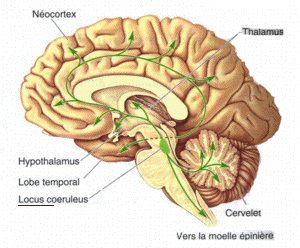
The locus coeruleus – a small bundle of neurons perched on top of the brainstem – is the only place in the brain where norepinephrine is produced. Look how far connections from it extend across the brain. (Image from Diego69 – Wikimedia Commons).
Norepinephrine is produced in the noradrenergic neurons on the locus coeruleus. It starts with tyrosine → (broken down into) DOPA → (broken down into) dopamine → (broken down into) norepinephrine.
When the sympathetic nervous system needs more juice, NE is released into the synaptic cleft between neurons. It’s then rapidly recycled back into the neuron via the NET transporter and metabolized into its byproducts before it ever gets into the cerebrospinal fluid.
If the NET transporter isn’t working well, high levels of NE will seep into the cerebrospinal fluid, making it look like NE activity is high when it’s simply not being recaptured by the neuron. Alternatively, if the NE transporter is in overdrive, transporting large amounts of NE back into neurons, the resulting low CSF NE levels do not necessarily indicate low NE activity.
We need a test that assesses how much NE is actually flowing through the neurons. Because once NE returns to the neuron, it is metabolized into DHPG and MHPG, these readings tell us how much NE is actually cycling through the system. That gives us a good readout of actual NE activity.
The main source of NE activity assessed in this study, however, is a metabolite called MHPG. Because MHPG is the final stage of NE breakdown or metabolism, it tells us the most about how NE cycles through the system. The authors referred to MHPG as “the predominant analyte” in their NE assessment.
They summed cerebrospinal fluid NE, DHPG, and MHPG levels to form a “norepinephrine pathway index.” They did the same thing with the dopamine and its metabolites.
- NE pathway index = NE + DHPG + MHPG
- DA pathway index = DA + DOPAC + HVA
Very Low NE Pathway Activity
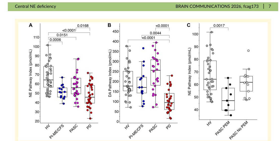
Notice the dramatic drop in the NE pathway in ME/CFS, long COVID (in the A diagram on the left), the normal dopamine pathway results for MECFS and long COVID but a big drop in Parkinson’s (B- center diagram), and a finding suggesting that PEM is affected (C – right diagram).
Only 16 ME/CFS patients participated in the study, but the probability factor, which assesses how likely a result was due to chance, was so low (p=.00055) that, even with the low participant numbers, it was VERY clear that the two groups were different. (There was about a 1 in 1,800 chance that the results were the result of random sampling.) Plus, the MHPG finding (P=.00044) was similar. (This was a group finding. Interestingly, not everyone with ME/CFS had dramatically lower NE metabolite levels: there are subsets here, too.)
We don’t know if these findings will translate into the ME/CFS population at large, but they show that at least with this group, the average NE pathway index was very different from that found in the healthy controls.
The norepinephrine index was reduced in the long-COVID patients, but the probability factor was not as extreme (p=.05). Norepinephrine metabolites were also dramatically reduced in the Parkinson’s disease patients (p<.0001).
Normal Dopamine Pathway Levels
The dopamine pathway index, on the other hand, was normal in both the ME/CFS and long-COVID patients. (It wasn’t in the Parkinson’s patients.)
In a way, the normal dopamine levels were encouraging because, if ME/CFS patients were simply stressed, inactive, or taking certain medications, both the NE and dopamine pathways might have been affected. Instead, the reduced NE findings and the normal dopamine findings zeroed in on something particular to ME/CFS and long COVID.
Handgrip Test
This NIH group loves the handgrip test, and Health Rising recently covered a prior handgrip study which suggested to the authors that the fatigue in ME/CFS is at least in part driven by the brain. The recent Australian study Health Rising covered also aligned with that finding. It reported a striking deficit in the supplementary motor area, which plans and carries out movement.
The researchers combined the ME/CFS and long-COVID groups and found that reduced NE pathway activity correlated with reduced ability to maintain handgrip force; i.e., the lower the NE metabolite levels, the shorter the time they could maintain a strong handgrip.
Note how essential a role norepinephrine plays in many of the problems associated with ME/CFS and long COVID: it’s needed for arousal, attention, effort, movement, autonomic nervous system regulation, sensory processing, and cognition.
We have a nice correlation, but the brain is a complex organ, and many factors could be preventing ME/CFS and long-COVID patients from being able to sustain muscular force, and causing them to become fatigued so quickly. They include inadequate motor drive from the brain (motor cortex, supplementary motor area), motivation, effort, autonomic nervous system, muscle metabolism, and nerve transmission, but the authors will focus on a particularly intriguing one: energy production.
The ATP Question
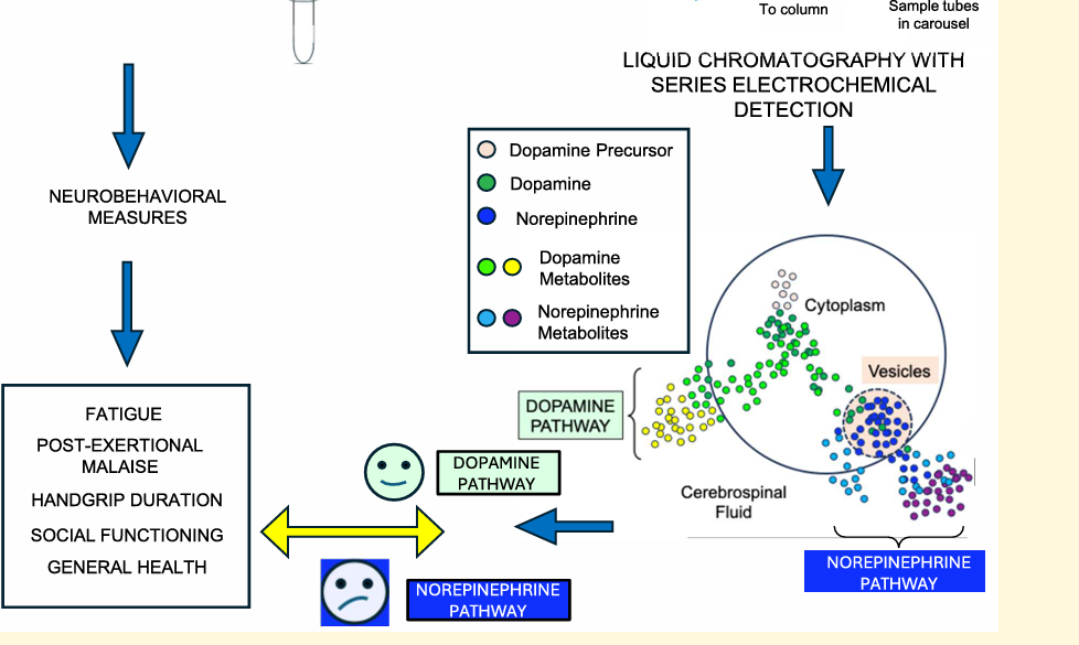
Notice how, on the left side of the image, dopamine flows freely into the cytoplasm of the cell, while norepinephrine has to be transported out of the vesicles – an ATP-intensive process.
Norepinephrine is the “arousal” neurotransmitter. Among other things, it gets the body moving and the mind concentrating. While it doesn’t generate ATP, it eats up lots of energy.
This is because the conversion of dopamine to norepinephrine takes place inside vesicles found in the cell. In order to get from the cytoplasm of the cell into the vesicles, norepinephrine has to be transported by an ATP-dependent proton pump called VMAT2 (vesicular monoamine transporter 2). That pump uses up a lot of energy.
Note that dopamine doesn’t require this step to be used. It’s already present and available in the cell. The authors propose that the normal dopamine levels, but low norepinephrine levels, suggest the cell simply doesn’t have enough energy (ATP) to replenish norepinephrine.
It’s a nice hypothesis, given that dopamine and norepinephrine are part of the same pathway (Tyrosine → DOPA → dopamine → norepinephrine), but only norepinephrine levels, which require ATP, were affected.
This all makes sense, but this study can only point to that possibility. We need more refined studies to determine that.
A recent study, however, does appear to bolster that hypothesis. The recent long-COVID study, which got Jarred Younger very excited and was covered by Health Rising, measured high-energy phosphate metabolism in the living brain.
While the study did not directly measure ATP, the reduced ATP/phosphocreatine ratios found suggested that low energy stores were present in the cingulate cortex – which just happens to be filled with neurons extending from the locus coeruleus (LC). Both the LC and cingulate cortex participate in many of the same features (effort, motivation, autonomic nervous system regulation, cognition, and pain) that plague people with ME/CFS and long COVID.
Effort Preference Redefined?
The findings cast the “effort preference” finding in Nath’s deep phenotyping paper, which sparked so much controversy, in a new light. Many people interpreted the effort finding to suggest that ME/CFS patients weren’t trying hard enough.
These findings, though, suggest a physiological basis for the “effort preference” issue. Parts of the brain we need to engage in exertion – no matter how large or small – may not have the energy they need to work well. That could make everything more effortful.
If Aregawi and Goldstein are right, that may all be due to that old bugaboo – a lack of energy production – that particularly kicks in during exertion.
Other Possibilities
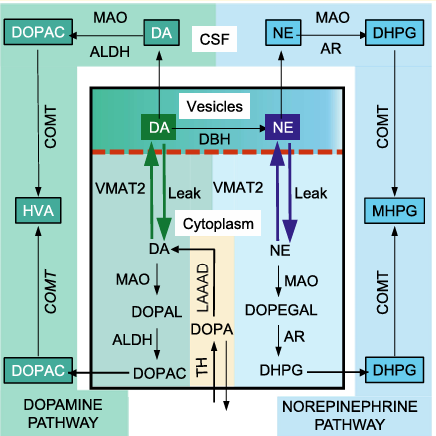
The catecholamine pathway – like everything in the body, it’s complex!
The energy production problem hypothesis is a nice fit given other study findings, but other possibilities exist. Less active or damaged LC neurons or problems with NE synthesis could reduce NE availability. Looking more broadly, neuroinflammation could be affecting the NE pathway. Or, as an earlier blog suggested, damaged muscles could be telling the brain to stop activating them.
There’s also Hwang’s 2023 WASF3 mitochondrial finding. A future blog will catch up on that.
Finding out which, if any, of these hypotheses fits is not rocket science. A follow-up study that included 31P-MRS brain energetics assessments (recently performed in long COVID), cerebrospinal fluid NE/DHPG/MHPG levels, a handgrip test, and an exertional challenge could tell us a lot.
If low baseline brain energy levels predicted reduced norepinephrine pathway levels after exertion, that could suggest energy production was the driver. A finding that a decreased ability to sustain a handgrip test was correlated with reduced brain energy and norepinephrine pathway levels would provide more evidence.
The NIH – Still Behind the 8-Ball
Nath said more papers would come out of the intramural study, and this is at least the third paper to do so. It’s good to see these papers; they are moving the field forward, but the intramural study was designed to give the NIH solid ground to dramatically increase ME/CFS funding.

With the NIH continuing its 30-plus-year pattern of neglecting ME/CFS, advocates are turning to Congress to compel it to fund ME/CFS.
These findings clearly call for larger, more comprehensive studies, but no grants have been approved for these researchers and, more importantly, no major grant packages exist.
While an intensive ChatGPT-aided search made it clear that Aregawi/Golstein/Walitt/Nath are continuing to purse the NE/motor cortex, there is no indication – and there should be an indication if such a thing was happening – that any large studies are underway.
Researchers can only go so far, no matter how dedicated, if they can’t secure the funding needed to conduct large-scale studies. It looks like the NIH is still pursuing its “50-year” plan for solving ME/CFS; i.e., it will fund a couple of large studies every year that can’t even begin to cover the gamut of possibilities, and it will engage in some small work on the side. What it will not do is take this disease seriously and fund the kinds of studies that move it forward in a timely manner.
As always, Congress is the hope, and ME/CFS advocates are pushing for $50 million in the 2027 appropriations bill to fund the ME/CFS roadmap. Accomplishing that would be a HUGE win. As the bill is usually due by the end of September, we will hopefully know its fate soon.
- Next up – Jarred Younger talks on ME/CFS





Now that is a rather interesting study! When beginning to read I had a ‘no way, we can’t be too low in norepinephrine (aka noradrenaline)!’. I imagine many patients will feel so.
When reading it is both the parasympathetic and sympathetic system ‘sitting at rock bottom’, it however made sense. In both biology and engineering many systems have a much more robust steering and control when two strong and opposing signals are used to control what happens.
I try and give an easy to understand example. If you ride a bicycle at high speed, you strongly hold both the right and left handle (especially on gravel). When you hold them equally strong, you go straight forward. When you make a slight turn to the right, you push a little bit more to the left handle and push a little bit less on the right handle. You still push on both with a decent force.
You could try and use much less force. Hold both handles with minimal force by resting your left respectively right pinky on them to go straigth forward. Result: the handlebars will flip over when you ride over the smallest pebble, turning the front wheel more then a quarter of a turn and landing you with your head in the ground. Trying to make a small well controlled turn will be very challenging, even without the slightest bump in the road. Each small change in force on the handlebars will have such a strong effect that your steering will be very erratic.
When one or two of the opposing steering signals have very little power, the other indeed needs to adapt (become weaker) in order for there to be any remaining control (regulation range left) of the system.
When that happens, any control will be rather jerky / unprecise and the smallest pebble / disturbance can topple the system over. That indeed sounds ‘ME/CFS’.
I wonder if my recent experience with my extended crash and my “reduced autonomic drive” speaks to this.
My symptoms – icy sweats on my face, rampant nausea – particularly after eating – and dramatically increased problems with exertion (including mental exertion) and my reduced resting heart rate, occasional dramatic jumps in heart rate variability – particularly after exertion, could reflect an unstable and weakened autonomic nervous system dominated by poor sympathetic nervous system functioning.
It was all brought on by pushing too hard – and being in the heat too much – two sympathetic nervous system stressors. (I think they should study me :)).
Hi Cort,
A crappy thing to go through; we are glad you’re feeling better now.
Thanks. I’m hopeful that another week or two of rest will get me back to baseline. 🙂
A recent blog of Cort showed there was reduced glymphatic clearance (drainage of brain waste via CSF) in ME/CFS. That got me looking for a link with norepinephrine as I felt there was a connection to be made here. See this paper https://www.cell.com/cell/fulltext/S0092-8674%2824%2901343-6
Mind the title: “Norepinephrine-mediated slow vasomotion drives glymphatic clearance during sleep”
This quote stresses its vital role in glymphatetic cleaning: “Norepinephrine oscillation frequency during NREM sleep predicts glymphatic clearance”.
I thought it helped explain the poor sleep (increased norepinephrine / noradrenaline at night to try and restore glymphatic cleaning levels) so often seen in ME/CFS. I thought our bodies tried to increase norepinephrine above normal, but with this ‘two signal steering’ thing explained in the blog and my comment above, it makes more sense now to say it looks more like norepinephrine being less deficient at night in order to remain some glymphatetic cleaning.
Important note: don’t just yet go and take meds to increase norepinephrine, read my reply!
Important note:
I feel that *both* the resting and activating (parasympathetic and sympathetic) systems are profoundly weakened. That is a fundamentally different view when it comes to treatments (versus seeing only the sympathetic system weakened).
When both are profoundly weakened even modest tweaking of one system’s strength can hammer the other side into problems. Both trying to unilaterally up either norepinephrine (activating) or GABA (desactivating / rest) signals is like using a crowbar on a steel door welded to its frame: expect something to break.
The reason why I think both are profoundly weakened, next to experience: GABA is (one of the) important resting chemcial signals. And according to dr. Robert Phair’s rather interesting Itaconate theory, GABA is one of the signalling molecules that gets ‘eaten’ (used as emergency fuel for making ATP) when itaconate blocks the mitochondrial citric acid cycle so much that the (for mitochondrial functionning) so important CoA cofactor gets locked up. When this happens (during our crashes according to the theory!), a main brain / neuron calming chemical signal as good as dissapears.
This seems to happen almost in lockstep with the shortage of ATP that is needed to pump dopamine into veshicles in order to produce norepinephrine.
I choose to stay away from forcing high levels of norepinephrine when key calming chemicals are near knocked out of existence *during the depth of PEM*.
Well, I probably once did inadvertely by using a NE reuptake inhibitor at modest dose when I was near my worst. It sort of energized me, I give you that. But it did so in a way that drove my heart to near out of control rates and gave me an adrenaline rush I didn’t even experienced (both intensity and duration) when someone was trying to rob me by trying to knock me with a bat on the head when I was healthy and travelling. Now I better understand how that comes ;-). Thanks Goldstein for researching and Cort for reporting this.
Before someone asks: No, upping both GABA and norepinephrine is not the solution to try and circumvent the risks of only upping norepinephrine.
Why:
* When both systems are depleted, even the fine processes of our body have a very hard time to keep a semblence of balance => dosing will be tricky
* There will be plenty variation over time especially when we get near exhaustion and into PEM. Many different processes will succeed at a fairly high rate. Each process will be influence differently by the chosen method of upping both NE and GABA => even if it worked at rest, it’ll be chaos when exhaustion and PEM sets in => dosing will get very tricky
* There are more activating and calming chemicals then only NE and GABA => trying to increase only those two will create an inbalance with the rest of them
Increasing Gaba is definitely not the solution for an overactive sympathetic nervous system and a fight-or-flight system in overdrive, as seen in ME. Benzodiazepines can actually help bring calm. A large part of ME patients benefit from this medication. The group that stays in an overactive state.
Do you include modest supplements of glutamine – gaba precursor- as a risk to creating this imbalance you warn about? If the GABA is being gobbled by the desperate ATP process, wouldn’t it make sense that it needs some form of replacement?
And Cort, sorry to hear that officiousness has reared it’s ugly head in your vicinity. Hope you’re settled comfortably elsewhere.
“don’t just yet go and take meds to increase norepinephrine”
Right. One of the recent studies noted that central acting drugs haven’t proven very helpful in ME/CFS. If Goldstein is right about the ATP problem – that needs to be fixed – and we would need to know what is causing THAT – in order to do that.
It could just be inflammation!
I think the theory about the muscles being damaged and telling the brain not to activate them is close to the truth. When the muscles are all knotted up, the brain can do nothing so must bring to our consciousness the necessity to treat them as the knots tie up a lot of our energy. This the brain does by changing the brain filters to let in more stimuli so that more substance p and glutamate gets to the brain and makes us wake up and do something about the trigger points. How we got the trigger points in the first place is the real question, I believe. I have always suspected damage to the hypothalamus, probably from trauma or infection.
Noradrenaline signals to the hypothalamus are critical to alert the hypothalamus to problems. If they are deficient, then the hypothalamus will not have information needed to address the problems adequately.
There are definitely signal loops going on and we still do not understand them. Our low oxygen usage during exercise (from 10 or 11 years ago) and more recently from last year the finding of muscle hypoxia during exercise, might well send signals back to the brain. If this issue is more widespread than just muscle, then the hypothalamus and other brain areas might be getting lots of signals.
One highly speculative idea of mine is that the hypothalamus or other regions might be reading our hypoxia and organ stress signals as signs of viral infection, and is deliberately inducing our problems as a kind of compensating mechanism … viruses need a lot of energy to replicate. This is related to the cell danger response hypothesis.
It would sure seem to make sense that low NE would impair glymphatic clearance. The recent Aussie study suggested glymphatic clearance was impaired….
Nice find.
Yes, taking meds to increase noradrenaline (norepinephrine) is risky and should await clinical trial results at least, and even then be under the supervision of a medical professional. Drugs boosting dopamine, a precursor, might be even more dangerous.
The fight-or-flight system is a major problem—whether primary or secondary—in ME/CFS/POTS. The cause is not psychological; rather, the system is impaired at a physical, biological level. It might even be the root cause!
I agree, the brain simply doesn’t have enough ATP available to do what it needs to do. The weakened and unstable fight-or-flight system may be downstream of that cellular-energy deficit—not the root problem itself. This could explain why trying to “retrain” the nervous system may sometimes reduce stress or conserve energy, but doesn’t fix the underlying illness.
Definitely is the root cause as far as I’m concerned.
The “Psychological” certainly is part of the process. It’s an integrative. There is no need to go back to Descartes, and try to separate the mind and body.
My two bits are that if you can reduce stress – psychological or physical – which eats up a lot of energy – that’s a good thing 🙂
Yes. well said.
BTW, your “2 cents” -‘ US, of course, ( Canadians eliminated the penny) are worth more than any of ours; you know a lot more, and you strive to be even-handed.
Best,
Harold
Kingston, Ontario.
Ha! Canada wised up about the penny about ten years before we did.
Thanks for your kind comment.
🙂
The psyche and thoughts are, in an objective sense, very vague concepts. If the psyche thinks its finger is broken, does that really mean it is on a physical level? Conversely, people do reason that way—your thoughts can make you sick… which is often claimed in cases of ME. I don’t believe in this.
I just keep wondering how humans can Harbour so many diseases.
Yes the animal world does also have illnesses but not in the hundreds of diseases like humans.
There has to be one common denominator at play.
What is befumbling is that all these so called entities that hold all the cards , all the funds and claim the top roost hav’nt come up with cures for any of these diseases.
I suspect the animal world has as much complex illness as we do, or if it doesn’t, it might be because of evolutionary selection. Most animals are prey. The weaker ones would die younger at a greater prevalence and therefore breed less, causing fewer predisposed genetic types to proliferate.
That might have been the case for humans as well until a time more of us could make a living while peacefully sitting in a chair. Maybe even the brightest, most talented and physically gifted in other ways even survived to carry the genes—autism and adhd for example. Neurotypical people have trouble fitting into society, but the ones who can, and even some who can’t, contribute at times exponentially more—Einstein for example.
So I’m suggesting many of us would have had a harder time surviving war, famine, natural disasters, especially those without a strong clan, family or tribe committed to supporting them. And the more useful ones would have likely been protected at a higher rate. This is me postulating because at 65, my health is going downhill and my world is getting smaller. I have to figure out a way to make that work for me. I can still think.
Great article. I also found it interesting in the context of the recent study about Dopamine injury, which aligns with much of what you wrote.
https://www.cidrap.umn.edu/covid-19/new-study-offers-clues-about-long-covid-s-brain-symptoms.
When I started taking Bupropion/Wellbutrin and Dextromorphan together (a cheaper version of Avuelity), it improved a number of my Long Covid symptoms, especially cognitive ones.
This combination uses norepinephrine reuptake inhibitors, increases the total amount of norepinephrine available to the brain, though the interaction together may be a bit more complex. I do think the community may want to explore whether this combination helps other people with brain fog or Parkinsonian-associated symptoms.
Bupropion has also been around for a while, so many providers are comfortable experimenting with it.
What an interesting combination! I looked it up and Bupropion inhibits CYP2D6, the enzyme that rapidly converts dextromethorphan (DXM) to dextrorphan. That substantially raises and prolongs exposure to DXM. Dextro was on the top of Jarred Younger’s list of drugs to fight neuroinflammation.
As I remember the trial did not work out but maybe DXM needed a kickstart? How much do you take?
Thanks for your interest.
100mg of Bupropion w 45mg of Dextromorphan, morning and night
The Dextromorphan is purchased OTC.
Also nice, is that this is a relatively inexpensive thing to try, and the treatment is relatively rapid-acting. I could feel the difference the first week I started.
Be careful! I took Bupropion/Wellbutrin for a month in an attempt to lift dopamine and my health spiraled dramatically. It ignited what I can only describe as a state of immense psychological distress out of nowhere and rendered me unable to walk = massive knock-on neurological impact on me. I could stand, but my gait was reduced to a shuffle. Very frightening. These symptoms resolved within days of stopping it.
Much like yourself Cort, I feel I may be in a neurological subset of ME/CFS. I too developed a heat intolerance situation over the last two summers. I have determined through painful trial and error, that I cannot tolerant ambient temperature over 75 F. When I overheat all sorts of symptoms flare up. Over the summer I have had to become very disciplined to check the temperature and move indoors to AC when it hits 75F.
Do you know if you have the slow COMT gene?
I just googled the symptoms of slow COMT and would say, no on the symptoms list overall.
I experience more problems with walking and unnatural gait including shuffle (very slow) which super slows me down. Pushing self to stretch in all directions often with some initial but not long lasting pain, eventually clears it out with frequent chiropractic but miss one week at chiropractor and my walking rapidly deteriorates.
Almost everybody I encountered in 2026 seemed to suffer from heat shock and complained of finding it very difficult to breathe outside. I don’t think that was unique to CFS ME community. People were having heat stroke, too. Dehydration was a major issue, but something was in the outside air that was making it very difficult to breathe outside for everybody. The air supposedly had Canadian smoke in 2026, but it wasn’t seen or smelled contrary to 2025 when the smoke and fire smell was visible with fire smell permeating everything for hundreds of miles. Perhaps nanoparticles from the fire were present, but would they travel beyond where smoke and smell fell off and disappeared?
This appears to be another in a growing series of studies pointing at the same thing. Dopamine beta hydroxylase deficiency, discovered in 1986, has nearly identical symptoms to ME. When neurological availability of noradrenaline (norepinephrine) is low, someone gets ME symptoms, especially orthostatic intolerance and exertion intolerance. Its not an absolutely perfect match possibly because these noradrenaline deficient patients lose noradrenaline in the whole body. Furthermore when you consider a slow NA/NA synthesis, our favorite strategies of pacing, switching and resting make total sense. We have a broken charger and are on a slow trickle charge.
Before proceeding, let me give some nuance to one comment. The LC in the brainstem is the primary source of the neurotransmitter noradrenaline but not the only location. The effect is more widespread. I have forgotten the details.
Finding low NA/NE end products in the spinal fluid gives us a cause. We have the symptoms. Occam’s razor suggests, but does not prove, that NA /NE deficiency is a primary driver of symptoms in ME.
The potential deficiency has been found in ME, post cancer fatigue, and long covid. It might be that most people with post infective fatigue syndromes, regardless of the label, are noradrenaline deficient.
This current study ties severity to probable NE/NA levels. That might be a dose dependent response, or evidence of causation. I know static muscle load damages my muscles, though of course that is anecdotal. However when I read your comments about handgrip endurance it got my attention.
On July 2 this year a paper came out only on long covid. It identified a depletion of the vmat2 transporter, there is not enough of them. I have not read the full paper but it used, I suspect, a tagged molecule to measure transporter numbers using a PET scan. They were low.
Oxidative stress is a known cause of this, but many other things could be in play.
Noradrenaline deficiency via vmat2 depletion has been my primary working hypothesis for what is driving the symptoms of ME for nearly two months now. Its not sufficient to determine detailed mechanisms of what is causing the problem, but its enough to give a drug target for the pharmaceutical giants to pay attention, with one drug and one supplement as candidates for helping, though only in rats so far.
If its fast tracked the first drug could be available in two years. If not then maybe three to five years. If a whole new drug is needed then ten to twenty years. Presuming, of course, all these findings continue to be replicated and expanded.
One last comment. The potential long covid population in the world might be 400 million. Drug companies are going to really, really want to capture that market. I hope they do.
Whoa! Very interesting! That’s the same transporter! That’s fascinating….
https://pubmed.ncbi.nlm.nih.gov/42431745/
“Findings of reduced VMAT2 binding may reflect reduced dopaminergic terminal integrity in long COVID. Loss of dopamine nerve terminals may be contributing to symptom correlates of apathy, motor slowing and memory decline suggesting improved function of dopaminergic synapses as a new therapeutic direction to treat long COVID.”
From a Toronto research group.
Yes. It completely changed my view on the subject. A specific causal mechanism, presuming its replicated and expanded, is a powerful target for drug development. There is a drug already being tested, though not for ME. It has some long letter code I do not recall, not a name, as its not nearly ready for market. However if there is loss of the nerve terminals, not just the transporters, its a whole new ball game.
I am deeply sceptical of the existing commercial drugs to treat noradrenaline disorders. They do not address this specific mechanism. I suspect the clinical trials they are planning will fail. I hope I am wrong.
From the paper Cort supplied: “Approximately 95% of VMAT2 binding in striatum is contained within dopamine releasing neurons22 so a reduction in (+)[11C]DTBZ BPND is inferred to represent a loss of dopamine releasing neurons. This interpretation is consistently applied”
That alligns with Cort’s quotes too. Now add your quote: “On July 2 this year a paper came out only on long covid. It identified a depletion of the vmat2 transporter, there is not enough of them. I have not read the full paper but it used, I suspect, a tagged molecule to measure transporter numbers using a PET scan. They were low.
Oxidative stress is a known cause of this, but many other things could be in play. ”
=> My guestimate becomes: Vmat2 being vulnerable to oxidative stress (if this is correct , I did not do the effort to check) is likely a *deliberate* and protective feature. Why? See https://www.nature.com/articles/s41467-024-51960-z, figure 1a (the magnified part). Vmat2 itself doesn’t consume ATP, but (uses the gradient of H+ inside the veshicle) 2H+. Also from the same figure, those H+ are pumped into the veshicle by V-ATPase.
The figure seems to indicate that V-ATPase only yields one H+ per H+ but I *think* that is a common simplification often used in figure to say ‘V-ATPase pumps H+ using ATP’ but disregarding stochiometry. From https://journals.physiology.org/doi/full/10.1152/physrev.00045.2003, figure 1 it says 2H+ per ATP (at least in renal V-ATPase).
Anyway, if it is 2H+ per ATP then Vmat2 indirectly consumes 1 ATP (converts to ADP) per monoamine transported plus some for losses. If the first paper would be right, double the ATP cost.
=> plenty of ATP cost to ‘use’ Vmat2. Basically the same as Cort’s blog said, but then specifically calculating it back to Vmat2.
=> Why is this important? Reducing Vmat2 in case of oxidative stress may be a way of the (dopaminergic) neurons to try and protect by conserving energy during oxidative stress. High energy consumption during oxidative stress can be ravaging. The body ‘making’ Vmat2 vulnerable against oxidative stress would then be a protective feedback strategy.
=> the really important part: trying to ‘restore’ normal Vmat2 levels with a drug without reducing the oxidative stress these neurons experience would be a rather good attempt to try and kill dopaminergic neurons (aka trying to trigger Parkinson Disease). I’ll stay away from being a guinea pig on this one.
This paper is interesting relating to this discussion: https://www.science.org/doi/10.1126/sciadv.adz5645
“VMAT2 dysfunction impairs vesicular dopamine uptake, driving its oxidation and α-synuclein pathology in DJ-1–linked Parkinson’s neurons”.
So too few Vmat2 can increase oxidative stress in dopaminergic neurons and increase PD symptoms. BUT the driving mechanism is increased dopamine oxidation which IMO is a function of both dopamine concentration (quadratic relationship?) and amount of oxidative stress. Increasing dopamine without helping Vmat2 in PD therefor may cause more oxidative stress. But if levels of dopamine and dopamine waste products are fairly normal in ME/CFS, this may not lead to increased dopamine oxidation in ME/CFS *IF* oxidative stress (comming from other source then dopamine oxidation) is sufficiently under control.
Dear Cort, I take for about 8 years now gaba 3 grams and Mucuna Pruriens highest dosis. Should it increase my exhaestion for the last 6-7 years. I take Gaba for stress and Mucuna for Resles legs
With love from Holland
Lenny
Hi Lenny,
Are you saying that the macuna and the gaba are helping you over that timeframe? GABA helped me for a while, but the effects decreased. However, I was only taking 500mg.
One additional caution. Raising brain noradrenaline levels is not sufficient to fix the problem, and might actually make things worse. The noradrenaline needs to be inside the synaptic vesicles, and the vmat2 transporter defect means this might not happen fast enough with high extracellular noradrenaline to be useful, and might be damaging. Caution is needed. The first drug trials of noradrenaline drugs are being considered now, but these drugs might be worse than useless. We need to fix the problem, not just noradrenaline levels in the brain.
“Dopamine beta hydroxylase deficiency” is THE problem?
Sounds too specific. It’s a multifaceted syndrome.
Hi, look up the symptoms. They are nearly identical, though some few are not the same. Also, DBHD is not THE problem. Latest data shows its the vmat2 transporter, which also results in low noradrenaline and almost the same set of symptoms, at least in Long Covid. So far ME and post cancer fatigue are only proven to be noradrenaline deficient.
What we do not know is WHY this happens. Many of the other brain hypotheses might explain that in time.
So nearly all symptoms explained; resting, pacing and switching explained; a known cause is present, it has a specific mechanism, though we still do not know why vmat2 is not expressed, or lost or damaged or something.
VMAT2 (though I prefer the acronym VMT2) is also a generalized monoamine neurotransmitter transporter, so we can expect some small impact from dopamine and serotonin deficiency. However, as of current research, neither of these is strongly implicated nor even necessary to explain the symptoms.
One unanswered question is where do different pathogens etc., or even the same pathogen in different people, trigger different patterns of deficiency, or even if such patterns exist, in different neuronal clusters in the brain.
It also does not take into account pathogen specific problems. This is about the post pathogen fatigue syndromes, and looks to be a common mechanism. Individuals will have other things going on as well.
For example I have salicylate sensitivity, which via two critical enzymes will impact synthesis of eicosanoids, the most important group of hormones, conserved in all muticellular organisms. This has at least two known causes now, different snps on at least two liver detox enzymes. How will that impact my specific symptoms? I do not know. I can bypass the issue with specific dietary strategies, especially arachidonic acid, EPA and DHA supplementation. What I do know is that EPA and DHA deficiency in the brain will disrupt neuronal cell membranes. Individual genetics and comorbidities are likely to result in slightly different symptom expression and severity.
The data is also strongest in Long Covid at this point.
When a treatment can restore VMAT2 capacity AND results in symptom remission we will finally know if its causal, or at least causing the symptoms. That is a minimum of two years away, and maybe twenty.
Finally, early data suggests that for about a tenth of us this explanation does not apply, they have something else wrong with them.
Yeah, the root causes/mechanisms remain unknown. Better to not mislead readers by suggesting that “Dopamine beta hydroxylase deficiency” one of those, given that you are simply speculating.
I get the temptation, but I only truly listen to reasonable scientists (with “educated” opinions). I take other opinions with huge grains of salt, as I do AI.
Scepticism is good. Its the foundation of science. However you seem to have completely misinterpreted my position.
I have called it a dominant working hypothesis. Not a fact. I have never claimed it to be dopamine beta hydroxylase deficiency. Go back and reread things. This rare genetic disorder has the same symptoms. These symptoms are due to noradrenaline deficiency. Most of us have noradrenaline deficiency. A genetic disorder gives us an independent glimpse at pure causation and what symptoms are driven via a specific mechanism.
As for scientists, let me give you a glimpse at my resume. I have two science degrees, one of which is in biochemistry, first class honors, a university medal, and was a PhD candidate when ME finally crashed not just my body but my brain. You cannot do a PhD when you cannot read, count to three, or even speak. Along the way I was briefly a university lecturer, and I was a debating partner with two PhDs involved in ME research for over five years. One of those was Martin L Pall, who had an influential hypothesis about ME two decades ago. Cort knows who I am.
I have a history of analyzing and building ME models. I had a successful ME treatment protocol with complete reversal of symptoms about the time I presented an ME paper at a medical conference in 1999. At that time I thought ME was probably hypoxia. I still think it is largely hypoxia, which noradrenaline deficiency might be able to induce, though I no longer think my mechanism was correct. My treatment protocol had to be abandoned due to cost and severe, even brutal side effects. The noradrenaline link might explain what went wrong, I am still thinking about that.
I have a special interest in ME and eicosanoids for obvious reasons.
Sure, let’s sit and wait 10-20 years for “reasonable” scientists to come up with answers. I’m too old to wait that long. The time is now.
In 2026, PubMed article only one form of a fat-based thiamine Vitamin B-1 when taken, went through the Blood Brain Barrier into the brain’s neurological cells and caused mood changes towards dopamine. This is not Benfotiame another fat-based Vitamin B-1, that is claimed to only go into the torso’s neurological system. This appears to be a synthetic but without a patent on the label, and synthetics often cannot break down the way a natural chemical would. It caused some very disturbing unhealthy problems in a ME CFS patient who only took 3 pills as directed on the bottle, but skipping one day over 4 days, but immediately discontinued those Allithiamine pills as soon as noticed it unexpectedly triggered a “high” with an edge. It triggered a perpetual “high” for nearly an entire month that was very disturbing and prevented sleep totally or limited it to 1-2-3 hours a night. This unexpected “high” was devastating and depleting energy, as the nervousness and anxiety seemed to eat up all energy present, leaving the patient with heightened energy and anxiety combined with such fatigue that disrupted any thought focus with heightened nervousness and anxiety, while the muscles in the body were fatigued of energy. It triggered an “edge” that was initially described as “pot-like high” but later re-described as not going in the same direction of “pot” to the extent it prevented sleep instead of allowing sleep to come easy. It behaved more like an amphetamine that speeded up the body to prevent sleep, when sleep was desperately sought and needed. It transitioned between heightened nervousness and anxiety combined with deep fatigue where thinking focus and concentration became almost impossible, and was disrupted with excessive intruding thoughts constantly. It interfered with the details of mathematical accuracy and logical side of the brain. It triggered more expansive creative spatial balance or 3D artistic thinking, which interfered with deadlines for logical thinking work.
An expired topical higher dose broad spectrum CBD with oil beading was put on a knee several times prior to using this allithiamine. It is strongly suspected that it interacted with the fat-based allithiamine that goes through the Blood Brain Barrier into the brain cells. Drug interactions between drugs are common and unpredictable and mostly unstudied. CBD is known for occasionally causing “highs” up to a month long, surprising the patient.
However, it is likely not a one-time event with the allithiamine. The manufacturer has went dark. The phone system does not work. No chat is available. No email address is listed. The dedicated product info phone line is no longer a working number, but is found on the website. Instead, the manufacturer without forwarding or without updating information has moved its products to a different website to continue same brand distribution under a new distribution name. This is the only manufacture known to make the exact allithiamne product found in PubMed.
Dopamine deficiency does appear to be speculation in ME, CFS, fibromyalgia, SEIDS, and similar chronic diagnoses.
I would think addressing the problem at the ATP generation level would be less hazardous and perhaps even less complicated than trying to regulate noradrenaline and Vmat2.
Trying to regulate noradrenaline without fixing VMAT2 is risky. All strategies have risk. That is why we need clear molecular targets, and clinical trials.
Whether fixing VMAT2 is even possible depends on what is wrong. However its a specific drug target. That is necessary for pharma to be interested.
Fixing ATP sounds good. I have been trying to do that for about fifty years. It is very problematic unless you know the specific mechanism. Which molecules and some idea of what is wrong. It also might be a problem if something the cell danger response is involved, as an ATP rise might potentially increase the suppression feedback. Again, we need mechanisms and clinical trials.
Many strategies have tried to restore ATP. Many had modest results, but no cure. We simply do not know enough. ATP is integrated into general metabolism, mitochondrial and immune and inflammatory response, genetics, nutrition, and neurological signals. Its like saying trying to fix fatigue. Great, what is the mechanism? If we can find one single mechanism, or several that are causally connected, and treat those, the ATP problem might fix itself automatically.
What I will say is that sometimes strategies to boost ATP do have modest affect on quality of life. Its often better than nothing. My shotgun protocol tried to do just that, combining more than 30 different methods to boost ATP. It worked, and then it became the problem. I call it a shotgun protocol because I had no specific mechanism in mind and tried to fix every one I could.
I understand what you mean about ATP not being a specific enough target. I had in mind some recent thoughts on a mechanism from Putrino, wherein he proposed a thickening of capillary basement membranes, causing both: a) failure to remove mitochondrial waste from tissues, and b) failure to properly oxygenate tissues (i.e., a form of hypoxia). That made sense to me and, if it can be demonstrated, it may be possible to address without directly targeting Vmat2 and noradrenaline (perhaps these would adjust on their own?) There is some discussion of reducing inflammation with antivirals and then 40 sessions of HBOT, but I suppose you have tried this combination in your 50 years.
Detailed mechanims help a lot in drug development. Findings need to be pursued until we can figure things out. Too many leads have been ignored.
I have not tried antivirals and oxygen therapy, aside from one brief test of oxygen. However I have personally known people who did. One functions, and stays in work, only on antivirals. However many don’t benefit. Oxygen therapies help some and not others.
Let me quote an old doctor of mine, “sometimes you just have to suck it and see if its a lemon”. For some its lemonade, for some a disaster.
Wise words from the old doctor (and from you).
Good help
Always enjoy reading your comments Alex – going way (way) back 🙂
Thanks Cort. I still keep reading Health Rising. I enjoy the work you do. I better stop here before this becomes a mutual admiration society.
Great blog, thanks Cort!
This area of the brain is fascinating and it’s getting even more interesting with Alex’s find. As I noted in the blog, NIH researchers are continuing to dig into these areas which is good. They appear to have found a good string to pull on. I would love to see a big study.
I have had CFS symptoms a lot longer than my fight or flight response has been disabled. I’ve had CFS symptoms since I was a kid, 70 years ago. The past 2-3 years I’ve become aware that I no longer have a startle response, and I am slow to respond to any kind of threat. I can barely escape danger and just keep going along as if nothing happened. Maybe something else about CFS causes the weak fight or flight response instead of the other way around?
Noradrenaline problems go far beyond fight or flight, that is as much an hormonal thing as neurological. Our noradrenaline deficiency is neurological, not hormonal, or it would have been detected by blood tests forty years ago. This can induce a generalized dysautonomia. It impacts all activity including exercise. It can induce memory loss and cognitive issues including brain fog, disrupt sleep and circadian rhythms, and even cause executive dysfunction.
Since there is no proven mechanism driving the noradrenaline deficiency or VMAT2 deficiency, we can only speculate on factors that are driving this at a deeper level.
What do people do about it? Just suffer?
As a long term ME patient, an encephalitis survivor, my three top strategies, as with many patients, are pacing, switching, and resting.
Pacing means reducing activities until you no longer exacerbate symptoms but not further. This is not easy as life forces us to do too much far too often.
Switching is about changing activities, not doing any one too long. A change in activity means we can do more without crashing.
Resting is obvious.
All three are informed by the noradrenaline VMAT2 hypothesis.
In case you have not read my other comments, a potential drug is in development, but will be two to five years away I suspect. There is also a natural supplement, but we may need to take far too much to be safe, so I wont mention it here.
In any case potential drug targets change how major pharma companies view things. The market is huge, the profit motive will drive them.
“Since there is no proven mechanism driving the noradrenaline deficiency or VMAT2 deficiency”
Would https://pmc.ncbi.nlm.nih.gov/articles/PMC4308505/ (sufficiently) fit that role?
Title “JNK inhibition of VMAT2 contributes to rotenone-induced oxidative stress and dopamine neuron death”
and quotes
“Here we report that the neural specific JNK3 isoform of the JNKs, but not JNK1 or JNK2, is responsible for this neuron death in primary cultured dopamine neurons.”
“JNK3 contributes to ROS generation and caspase activation in dopamine neurons upon rotenone treatment”
“JNK is activated by microtubule destabilization
Previously, we demonstrated that rotenone induces microtubule depolymerization, followed by accumulation of cytosolic dopamine and ROS that lead to dopaminergic neuron death” => microtubules are quite prone to oxidative stress
And from https://en.wikipedia.org/wiki/C-Jun_N-terminal_kinases (I know, not the very best of referrences but I am getting tired so it’s a starting point for this kind of general information) JNK are “They belong to the mitogen-activated protein kinase family, and are responsive to stress stimuli, such as cytokines, ultraviolet irradiation, heat shock, and osmotic shock. They also play a role in T cell differentiation and the cellular apoptosis pathway.” => That points to stress activated kinases.
Thanks for this. However we still do not know if the issue is cell death or some other depletion of the transporter. This is something that really needs to be investigated further. We need more research has been my mantra for like seventeen years, ever since the XMRV finding.
However there is a very long history of partial or temporary remissions in many patients, and my shotgun protocol from 1999 reversed ALL symptoms until it created new ones. I went from a slow shuffle walk across a room, to brisk five hour hikes and feeling great at the end, in five days of treatment.
I had another partial remission on another protocol, which I will not name, because the crash that followed was very bad. Every three days I needed to double my dosage, and when I finally stopped at a ridiculous dosage I went into a severe crash that lasted months. I suspect some treatments that seem to work might result in the same kind of eventual crash, though I have no idea which one.
A third and fourth protocol got me through my biochemistry degree. I had to stop both after months due to intolerable side effects. I thought I would have to quit my degree in the third year as my health kept declining without my protocols.
The temporary remissions occurred for me after being on the shotgun protocol, lasted about six hours, and happened maybe a dozen times over several years.
If the nerves or nerve endings were destroyed, and this is the primary cause of ME symptoms, then anyone with partial or full temporary remission would have to have another cause. The nerves are not likely to easily regrow in part or in full, though some nerve growth is possible.
Were side effects of some of these protocols by chance either mimicking PD (PD symptomps but with permanent damage, seems not in your case)
or mimicking mainly part of PD (PD symptoms but of the rather strong inhibition type (temporary, hibernation or Dauer of motor nerve system, so mainly (very) severe (at times, fluctuating) loss of strength and responsiveness of motor function but temporary and with very few shaking or jerky movements; I guess this one)?
One was brutal headaches for most of every day that laid me up in bed, painkillers did not touch, but there was no aura. At the time I suspected B6 overdose. This was my shotgun protocol that reversed all symptoms.
The next was a massive loss of energy for the protocol that doubled every three days. I went from long walks to struggling to cross a room, and took six months to recover.
The third was a whole body painful burning sensation, that later happened again when I was on blood thinners after surgery. This was a glutathione therapy that really helped for about a year and a half.
The fourth was a massive, and I mean drastic, change in psychology and outlook (no, not depression, but I will not detail this protocol, its dangerous). As soon as I figured out it might be the protocol, that took about three days, I stopped using the protocol and recovered in about a day. My choices in that time were a disaster that persisted however.
By PD I presume you mean Parkinsons? My uncle died of that.
Let me try and look from another angle on this. Take this attempt with a large pinch of salt. Brainstorming quality only.
case 1: B6 is a https://en.wikipedia.org/wiki/Aromatic_L-amino_acid_decarboxylase cofactor; would / could increase dopamine (production, in cell but not in veshicles if Vmat2 is low) so also chances for dopamine toxicity if vmat2 is / remains low.
Also, drug or other addiction withdraw often involves brutal headaches. Some researchers blame the combination of (after stopping the stimulant) low current dopamine in combination with rather high toxic dopamine metabolites from prior addiction phase.
Potentially in your case (if Vmat2 rose less then dopamine synthesis): increased dopamine toxicity (as per previous comment) while still little extracellular dopamine (seen to cell as ‘low dopamine triggering receptors on cell surface’); combination resembles ‘post addiction stage’.
case 2: seems to be clear Dauer: the body was trying to abbruptly undo the increase in energy production and consumption you forced. The longer you tried to enforce it, the more fierce the body tried to inhibit ‘no matter the cost’. I would expect a very deep PEM and it lasting for rather long with rather hard and wonky recovery. That seems to be in line with what you experienced if I get your comment above.
case 3: glutathione (if getting ‘functional’ into the bloodstream; passing whole through the bowel into the bloodstream is IMO not supposed to happen in a healthy gut; but is an ME/CFS gut healthy??) would not only reduce the oxidative stress neurons and muscle cells see, but also the oxidative stress that IMO acts as an inhibitory feedback signal to weaken and reduce a large number of immune cells. If the original stressor remain then that allows for an overactivated immune system with plenty of colateral (in part temporary) damage like burning neuropatic pain. Would it by chance be a period with accelerating food and other sensitivities?
case 4: https://en.wikipedia.org/wiki/Aromatic_L-amino_acid_decarboxylase_inhibitor also is involved in seratonin synthese; see https://en.wikipedia.org/wiki/Serotonin; biosynthese picture; step from 5-HTP to 5-HT. Messing with serotonin can cause ‘psychologocial changes’. Potential for crosstalk in this dual use enzyme.
Yes, with PD I meant Parkinsons.
As said, take everything for what it’s worth; I have no official medical schooling.
case 4 is interesting. I just remember the metabolic trap concept from dr. Phair https://www.omfcanada.ngo/metabolic-trap-study/: a ‘stuck’ feedback loop producing too much seratonin from tryptophan instead of sending tryptophan into the NADH producing branch.
Not only has this NADH producing branch some rather (neuro)toxic chemicals in it (causing problematic levels of oxidative stress), but ‘trapped / excessive’ seratonin production would draw not only tryptophan away from the NADH producing branch to the seratonin producing branch but it would also have quite an influence on the AACD enzyme.
IDK if dr Phair hypothesized a strong increase in AACD or a diversion from dopamine production to seratonin production. Excess dopamine is a ‘pro oxidant’ while seratonin and following melatonin are rather potent anti-oxidants. Together with this ‘trap’ reducing NADH and with it energy production and potentially ROS, it might be more then a simple unlogical flaw in our genes…
Yes, in case 1 B6 can be an issue. I recently started a new protocol using it and other things to boost dopamine production, hoping that it would push dopamine beta hydroxylase activity. As soon as I read about the transporter issue I stopped. Too dangerous.
Case 2 I am less sure about, but it kind of fits.
Case 3 was not raw glutathione. It was cold processed whey protein. i was talking to a researcher in Canada who went through the case of whey protein increasing glutathione unless the whey protein was denatured, which pasteurization can do. This stuff works but too many products are only claimed to be cold processed.
Case 4 fit low serotonin, I exhibited compulsive behavior but I wont go into detail.
An enquiring mind and lots of effort can go a long way to making up for lack of formal training. I was deep into the biochemistry before I finished my biochem degree. It gave me focus and I learned much more because of that. Most disciplines are like learning a new language. Once you know the language things become easier.
Another related one: https://www.nature.com/articles/s41388-018-0582-8
Title “The ERK and JNK pathways in the regulation of metabolic reprogramming”
quotes
* “Of the three types of MAPKs, ERKs and JNKs have been recently shown to regulate the redirecting of energy harvest to glycolysis in both malignant and highly proliferative cells by affecting the activity of key metabolic regulators.” => from cancer research, but should be redirecting ATP production away from OXPHOS towards glycolysis under other conditions too.
* Figure 3: “The shunting of pyruvate into the mitochondrion is regulated by the activity of pyruvate dehydrogenase (PDH)” and from the figure: JNK1 is a direct inhibitor of PDH (pyruvate dehydrogenase). => PDH is seen to be inhibited in ME/CFS too; this is the enzyme shuffeling pyruvate into the mitochondria. This paper says JNK1 but
search result:
“PDK3 – [Pyruvate dehydrogenase (acetyl-transferring)] kinase … – UniProt
Inhibits pyruvate dehydrogenase activity by phosphorylation of the E1 subunit PDHA1, and thereby regulates glucose metabolism and aerobic respiration. Can also phosphorylate PDHA2.”
=> Seems to me that JNK3 inhibits both Vmat2 AND OXPHOS (mitochondrial ATP generation, high source of oxidative stress in cells) *simultaneously*
=> So both the ideas: “if enough ATP, then too few Vmat2 might cause toxic oxidative stress due to dopamine self oxidation” and “if too much oxidative stress produced by wonky mitochondria, activate JNK3 to inhibit both mitochondrial ATP production and Vmat2 indirect ATP consumption” may still hold.
Too much or too few Vmat2 may be highly depending on exact conditions (external stressors and mitochondrial damage).
See also figure 4. ERK seems to activate and JNK to desactivate OXPHOS.
Nice find. I was considering dopamine oxidation as well, though its still speculative. However normal dopamine byproducts in the spinal fluid might not reflect localized oxidation problems such as at the synaptic vesicles.
I am just starting to recover from a ten year crash. My brain is not back yet. I keep hoping to do more investigation but real life is pulling me away for now. I am already working on blogs and one for legal advocacy for the aged and the disabled in Australia.
I was super interested in mitochondrial dysfunction including hypercitricemia at one point (1999, care to guess the name of my paper?). Later I moved to glutathione depletion and protein folding problems. Some of our mitochondrial enzymes are nucleus encoded, imported as a protein string, and then folded inside the mitochondria. Most enzyme quantity assays will show adequate enzymes, but activity can be massively depleted, or the mitochondria might fill with dysfunctional enzymes.
“Hypercitricemia in CFS”?
Seems to be lost in the archives of time.
Guessing by the title, you discussed the observation of too much citrate production / waste seen in ME/CFS? From some other info you seemed to focus on deficient aconitase and its vulnerability to oxidative stress?
If so, I guess that’s what led to your very quick improvement with your rather harsh and deep fall?
Extra citrate is produced at rapid pace in hypoxia in neurons, liver cells and mitochondria among others. It’s used to produce (paradoxically) fatty acids during hypoxia and is correlated with protective outcomes.
If my guess is right, your tried and restore aconitase quite aggressively in order to get the citric acid cycle working again and it producing ATP again.
From my point of view, our genes are tuned such that the dose of oxidative stress going with hypoxia stops citrate being used to make more ATP (and produce more NADH and FADH that can’t be oxydized due to lack of enough oxygen, causing even more oxidative stress produced by the ETC due to ‘ETC inbalance’) by deliberately using an enzyme (aconitase) that is quite vulnerable to oxidative stress. Aconitase acts as a feedback chemical in this view. It yields less charge imbalance and with it less oxidative stress and in addition it provides substrate for (paradoxal but protective) fatty acid production during hypoxia in these cells and the mitochondria. That fatty acid production would also inhibit fatty acid oxidation since fatty acid oxidation and synthesis are blocking each other. If you then tried to increase fatty acid import / oxidation in the process, as it sounds like you did… then your outcome starts to make sense.
I know, I make many assumptions about what you tried to do (even if you discussed part of it elsewhere).
Yes, Hypercitricemia in CFS, which I cannot find anywhere, however I discussed it here –
https://forums.phoenixrising.me/threads/citric-acid-to-treat-fatigue.12218/
I was at the time not directly trying to address aconitase, that came later with my whey protein protocol, after I started talking with my debating partners.
I had two investigative strands at the same time. One was hypercitricemia. The other was my shotgun protocol. In a nutshell I spent a year figuring out dosage for every supplement that boosted energy that I had available, using a protocol I called pyramid dosing, then started taking all of them, every day, a week before the conference. This was mostly an empirical find. The day I got headaches, or the day before, I ran out of B complex and got another from the pharmacy. I do not recall if I checked the B6 dosage.
As a result of that conference I was invited to a private email forum where researchers debated CFS research findings, which I don’t think exists any longer, called CPAR. The archives might still exist though, I am not sure.
My theory was that citrate might block an enzyme that made a substance that red blood cells needed to release oxygen to tissues on demand. This was probably wrong. It was about pH and citrate affecting enzymatic activity.
However my protocol might well have boosted dopamine, with consequences we can understand better now.
How does acetylcholine fit into this picture?
I have known for twenty six years that acetylcholine is important. Look up the work by Vance Spence, who used to be in Glasgow I think. However there was not enough long term follow up research.
A commenter on a previous blog in this forum mentioned (without citing the relevant research) that since cholinergic neurons use ATP for acetylcholine synthesis, the latter is compromised by low levels of the former; but also that drops in acetylcholine are not uniform across systems. I’m not sure how exactly this relates to noradrenaline but it could be a vital link, and one that Mestinon could address.
Wasn’t me, but:
* Acetylcholine requires acetyl-CoA. According to dr. Phair’s itaconate shunt hypothesis, mitochondrial CoA is during a crash in such short supply that it already cripples aerobic energy production (let alone have some spare for other things).
* When AcH would be produced in the mitochondria (IMO unlikely), acetyl-CoA is produced by making it from pyruvate by the enzyme PDH, which is strongly inhibited in ME.
* In the cytosol, AcH is produced from citrate, using an ATP requiring enzyme.
* like dopamine, AcH is best pumped in vesicles for use (problems if not?). Like with dopamine, it consumes ATP. Better said H+ gradient that cost (one ATP plus losses per AcH pumped?) ATP to produce. https://en.wikipedia.org/wiki/Vesicular_acetylcholine_transporter
Many things in the process do cost energy. Enough to be crippled? Likely during crashes at least I’d guess.
I think this is interesting research from Japan linking HHV-6, the brain and acetylcholine to long covid:
https://www.frontiersin.org/journals/pharmacology/articles/10.3389/fphar.2026.1807203/full
Darn, seventy years is a long time. I am sorry you have been ill this long. I have only been ill 58 years, there are not many of us who have been ill that long. Best of luck.
My earliest symptoms that I remember were EDs symptoms. I was 5 and they took me to a chiropractor. Then followed the CFS symptoms and then MCAS symptoms, although there was nothing named MCAS at that time.
EDS has a close association with ME. I personally know two ME patients with EDS. I first became aware of the association between EDS and ME when talking to one of my biochemistry lecturers, whose daughter had both ME and EDS.
Genetic disorders (and presumed genetic disorders) create even greater complications in understanding what is going on. In my case its salicylate sensitivity, though I have yet to have a genetic test to identify which enzyme is the problem.
Hi Cort! This morning, I’ve been down the rabbit hole of Jarred’s investigation into using dextramethorphan to calm NMDA receptors in the brain to reduce over excitability. Do you know if Jarred was able to use DXM as an active ingredient in his study without the addition of guaifenesin? I am very curious.
Gastrodin for VMAT2?
https://www.sciencedirect.com/science/article/abs/pii/S094471132400477X
Maybe, maybe not. Something like that would require a clinical trial. We would need to know how it works, but for a full cure the VMAT2 levels would have to be restored. There is a small chance it may help management of symptoms though. Now if that leads to changes that result in cure, so much the better. Clinical trials could test that.
Recently, my thinking has been that LC and ME-CFS are fundamentally different in driving pathology, but with similar and overlapping presentations. (Though in some cases the Coronavirus may trigger CFS). What connects the two might well be compromised ATP production, as Goldstein seems to imply. I can’t escape the notion of inflammation in the blood vessels in LC, which may not be the case in ME-CFS.
Noradrenaline deficiency drives most of the symptoms. LC and ME have that in common. That does not mean there are not additional factors. However the fact that three fatigue syndromes have noradrenaline deficiency gives us a single common pathway, and depending on the detailed mechanism its potentially treatable.
Many LC cases may not be a fatigue syndrome, but other Covid damage. The same applies to some percentage of ME patients.
Let’s hope you are onto something at least for the sake of biomarkers if not treatment. It is also interesting to consider viral reservoirs. Whereas this idea has gone out of fashion with ME-CFS, it is increasing in its likelihood with LC pathology. I’m not well versed enough in infectious disease biochemistry to know whether lingering viral fragments can redirect/reprogram ATP toward their own survival, but ideas along these lines (and thus anti-viral courses) are being taken seriously by leading researchers. Some patients have had success with Rapamycin, suggesting mitochondrial cannot clear waste from muscles into the capillary system.
Biomarkers make all this research much easier. We need them.
I have a broader interest in rapamycin as it suppresses mTOR, which is often elevated in ME. Are you aware that after six months of bed rest with ME patients there is often little if no deconditioning? Something causes that. In everyone else three weeks of bed rest causes substantial deconditioning. I suspect LC might be the same, but we need real data.
Hidden infections seem to be problematic. Some, especially viruses that integrate into DNA, seem to have a long term impact. Others are found in some studies, not replicated in others. It would be interesting to prove, but is not popular because of its history of failure. Now older versions of this hypothesis had replicating viral fragments, not whole viruses. I am not sure that has been substantially debunked, though its also not looking good.
I believe several groups are working on this matter, e.g.,
— https://polybio.org/projects/long-covid-defining-the-viral-rna-reservoir-in-the-gastrointestinal-tract/
— https://www.biorxiv.org/content/10.64898/2026.08.07.743616v1.full
“Are you aware that after six months of bed rest with ME patients there is often little if no deconditioning? Something causes that. In everyone else three weeks of bed rest causes substantial deconditioning.”
Sounds like a carefully orchestrated protective mechanism aka inhibition aka Dauer to me. Trying to break the inhibition (by any intervention that normally should help) would then risk to leave the body disorganized and require even a stronger inhibition to restore a measure of safety.
At least the psychomatic school might try and start to admire our extraordinairy mind control that allows us to protect our bodies an order of magnitude better from deconditioning compared to the population at large ;-).
I have had the same thought about psychosomatic claims and deconditioning.
A friend of mine, Samual Wales, who had/has an ME website called the Kafka Pandemic, used to write about the craw (this recollection is probably a memory distortion of mine, and it was actually the claw, I don’t recall).
Our minds are so powerful (?) that if we imagine we have a claw, or craw (a Get Smart reference I think) and do it long enough and hard enough, our hands will turns into craws. He has tried and tried. Obviously his mind was not powerful enough.
The brain is powerful but only works within narrow channels and mechanisms, which we are still figuring out. Anything more is fancy unless it can be proved.
I am dead set against psychogenic medicine. Psychosomatic medicine has a little legitimacy, not much, but psychogenic medicine, after way more than a century, has yet to proven ONE SINGLE CASE. Its all supposition and unproven claims.
No one doubts that behavior and thoughts can increase stress and other things, but these are biological and indirect causes of disease. The mind does not directly cause disease.
Whenever you hear or read a psychosomatic or psychogenic claim, replace “mind” with “brain” then ask yourself if it still makes sense. Rarely if ever. If it makes sense its nearly always neurology not psychology.
I guess this golbal wishfull thinking concept originates around 1986 with the book ‘The Power of Positive Thinking’ from Norman Vincent Peale. What a bullshit reasoning. Like if thinking positive would heal anything (and for sure not ME or LC). Of course, if you feel bad and you tell yourself every morning that you are a piece of shit, you are not helping yourself but that’s about it… We now have plenty of biomarkers identified by Dr.Alain Moreau from CHU St-Justine in Montreal as well as many other researchers to be fully convinced that something physical is going on (not psychosomatic).
FYI, the book you reference came out in 1952. 76 years ago.
Thanks, my mistake ! This being said, I still think this book influenced a lot the supposed power of positive thinking and later on the very easy assumption by the medical field (which is part of the global society) that everything not understood by them was necessarily of psychological origin.
What also needs to be mentioned is that the orexin system is intimately intervowen with locus coeruleus function. Orexin neurons send dense projections to the LC, where they act as a potent excitatory modulator, increasing neuronal activity and triggering noradrenaline release.
So, Orexin-Agonists can significantly influence how the locus coeruleus (LC) releases noradrenaline (norepinephrine).
Probably about time to study Orexin agonists for ME/CFS
There’s the brain, again….
I am REALLY interested in Orexin. The results of a study are just about due on Orexin and ME/CFS, should be interesting.
The major Japanese pharmaceutical Takeda has recently had an orexin agonist approved by the FDA.
Thank you, Curt as always for all of the hard work. I have a question related to your intro. I always thought that the lower the resting heart rate the better and the higher the HRV the better but you’re saying that that’s actually a bad sign. Would you mind elaborating? I’m really trying to figure it out because I have also noticed that my HRV goes up at night when I’m really exhausted. Thank you 🙏
That’s just for my circumstances. In general, a low resting heart rate is better. The thing was I would exert myself and instead of my heart rate going up as it should it went down. That indicated I had low autonomic nervous system drive. As I’ve gotten better it’s doing what it should. When I overexert myself, it’s going back up again.
AI suggested that a sudden significant increase in HRV after exertion indicated that the parasympathetic nervous system is trying to rein in an unstable sympathetic nervous system. I don’t know if that’s correct or not but it seemed to make sense for me
Hi:
In the “Gist” one of the bullet points states: Goldstein could not answer that question, but a recent long-COVID study that found low energy availability in neurons with high NE levels suggested he may be on the right track.
Should that read something like “… low energy availability in neurons with high DOPAMINE levels… ” Isn’t low NE the possible culprit when there is high Dopamine?
There is something the matter with that sentence. I can’t figure out what it means!
From reading Dr Goldstein’s finding my understand is that some neurons in the LC do not produce NE despite having sufficient Dopamine, and that the low NE is possibly caused by low ATP in the corresponding proton pump where Dopamine -> NE happens.
So in the GIST your bullet point reads ” *Goldstein could not answer that question, but a recent long-COVID study that found low energy availability in neurons with high NE levels suggested he may be on the right track.” Shouldn`t this read “… low energy availability in neurons with LOW NE levels” ? This connects the low NE levels with low ATP levels.
This is an interesting possibility that could do with more attention.
I was recently on a bus that had a tire blowout while travelling at high speed, causing the bus to slide off the road into a rocky field in the middle of nowhere.
I heard the large bang, saw that we were going of the road, and could not have been more relaxed about the situation.
This does not fit in with what I would expect from a heightened Fight/Flight situation.
I also haven’t felt anxiety since getting Covid, but doctors don’t want to hear that.
(No injuries and the field was full of desert flowers due to a very wet season. Got to photograph a lot of flowers I hadn’t seen before, so it wasn’t too bad.)