


Geoff’s Translation
The GIST\
Something in the Blood…
This was another “passive transfer” study where researchers transferred something in the blood – in this case, IgG antibodies- into laboratory cultures and watched what happened. It has ramifications
This study suggests that IgG and the immune complexes associated with it are whacking the mitochondria, impairing blood vessel functioning, and harming the connective tissues. in ME/CFS.
By focusing on IgG antibodies, the mitochondria, and the metabolism. the “Immunoglobulin G complexes from post-infectious ME/CFS, including post-COVID ME/CFS disrupt cellular energetics and alter inflammatory marker secretion” the study hit some hot topics in ME/CFS research. Thanks to Matthias for pointing it out.)
The mostly German/US study included Bhupresh Prusty, Carmen Scheibenbogen, Charite University, the University of Wurzburg, Stanford, UCSD (Bob Naviaux), and Latvian researchers. It underscored the fact that as ME/CFS, long COVID, etc., researchers dig deeper into these diseases, we can expect some surprises to pop up.

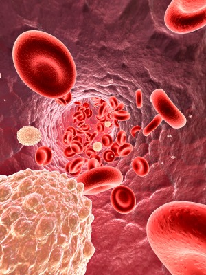
This nice-sized study (n=106) took IgG antibodies – the ones that usually do the mischief in autoimmune diseases – from people with ME/CFS (Canadian Consensus Criteria), long-COVID patients with ME/CFS, multiple sclerosis, and healthy controls, and cultured them with endothelial cells and human foreskin (!) cells. (The foreskin cells were a stand-in for a general cell type. The endothelial cells were chosen because of the apparent blood vessel problems.)
Stressed Mitochondria in…
While the IgG from every group (ME/CFS, MS, and healthy controls) was unable to enter general cells (human foreskin), it readily penetrated endothelial cells. Once the IgG entered the endothelial cells, though, things changed: only the IgG from ME/CFS and long COVID patients caused mitochondrial fragmentation, leaving more small mitochondria.
The GIST
-

Identifying what the IgG antibodies are attacking could result in targeted treatments and more successful clinical trials.
This is yet another “something in the blood” study in which researchers transfer something in the blood – in this case IgG antibodies – into a lab culture to see what, if anything, those antibodies are doing.
- If this study were successful, it would point another arrow at autoimmunity in ME/CFS and/or long COVID. It turned out that it was.
- The study found that IgG antibodies from ME/CFS and long COVID patients (but not healthy controls or MS patients) are fragmenting the mitochondria in the endothelial tissues that line the blood vessels in a substantial subset of patients. Mitochondrial fragmentation typically occurs when the mitochondria are under stress and start splitting off damaged sections.
- Interestingly, mitochondrial ATP production was not significantly affected, but the mitochondria were left in a damaged, hunkered-down, protective state that the authors believe likely affected blood flows.
- Further analyses suggested that the antibodies were inhibiting mitochondrial functioning but that the entire immune complex traveling with the antibodies was necessary to fragment them.
- An analysis of these immune complexes suggested that they damaged connective tissues (including blood vessels) in ME/CFS and led to increased clotting in long COVID.
- The authors proposed that long COVID presented an earlier form of ME/CFS and that long COVID patients would eventually look like ME/CFS patients; that is, the inflammation and clotting would eventually produce the connective tissue issues present in ME/CFS.
- The potential treatment ramifications of these findings are substantial. For one, the study suggests that a significant subset of ME/CFS/Long COVID patients (with mitochondrial fragmentation) might be more amenable to treatments that remove or block IgG and/or which improve blood vessel functioning. Identifying that subset should lead to more targeted and successful treatment trials.
- Because this was a lab study, it will be important to move into human studies that assess the effects of mitochondrial fragmentation on blood flows, symptoms, etc. and are able to identify patients that could benefit from treatments.. Identifying what the antibodies target will be critical to determining which treatments can stop them, and many options are available. Treatments that increase blood flows could be helpful as well.
- In short, this study identified a possible autoimmune process that causes mitochondria in the cells lining blood vessels to fragment, thereby affecting blood flows and opening potential treatment options if the findings are validated.
Support Health Rising and Keep the Information Flowing!
Health Rising is not a 501 c (3) non-profit
Mitochondrial fragmentation usually indicates that the mitochondria are under unusual stress. The mitochondria become smaller when they cut off damaged parts of themselves to survive.
…Stressed Endothelial Cells =
Indeed, the increased respiratory rate observed in endothelial cells exposed to ME/CFS IgG suggested that mitochondria were under increased stress. The finding that the endothelial cells in ME/CFS exhibit greater “glycolytic compensation” (i.e., are relying more on glycolysis (anaerobic energy production)) fits with the idea of that something has gone awry with the aerobic energy production system, which may result in more oxidative stress, etc.
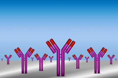
The antibody arms attach to the target. The fact that the arms of antibodies from ME/CFS and Long COVID patients affected mitochondrial fragmentation suggested that the antibodies were attacking the mitochondria.
Interestingly, ATP production was not reduced. The fact that it was not may have something to do with how endothelial cells operate. They get most of their basal energy from glycolysis – not aerobic energy production. They use the mitochondria for other activities (blood vessel production, stress response, etc.).
The authors of the paper believe the immune complex associated with ME/CFS and long COVID igG puts the mitochondria in endothelial cells into a “chronic protective stress response” state that sounds very much like Naviaux’s cell danger response.
Next, in an attempt to learn which part of the IgG antibody was affecting the mitochondria, they broke it up into its two major sections – the Fc and Fab regions – and assessed them in culture. The Fab (fragment antigen-binding) regions are represented by the two arms of the antibody. They are what the antibodies use to bind to pathogens, tissues, etc. The Fc region (the stem of the antibody) recruits immune cells and determines the strength of the immune response.
The ability of the Fab region to affect mitochondrial metabolism suggests that antibodies were attacking the mitochondria. Because it points an autoimmune arrow straight at the mitochondria/endothelial cells, this is potentially a big deal in this disease.
The fact that the entire antibody/immune complex was needed to fragment the mitochondria suggests that antibodies plus the immune complexes are needed to complete the picture. Note, though, that this part of the paper is based on a very small sample size.
Stressed Connective Tissue?
The researchers performed proteomic analysis on the immune complexes associated with IgG isolated from ME/CFS and Long COVID ME/CFS patients. Now, the researchers wanted to know what those immune complexes consisted of and if they were having an impact.
Immune complexes can hold the key to what’s going with autoimmunity and IgG because they’re clusters of cells that form when IgG binds to an “antigen” or target. They hold keys not only to what the antibody is attacking but the effects its having.
Look at the clean separation between the healthy controls (red) and the ME/CFS, long COVID ME/CFS, and MS patients’ proteomes. The ME/CFS patients’ proteomes (green) are clustered near but still nicely separated from the long COVID patients’ (purple) and the MS patients (blue).
The results suggested that the immune factors traveling with the antibodies clearly play a role, and they appear to play somewhat different roles in ME/CFS and long-COVID ME/CFS.
The immune complexes associated with the ME/CFS IgG showed increased levels of extracellular‑matrix (ECM)–related proteins, i.e., connective tissue proteins. Note that the blood vessels are connective tissues, and endothelial cells are embedded in an extracellular matrix. The increased levels of extracellular matrix proteins were an intriguing finding, given recent findings suggesting that increased basement membrane production may be preventing blood from flowing to the tissues.
Increased levels of an intriguing protein called SPEG, which plays a major role in muscle development and maintenance, provided another possible lead. Because SPEG is not usually found outside of cells, the increased levels suggested that some sort of muscle injury had occurred, and suggested that autoantibodies to it had been formed. IgG binding to SPEG could result in disturbed muscle calcium handling (aka Wirth and Scheibenbogen?), fatigue, exercise intolerance, and post-exertional malaise. Other proteins that popped up also provided potential leads.
Long COVID + Time = ME/CFS?
The long COVID ME/CFS IgG contained more proteins associated with clots. This might have been expected, as microclots, while present in ME/CFS, appear to be more evident in long COVID. While the end result – poor endothelial functioning – may be the same in ME/CFS and long COVID, it may be produced in different ways. In ME/CFS, the connective tissue is more affected, whereas in long COVID, clotting is a bigger issue.
The authors raised the intriguing idea that the increased inflammatory profile in long-COVID ME/CFS simply reflects an early stage of ME/CFS, which ultimately reverts to a more metabolically challenged state with greater connective tissue damage as the disease progresses.
The treatment ramifications of these findings are substantial. For one, the study suggests that the subset of ME/CFS/Long COVID patients with mitochondrial fragmentation might be more amenable to treatments that remove or block IgG and/or which improve blood vessel functioning. Identifying that subset should lead to more targeted and successful treatment trials. As the last blog pointed out, targeting subsets has become a central theme in ME/CFS and long COVID research.
Conclusion
This was yet another intriguing paper from Prusty and colleagues. This study very nicely potentially tied together two hot topics – IgG antibodies/immune complexes and blood vessel problems – plus, it added a new player to the mix – immune complexes – and boosted the idea that an autoimmune process is present in at least a significant subset of patients.
Increased levels of fragmented mitochondria, which appeared to be hunkered down, suggested the mitochondria in the endothelial cells from ME/CFS and long COVID patients were in a protected, stress-response state.
In the lab, at least, the IgG antibody/immune complexes from ME/CFS and long-COVID ME/CFS patients fragmented the mitochondria, putting them under stress. ATP production was not significantly affected, but the mitochondria were discombobulated and were not working well.
This is the second time, by the way, that Prusty, Naviaux, et al. have been able to fragment ME/CFS mitochondria in the lab. In a small 2023 study, they found that transferring ME/CFS serum in the lab could induce mitochondrial fragmentation in certain cells.
The immune complex analysis – which highlighted coagulation and connective tissue problems – suggested that researchers need to start assessing the immune complexes in which the antibodies are embedded. In fact, it’s possible that immune complexes play a larger role than the antibodies themselves.
Damage to the dynamic endothelial cells that line our blood vessels could translate into, among other things, reduced blood flows (particularly in the microvasculature) and inflammation. The paper identified several proteins that could play a role in these diseases.
The paper had its limitations. Because it was a lab study, it couldn’t provide evidence that its findings – no matter how intriguing – are actually happening. Some of the analyses were also done on very small sample sizes. Some ME/CFS participants’ IgG did not appear to induce high levels of mitochondrial fragmentation.
Next Steps
It also provided some nice leads. Bigger studies are needed to validate and expand its findings. For instance, future studies assessing endothelial cells from other sites in the body (e.g., the brain and skeletal muscle) could determine how widespread this process is.
Getting out of the lab and into human studies is critical. Studies that assess the extent of mitochondrial fragmentation, and its effects on blood flows, inflammation, orthostatic intolerance, handgrip strength, etc., would provide strong validation that mitochondrial fragmentation is playing a major role, and let us know which patients it’s affecting and how it’s affecting them.

Attempting to identify what the IgG antibodies are attacking is a possible next step.
Identifying what the antibodies/proteins are targeting or attacking would play a key role in uncovering treatment options. Once the target is found, many approaches (plasmapheresis/immunoadsorption, IVIG, efgartigimod, complement inhibitors, Rituximab, decoy antigens, etc.) that block those antibodies/proteins from attacking the mitochondria/blood vessels are potentially possible.
Other treatments (statins, ACE inhibitors, ARBs, sildenafil, and more) that attempt to increase blood flows and improve blood vessel health are a possibility. Reports indicating that, in some cases, hyperbaric oxygen therapy (HBOT) has returned some patients to health suggest that, over time, oxygenating tissues might be enough for some. HBOT may be doing this by activating repair programs, creating new, healthier capillaries that increase perfusion, turning off established inflammatory/oxidative stress loops, and even increasing blood flow, thereby promoting neuroplasticity in the brain.
The key will be identifying the patients who are more likely to benefit. Indeed, finding treatable subsets has become the theme of the day. (Thanks to Matthias for the tip about the study.)
- Coming up – “Nothing in the Blood”? A validation study fails….
Support Health Rising and Keep the Information Flowing!
Health Rising is not a 501 c (3) non-profit


TIRED OF LOOKING AT OUTCOMES. AFTER ALL THIS TIME, I WANT TO SEE SOME SERIOUS EXPLANATION OF CAUSES. HOW ABOUT MERCURY.
several researchers and clinicians have identified elevated mercury levels in subgroups of patients with Myalgic Encephalomyelitis/Chronic Fatigue Syndrome (ME/CFS) or related conditions like Multiple Chemical Sensitivity (MCS), often correlating these findings with dental amalgam fillings or fish consumption.
ScienceDirect.com
ScienceDirect.com
+2
Key findings from research and clinical observations include:
Elevated Mercury Levels in MCS/CFS Patients: A study on Multiple Chemical Sensitivity (often comorbid with ME/CFS) found that 81.2% of patients had elevated mercury levels in their biological matrices (blood, urine, hair, and saliva).
Dental Amalgam Correlation: Research has shown a strong association between the number of mercury-containing dental amalgam fillings and elevated mercury levels in ME/CFS-like patients, particularly in saliva and urine.
Fatigue Improvement After Removal: Studies have documented that removing mercury amalgam fillings, combined with appropriate treatment, resulted in significant reductions in fatigue, memory loss, and depression in patients previously diagnosed with chronic fatigue.
High-Mercury Fish Consumption: A study at Stony Brook University found that patients with high-mercury seafood consumption (e.g., tuna, swordfish) were significantly more likely to suffer from fatigue (64% vs 31%) compared to those who did not, suggesting a link to mercury bioaccumulation.
Immune Sensitization: Research by Dr. P. Pigatto and colleagues found a high prevalence of metal allergies (92.3%) in MCS patients, with a 50% allergy rate specifically to mercury compounds.
Genetic Factors: A significant correlation has been found between chronic mercury toxicity (presenting as fatigue) and the Apo-lipoprotein E4 genotype, suggesting some individuals are genetically more susceptible to mercury toxicity.
Fatigue to Flourish
Fatigue to Flourish
+5
Important Context: While these studies indicate a potential connection between mercury and symptoms of fatigue, many in the scientific community state that there is not yet enough evidence to suggest that mercury toxicity causes ME/CFS in all cases, but it may be a significant contributing factor for specific subgroups.
Fatigue to Flourish
Fatigue to Flourish
+1
High mercury seafood consumption associated with fatigue at …
Highlights * • We examined seafood consumption and symptoms related to mercury toxicity. * 118 patients from several specialty med…
ScienceDirect.com
Allergological and Toxicological Aspects in a Multiple Chemical …
Dec 3, 2013 — We ascertained the prevalence of allergy to metals and toxicological aspects in MCS patients. Methods. We conducted a retrospectiv…
National Institutes of Health (.gov)
Mercury Toxicity and Treatment: A Review of the Literature
One hundred six probands completed the fourteen-day trial with oral DMPS 400 mg per day. The only complication was an allergic ras…
PubMed Central (PMC) (.gov)
Show all
From my years of reviewing the research what’s at the bottom of most of the rabbit holes is toxins and pathogens for all illnesses so you are probably correct. Have you gotten tested? I also think it’s important to take charge of the science along with our health and start removing potential sources and see how you feel along the way.
I find it interesting that Carmen S is going to study GLP-1 agonists in ME/CFS. I struggle to see the link between her primary theoretical propositions and the mechanistic action of GLP-1 agonists.
As I have said before I am somewhat skeptical on GLP-1 agonists in ME/CFS and it concerns me a bit when researchers jump into such fads.
Of course I hope my skepticism is misplaced and these drugs do offer genuine promise for ME/CFS.
I believe she is doing an online presentation in April, I will be interested to hear her rationale.
https://solvecfs.org/february-2026-catalyst-awards/
I assume that she’s seen too many patients for whom it worked for her not to put this trial on. She is, after all, embedded in quite a large ME/CFS network at Charite. I’m sure she’s regularly hearing from people with ME/CFS.
I think this is great. There’s certainly not another treatment option which has generated this kind of excitement. Two large and expensive GP1-agonist trials (UCSD/RECOVER) are being done in Long COVID and I imagine they funders felt pretty good about GLP-1’s chances there. Glad to see we’re getting an ME/CFS trial so quickly 🙂
I will be interested to hear her take on it.
It shouldn’t be forgotten that there’s huge $$$ behind the GLP-1 agonist industry. Not necessarily a bad thing – it could work in a positive way – but one should keep in mind that the companies’ valuations stand to benefit from wider applications.
Again, I am not dismissing the possibility of value in treating ME/CFS but just making a cautionary note.
I will register for the session on 18 April.
What I understand is that she also suspects a problem with general energy management in the diseases and those drugs are known to heavily influence these targets. So there is a chance they might help a subset I assume.
Hopefully, I’m proven wrong, but given that GLP-1 agonist are among the more readily available drugs to patients (at least in some countries), I’d be surprised if they proved to be a breakthrough treatment, purely from an anecdotical standpoint. These are among the drugs that many people on the forums/subreddits have reported trying long before ME trials, and while you do see the occasional report that they proved to be somewhat helpful, they don’t seem to be a gamechanger for the field so far.
GLP-1 agonists were hyped up on several medical fields these last years, like for example for treating dementia, where studies have now come back with negative results.
Personally, I found them to be helpful in weight management in this new involuntarily sedentary life, but other than that, I haven’t experienced any beenefits. Of course, ME patients are very heterogenous, and I’m all for the trials – I’d just be surprised if this drug turned out to be a silver bullet.
An wen kann man sich wenden, um bei sich selbst zu identifizieren, zu welcher Gruppe an ME/CFS-Erkrankten man gehört, um zu wissen, welche Behandlung überhaupt anschlagen bzw. nicht schaden würde? E-Mail und Ansprechpartner wäre toll.
Google Translate
Who can you contact to identify for yourself which group of ME/CFS patients you belong to, in order to know which treatment would actually be effective or not harmful? An email and a contact person would be great.
Groups are working on finding subsets and some studies have elucidated what they believe are subsets (Fluge/Mella) but they’re pretty experimental at this point. It’s a long road but I hope over the next year or so that we get some more established subsets. The OMF’s Bioquest project, and PrecisionLife’s work are two that come to mind.
GLP-1 agonists at low doses have been used successfully in smaller studies by others to ameliorate mast cell reactions where nothing else has helped.
Considering that energy (glucose) is an issue with ME/CFS there is likely more going on.
This is not following a fad but following convincing indications that there is something widely available that may help and that is somewhat affordable.
So it is unlikely to target a primary issue, imho, but very likely to target secondary issues that are relevant to pwME
I think that’s a dangerous path. Even if it is effective, the tradeoff will not be worth it because of the muscle loss. Not every person will be able to begin a strength/resistance training program or increase protein intake to adequate levels to counteract the effects.
The paper that is the topic of this blog says:
“Elevated glycolysis could potentially lead to oxidative stress, increased ROS production (Shankar et al., 2025), and impaired energy metabolism, ultimately contributing to mitochondrial dysfunction (Hu et al., 2012; Bhatti et al., 2017).”
And https://journals.lww.com/jasn/fulltext/2024/10001/glucagon_like_peptide_1_receptor_agonist__glp_1.780.aspx says:
“Notably, we found that treatment with Semaglutide 1) decreased glucose uptake, ATP generation and glycolysis characterized by a decrease of the expression of the key glycolytic enzymes, and 2) normalized the structure of mitochondria and the expression of the genes involved in mitochondrial metabolism, in Pkd1 mutant renal epithelial cells and kidneys.”
So the paper of this blog says that increased glycolysis *may* contribute to increased ROS and increased mitochondrial stress and the link I provided says that GLP-1 agonist Semaglutide does reduce glycolysis by reducing its key enzymes and normalized mitochondrial structure and metabolism in *some* cell types.
=> So it’s not that a far fetch. But for it’s use as a cure or even a helpfull (side-effects versus benefits) a lot more research has to be done to see it’s viable. So I am for it, even if there are plenty of IFs left.
GLP-1 agonists have profound impacts on immune signaling, function, and even can stabilize mast cells.
One of their impacts is to bloom amounts of Akkermansia muciniphila in the gut, which causes release of endogenous GLP-1, can increase Treg cells for immune stabilization, and protects integrity of the gut barrier. There may be some downstream impacts on NF-kB, mTOR, et al. Lowering insulin resistance seen in earlier studies for ME. Potential impacts on microglial activation.
Drs. Ruhoy and Kaufman have had some pretty profound results with them. In my personal case, they have modulated my improved gut to produce Butyrate again, as well as allowed me to reintroduce lost foods – something that IVIg treatment, nor Xolair really moved the needle on and medicine couldn’t evoke over multiple decades. I’ve been able to discontinue or lower medication doses and supplements in the process, too.
That said, Akkermansia is chronically absent from my gut on numerous tests along this journey, so I am likely not seeing the entire benefit. I’m currently running an Akkermansia engraftment protocol, to see if this favorable milieu will be successful.
Hi:
I recall that Prof. Bhupesh Prusty had already identified the role of IgG in LC and ME/CFS during his work in Würzburg in 2022.
He has since moved to Rīga Stradiņš University in Latvia.
After a brief introduction in German, his presentation at the ME/CFS Conference 2025 (organized by Fatigatio e.V.) continues in English from minute 2:39 onward.
https://www.youtube.com/watch?v=aDVfpsGo0B0
THANKYOU for these many years of bringing these studies to our attention and making them more understandable.
Thanks! 🙂
Cort, you are the best! Great article!!!
Thanks for a well written summary of this Scheibenbogen-Prusty paper. I really enjoyed reading it. I look forward to Prusty-Scheibenbogen papers so much.
I am also looking forward to the Xist studies, and seeing how that plays into the AutoImmunity hypothesis in ME/CFS.
In ME/CFS, it always feels like there’s so much at play: AutoImmunity, Viral Reactivation, Viral Persistence, T and B Cell Immunity- I wish we had the funding needed to nail which is the chicken and which is the egg. Who is the main driver, and what is the process of cross play between them all?
Again: would be nice if researchers investigating a disease defined by PEM would drop a word about PEM, wouldn´t it?
Possibly some antigen associated with exertion could induce the formation of immune complexes which in turn would fragment mitochondria?
Possibly then a good idea would be to do the same experiment and stratify patients by their “ME/CFS status” (non-PEM versus PEM)?
Thanks for the summary!
What exactly do you mean by “a word about PEM”?
Are you interested whether the research question has a link to an explanation of PEM or do you ask for a study that seperates patients according to “having PEM” or being in remission?
Here a very interesting study by Jacqueline Cliff at Brunel Uni is underway where they test for HHV-6b reactivation in saliva in ME/CFS patients.
This is very important for me because I can fight my ME flares with a herpes drug but it’s also a very well designed study. I think that the team understands better than everybody else the remitting-relapsing nature of ME/CFS with an acute phase where inflammation is going on and a calming down and stopping of this where people are not recovered but in still in a kind of remission. They let patients give saliva and fill out a questionnaire about symptom severity every time they test.
I wonder whether you are asking for that?
When you say PEM are you interested in the connection between the causation and aggravation of flares through exertion or do you point to the acute and remitting phases and the idea that a different biochemistry could be expected during the different stages?
Thank you, Lina, this is a very good summary of my ideas. I am not a immunologist/virologist so I cannot comment on the kind of tests they should do but I know that Eugenia Ariza at Ohio state would be the top expert in this field, she often works with the HHV6 foundation. At a minimum they ought to follow dUTPases ((https://hhv-6foundation.org/all/the-dutpases-of-hhv-6a-and-ebv-may-encourage-autoimmunity-in-me-cfs )). Hope they don´t squander their chance, looking at virus abundance in saliva alone dosen´t cut it. Also this ist very dynamic, activation happens within a short window of hours after exertion so the typical typical schedule “get labs before exercise then 24 hours later” is not a good idea. As a side note – it is a great weakness of so many exercise studies that they leave out the window of 0-2 (-6) hours after exertion. The 24-hour interval may capture the peak of PEM symptoms, but the early hours are critical for studying the pathophysiology of what triggers it. Light et al have demomstarted how to do it 15 years sago ((https://pmc.ncbi.nlm.nih.gov/articles/PMC3175315/ )). If you need Eugenias e-mail adress let me know.
Maybe also of relevance when discussion this experiment: while interesting, the immune complex formation seems to be of relevance (in terms of mitochondrial changes) only for females. Also, there is no correlation with clinical severity of the ME/CFS donors.
This questions if immune complex formation is really a core element underlying the clinical picture of ME/CFS.
I am wondering if autoimmunity is an issue in my case. And given the study you just wrote about I’m wondering if I should be taking SCIG or not. It has been offered.
IVIg has changed my life. If you can try Ig, I would suggest it. So far, it is the cornerstone of my medical treatment. I hope that someday I can move on, because it isn’t remission, but it definitely improved fatigue, PEM, POTS, pain, neuropathy, gastroparesis. I’ve been receiving it for over 7 years now.
Thank you. Perhaps we can chat?
My email is helenfcg@gmail.com
This antibody/endothelial-cell angle helps explain why so many patients feel the illness is physical even when routine tests do not catch it. It also argues against one-size-fits-all advice. When immune complexes, blood vessels, mitochondria, and inflammation may all be part of the picture, patients need a way to organize recovery-support decisions carefully instead of chasing a new cure headline every week.
Thank you for shedding light on the intriguing connection between antibodies, mitochondria, and blood vessels in ME/CFS and long COVID. It might be interesting to explore how these interactions influence energy levels in patients. Have there been any discussions about lifestyle interventions or therapeutic approaches that could harness this knowledge?
I found it fascinating how the study showed IgG antibodies from ME/CFS and long COVID patients fragment mitochondria in endothelial cells, a detail that ties directly into the triad of antibodies, mitochondria, and blood vessels highlighted in the title.
Great read on how IgG antibodies from ME/CFS and long‑COVID patients cause mitochondrial fragmentation in endothelial cells—such a clear link to the blood‑vessel issues we see clinically. It really underscores how immune complexes are disrupting cellular energetics. If you’re into detailed research tools, check out my Fortnite Sprite checklist. insanity!