




Geoff’s Narration
The GIST
“Together, these findings demonstrate that WASF3 suppresses oxidative metabolism in the brain and contributes to cognitive dysfunction, providing mechanistic insights into the “brain fog” reported in ME/CFS.” Hwang et al.
WASF3 – a mitochondrial-inhibiting protein – an already very interesting potential factor in ME/CFS – just got more interesting. (Thanks, Matthias, for the tip about the new study.) You can read about Paul Hwang’s fascinating saga with the mitochondrial inhibitor WASF3 in a past blog (see below).
Hwang, a senior intramural researcher at the NIH, first got interested in WASF3’s effects on the muscles. His finding that elevated WASF3 activity in ME/CFS was linked to disrupted mitochondrial activity could help explain the muscle fatigue found in ME/CFS.
He also found that reducing ER stress with a drug called salubrinal improved mitochondrial functioning in patient-derived cells. It appears that endoplasmic reticulum stress is the trigger that sets off WASF3, which then impacts the mitochondria.

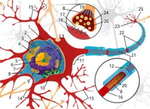
The MAMs finding suggested WASF3 could be affecting neuronal functioning as well as energy production.
THE GIST
- WASF3 – a mitochondrial-inhibiting protein – and an already a very interesting potential factor in ME/CFS – just got more interesting.
- Hwang found that elevated WASF3 levels were associated with disrupted mitochondrial activity, which could help explain the muscle fatigue found in ME/CFS.
- In this new study, he examined WASF3’s potential effects in the brain using a mouse model. His findings supported his earlier studies and expanded on them. Right now, WASF3 appears to have an even larger effect on the brain than on the muscles.
- Hwang’s new study validated a crucial idea – that it takes cellular stress plus WASF3 elevation to create problems. The cellular stress + idea makes perfect sense, given that many ME/CFS studies show increased oxidative stress, membrane damage, and problems with energy production.
- Hwang found WASF3 throughout the mouse brain but then zeroed in on the seat of short-term memory retention, emotions, learning, and others – the hippocampus – and subjected hippocampal tissues to varying WASF3 levels.
- Increasing WASF3 levels in these tissues reduced their oxygen levels by a startling 50% and put them under dramatically increased stress.
- A metabolomic analysis of these tissues suggested that widespread problems with energy production had occurred.
- Hwang found WASF3 present in more parts of the brain cells than he had in the muscle. Besides the mitochondria, he found it in spades in the endoplasmic reticulum and the “mitochondria-associated membranes (MAMs)” in mouse brain cells.
- Finding WASF3 in the “MAMs” was particularly noteworthy because these membranes regulate a lot of neuronal functioning. This suggested that if WASF3 is indeed having negative effects, it could be doing so in several ways.
- Hwang also uncovered more ways WASF3 could be affecting the mitochondria. He demonstrated three different ways WASF3 could be negatively affecting mitochondrial activity in brain cells.
- The fact that WASF3 appears to have particularly detrimental effects when cellular stress is present fits nicely with what happens in ME/CFS; i.e., that stress – whether it comes from physical or mental activity or emotional issues – makes things worse. At its core, ME/CFS seems to be a stress response disorder.
- In this scenario, increased cellular stress increases WASF3, which then inhibits mitochondrial energy production. Fatigue and other symptoms result. Post-exertional malaise could be explained by a long-term reduction in the mitochondria’s ability to produce energy, accompanied by increased oxidative stress, and so on.
- Some really interesting work is going on in the brain in the NIH right now. It’s mostly being driven by internal or intramural researchers who have latched onto the illness in different ways.
- We have two senior intramural researchers (Hwang and Chung) digging into the mitochondria and the brain in their labs. We have an emeritus, highly accomplished neuroscientist (Goldstein) looking at norepinephrine and energy production, and a neuroscientist (Bedard) focused on the brain-muscle interaction.
- Most intriguingly, they could all fit together. Every study suggests that energy production problems and the inability to tolerate “stress,” whether at a physical (handgrip test) or cellular level, play critical roles.
- WASF3’s role in these problems remains conjectural, as studies to date have been small, but so far the evidence suggests it could be a major player in ME/CFS. We vitally need larger studies that can assess its relevance across broader ME/CFS groups.
- Thankfully, an ME/CFS drug trial is in the works. I have a date to speak with Paul Hwang when it’s ready to go – probably sometime around December.
Support Health Rising and Keep the Information Flowing!
Health Rising is not a 501 c (3) non-profit
The Study
WASF3 is also found throughout the brain, and Hwang earlier suggested the WASF3 problems he was seeing in the muscles might be present in the brain as well. His recent study, “Stress-inducible WASF3 regulates brain metabolism and cognitive function“, in the venerable Proceedings of the National Academy of Sciences (PNAS), suggests WASF3 could have a major impact on the brain.
Hwang used two different genetic techniques to produce mice without WASF3. One technique used CRISPR to delete the part of the gene that produces the WASF3 protein, but it did not find that deleting WASF3 impaired the mice.
Using something called “Cre-lox”, which also creates cellular stress, they found that increasing cellular stress in mice with WASF3, by contrast, resulted in altered “fear conditioning behavior” and learning. However, when they removed WASF3 from the mice, increased cellular stress did not reduce cognition.
This suggested that the key factor is not just increased WASF3 levels but cellular stress plus increased WASF3 levels. This fits with Hwang’s prior finding that increased endoplasmic reticulum stress was associated with increased WASF3 levels and impaired mitochondrial functioning.
A Close Look at the Hippocampus

Hwang found that increased WASF3 expression dramatically reduced oxygen levels in hippocampal tissues.
Hwang found WASF3 throughout the mouse brain, but WASF3 levels were particularly high in three brain regions associated with ME/CFS: the amygdala, hippocampus, and cerebellum. Hwang then subjected hippocampal tissues to differing WASF3 levels.
The hippocampus plays an important role in short-term memory retention, emotions, spatial navigation, and learning.
Increasing WASF3 levels in those tissues reduced their oxygen levels by a startling 50%. The tissues were clearly under dramatically increased stress, as markers of mitochondrial oxidative metabolism, including ATPIF1 and MTCO1, and oxygen consumption rose.
The more WASF3 present, the worse complex IV of the mitochondrial electron transport chain functioned. Conversely, reducing WASF3 improved mitochondrial functioning.
Hwang and company next did a metabolomic analysis of those tissues. The fact that increasing WASF3 levels generally produced lower levels of several metabolites suggested WASF3 had put the brain into a hypometabolic or low-energy state – as some brain ME/CFS studies have suggested
For instance, decreased levels of glycolysis metabolites in high WASF3 mice suggested that: a) glycolysis wasn’t proceeding normally; and b) an attempt to compensate for reduced mitochondrial ATP production was underway.
Similarly, fats provide an important energy source, and some fat metabolites were reduced. Plus, increased levels of some fatty acids (medium-chain dicarboxylic acids pimelate, suberate, and azelate) suggested fatty acid metabolism had been disrupted.
Even peroxisomes, for those who remember Ian Lipkin’s big paper, got into the act. Lipids expected to accumulate when peroxisomes are dysfunctional increased. WASF3 also inhibited a histamine-derived metabolite called IAA, which triggers activity of the feel-good compound GABA.
Note that these studies are still very small – just five or six mice – but Hwang’s findings suggest that increased WASF3 levels are highly detrimental to the mitochondria.
The Real Target? The Brain…
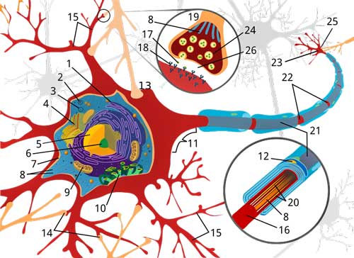
The MAMs finding suggested WASF3 could be affecting neuronal functioning as well as energy production.
While Hwang found WASF3 in mitochondrial membranes in muscles in his earlier study, this time he found it in spades in the endoplasmic reticulum and the “mitochondria-associated membranes (MAMs”) in mouse brain cells.
Finding WASF3 in the MAMs surprised Hwang and is potentially important because it suggests WASF3 may affect much more than energy production. Besides energy production, these mitochondrial-associated membranes, or MAMs, also affect calcium transfer, lipid metabolism, stress signaling, and coordination of mitochondrial activity.
This suggests ER stress/WASF3 could also affect many aspects of neuronal functioning. Cognition, learning, and the ability to sustain mental activity could all be affected.
More Ways to Pummel the Mitochondria
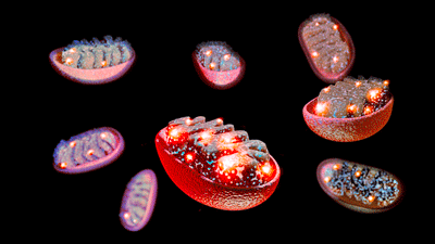
Hwang also found more ways WASF3 could be impacting the mitochondria.
As they dug deeper into WASF3’s effects on mitochondria, they found that increased WASF3 levels reduced levels of a compound called ATPIF1, which regulates how much ATP mitochondria produce. (WASF3 does this by increasing the levels of another protein that degrades ATPIF1. Reduced ATPIF1 was recently found in ME/CFS.)
Hwang’s findings now suggest WASF3 could be whacking the mitochondria in three ways:
- by interrupting the electron transport chain at complex IV.
- by reducing how much ATP the mitochondria are asked to produce (low APIF1).
- by impairing mitochondrial movement. There’s ATP production, and there’s getting the ATP to where it’s needed. Mitochondria should be able to move closer to where the cell needs energy. WASF3 also appears to impair that.
Hwang further tested WASF3’s ability to impact the mitochondria by introducing a chemotherapy agent known to produce “chemo-fog” and found that, in the presence of that agent, WASF3 inhibited complex IV in the electron transport chain in mitochondria of brain cells as well.
Interestingly, Hwang’s new paper also indicates that different parts of the WASF3 protein have different effects. He demonstrated that removing one part of the protein resulted in mitochondrial movement being restored, but complex IV in the electron transport chain was still affected.
The findings suggest that Hwang will likely dive deeper into WASF3’s molecular structure to understand the effects of different parts of the protein. These findings could have important treatment implications.
The Key – Cellular Stress
The fact that WASF3 appears to have particularly detrimental effects when cellular stress is present fits nicely with what happens in ME/CFS; i.e., that stress – whether it comes from physical or mental activity or emotional issues – makes things worse. At its core, ME/CFS seems to be a stress response disorder.
In this scenario, increased cellular stress increases WASF3, which then inhibits mitochondrial energy production. Fatigue and other symptoms result. Post-exertional malaise could be explained by a long-term reduction in the mitochondria’s ability to produce energy, accompanied by increased oxidative stress, and what have you.
Cautionary Note
The fact that Hwang continues to explore WASF3 in mice is an encouraging sign as they’re probably the next logical step for many researchers who want to understand what’s going on. The mouse findings cannot prove, though, that increased WASF3 levels cause mental or physical fatigue or cognitive problems in people with ME/CFS. We don’t know, for instance, that WASF3 levels are increased in the brains of people with ME/CFS.
The mouse studies suggest that if they are, they could be having quite an impact.
Almost In Spite of Itself, the NIH Turns Interesting
Say what you will about an NIH administration that has refused to sufficiently fund ME/CFS for decades; some really nice work is going on in the brain right now, and it’s mostly being driven by internal or intramural researchers who have latched onto the illness.
We have two senior intramural researchers (Hwang and Chung) digging into the mitochondria and the brain in their labs. We have an emeritus, highly accomplished neuroscientist (Goldstein) looking at norepinephrine and energy production, and a neuroscientist (Bedard) focused on the brain-muscle interaction.
These researchers got to ME/CFS in a variety of ways. Hwang came via a person with ME/CFS who happened to be in a family cancer study, Chung via a Solve M.E. board member whom he met at a wedding (!), and Goldstein and Bedard via Nath’s intramural study. Thanks to a Solve M.E. grant, an ME/CFS mouse study is underway in a most interesting drug.
As Hwang was uncovering WASF3’s impact on muscle mitochondria, Chung was producing a potentially powerful mitochondrial compound (IVO-21) that may affect WASF3. (A Solve M.E. grant is exploring that.)
Meanwhile, quite apart from the others, Goldstein proposed that reduced ATP production (energy again) caused the reduced norepinephrine activity he found.
Hwang’s latest finding suggests that WASF3 is not only present in more places in brain cells than in muscle cells, but could also interfere with mitochondria and ATP production in more ways than previously thought.
Plus, Bedard’s finding that ME/CFS patients’ muscle force declined rapidly during the handgrip test suggested that the brain-muscle system gets plastered under physical stress. Whether triggered by physical or mental activity or emotional distress, we’re talking about problems with energy production and stress at the cellular level. The big question right now is how much of that is due to WASF3 and ER stress.
Of course, Hwang examined mice, not humans, and we don’t know that WASF3 levels are increased in ME/CFS and long-COVID patients’ brains. Still, the signs, at least for now, are positive.
The big fly in the ointment right now is that the studies are still quite small. Before we can say WASF3 and ER stress are major factors in ME/CFS, these findings need validation and expansion in much larger studies.
The potential certainly seems there for a great deal of collaboration, but we don’t know if these groups are working together. Because these are intramural efforts, we don’t know how much funding is going into this research or what the next steps are.
As far as I can tell, there are no signs that a major study is currently underway. One would hope some grant applications are in. If the NIH has been looking for a reason to fund a major grant effort (a big if after all these years), I wonder what more they could want? These findings, after all, come from internal NIH researchers, mostly senior NIH researchers. If they don’t trust their results, who can they trust?
In any case, regarding Hwang’s latest finding, some further studies could determine if physical or cognitive stress really does increase WASF3 activity in ME/CFS, and if increased WASF3 levels are associated with an inability to sustain a handgrip test, aka Bedard, and/or reduced NE functioning/synthesis, aka Goldstein. A positive result to those studies would suggest researchers have identified a major player in ME/CFS.
Drug Trial Coming
A drug trial showing that reducing WASF3 levels reduces symptoms would, of course, be huge, and the good news is that Hwang told me an ME/CFS trial is in its final stages of approval. The only thing we know about that drug is that it has not been tested in ME/CFS before. (Paul Hwang has promised a Zoom interview sometime, probably in December, to discuss it.)
Support Health Rising and Keep the Information Flowing!
Health Rising is not a 501 c (3) non-profit





You’re welcome, Cort. We all need to work together!
You are right to point out the limitations of this work for now, but that doesn’t negate that I think this is very interesting and hopeful (and I am not implying that you think that btw!)
.
Dr Hwang very kindly responded (some researchers don’t) to me a year or two ago on his research. He confirmed my thinking that the elevated WASF3 and ER stress are downstream of something else. But again that does not negate the potential significance of WASF3.
It seems to me, that if elevated WASF3 really is significant *clinically* in ME/CFS, then the ideal would be to address what is upstream of it. Having said that, if elevated WASF3 is contributing to symptomology, then even addressing that itself could potentially help. A
Personally, I think it is chronic low-grade inflammation (CLGI) that is the upstream factor. I think CLGI is central to the illness. The BIG question then for me is what is sustaining that CLGI?
As you know, I’ve been skeptical on reactivated viruses playing a central role in ME/CFS. I’m more open to that theory now. There’s been some really good papers in the past 3-4 years on T cell exhaustion. Nath suggested in his NIH study that antigen persistence could be a root cause.
One of the problems I always had with T cell exhaustion is that it comes later. So if it is central to ME/CFS, why the symptoms earlier in the illness (when there isn’t necessarily any T cell exhaustion)?
I’ve worked out my own kind of rationale on that. That is, it’s not T cell exhaustion per se that is causing the condition and its symptoms. Rather, T cell exhaustion is the by-product of an inflammatory, activated immune state. If that activated state is triggered by reactivated viruses (for example) that can’t be cleared, then once we get the T cell exhaustion kicking in it becomes harder to clear the reactivated virus. So it remains there, spitting out inflammatory factors, and perpetuating the illness. And the T cell exhaustion is perpetuated. A perpetual cycle.
Under this theory, slamming down the reactivated virus could help! It could reduce the activated state, and improve the T cell exhaustion. The CLGI cycle would be interrupted.
That’s why I am quite interested in PolyBio’s study on Truvada, which is known to have anti-viral properties against a number of viruses that *might* be re-activated in ME/CFS and Long Covid.
Another possibility is that it is something other than a viral factor that is perpetuating the CLGI. The possibility there, I think, is some sort of systemic feedback loop. Of course, this could also be happening WITH a viral factor.
I’m really looking forward to the video with Paul Hwang!
“Rather, T cell exhaustion is the by-product of an inflammatory, activated immune state. If that activated state is triggered by reactivated viruses (for example) that can’t be cleared, then once we get the T cell exhaustion kicking in it becomes harder to clear the reactivated virus.”
Nice catch on the extended time to T-cell exhaustion. I hadn’t thought of that before. If I have it right what you propose is Ruiz-Pablo’s idea as well. 🙂
Only heard of Ruiz-Pablo’s work in passing, will have a closer look
It’s fascinating research indeed. I agree the problem likely needs to be dealed with upstream, not just inhibiting WASF3. WASF3 and the downstream effects show many signs of what is needed to protect the mitochondria in case of excessive ER / cellular stress.
One of the big dangers to cells and mitochondria during excessive oxidative stress is self-accelerating destruction.
A ‘classic’ cell death route is oxidative stress releasing cytochrome_c from the mitochondria and that then triggers an accelerated death of the cell. See https://www.nature.com/articles/4401950 saying “Mitochondria-generated ROS play an important role in the release of cytochrome c and other pro-apoptotic proteins, which can trigger caspase activation and apoptosis.”
Note: apoptosis is organised cell death, something that one does not want too much of especially in the brain.
How WASF3 is said to effect mitochondria:
1) by interrupting the electron transport chain at complex IV.
2) by reducing how much ATP the mitochondria are asked to produce (low APIF1).
3) by impairing mitochondrial movement. There’s ATP production, and there’s getting the ATP to where it’s needed. Mitochondria should be able to move closer to where the cell needs energy. WASF3 also appears to impair that.
Now mitochondrial ROS is controlled by how polarized (electrically charged) the mitochondria are. Both too much and too few is disastrous. https://www.mdpi.com/2073-4409/13/6/493 gives a good overview on that. The figure called “graphical abstract”, below the abstract, shows that “uncontrolled respiration” is the big danger to apoptosis (cell death). That means both too low polarization (depolarized) as too high polarization (maximum polarization) lead to cell death.
By interupting complex IV and by reducing ATP demand, one moves away from the risk of depolarization. (Note: every production of ATP from ADP costs charges and decreases polarization; production of new charges needs to keep up or polarization collapses).
1) saves these charges by reducing conversion speed from charges to ATP.
2) saves charges https://en.wikipedia.org/wiki/ATPIF1 “It is thought to inhibit ATP synthesis by preventing the release of ATP.” by decreasing the demand / signal to convert more charges to ATP.
3) saves charges by reducing how easy ATP gets to the place of usage, increasing ATP inside the mitochondria as they don’t release it as much. This in turn reduces the need to convert more charges into ATP.
=> WASF3 has a rather strong potential to help prevent collapse of mitochondrial polarization and with it cell suicide.
The other side, hyperpolarization, has to be prevented too. That can be done for example by blocking Succinate Dehydrogenase. https://en.wikipedia.org/wiki/Succinate_dehydrogenase is also called respiratory complex II (see link). That is a respiratory complex sitting before complex IV (Roman notation for four) in the mitochondrial ETC. Blocking complex II is a rather potent way to reduce the production of charges (helping to prevent building excessive amounts of charges).
Blocking Succinate Dehydrogenase can be done rather well by *Itaconate*. See https://pubmed.ncbi.nlm.nih.gov/39406969/ with title “Itaconate drives mtRNA-mediated type I interferon production through inhibition of succinate dehydrogenase” (throwing in the Itaconate shunt hypothesis here). Other oxidative stress chemicals help here too.
=> Itaconate has a rather strong potential to prevent building up too many charges (helps preventing mitochondrial and cell suicide due to overpolarization).
=> Combined: a ***carefull*** balance between WASF3 and Itaconate (plus other chemicals!) is able to stave of a long list of dangers (no time or space to write on it here) *while* preventing the mitochondria and cells to go suicidal due to too much or too few polarization.
With our strong metabolic (and blood and oxygen delivery) swings during overexertion and crashes, it’s a near miracle that the cells and mitochondria are able to keep away from these two (hyperpolarization and depolarization) very critical danger zones. And that is what IMO WASF3 and Itaconate help to do:
A) Strongly reduce risk for depolarization
B) Strongly reduce risk for hyperpolarization
C) Keep a small and narrow zone of functioning left (we have a rather narrow zone where we function ‘sufficiently well’ (in order to survive at least) without crashing).
D) Do so at the double whammy of reduced ATP production and ATP release to the cell, very strongly limiting our ability to function beyond the bare minimum.
E) Provide our ‘astonishing’ ability to survive with such poor blood and oxygenation and ATP production levels. Why? IMO because mitochondrial depolarization and hyperpolarization are a stronger cell death signal then poor blood flow, oxygen delivery and ATP production. Cells seem to adapt better to very low ATP levels then to depolarized or hyperpolarized mitochondria.
In addition, the increased ROS produced by mitochondria constantly showers and attacks pathogens with chemicals that kill or at least inhibit plenty of pathogens too.
=> Dauer at work?
I try and stay far away from messing with these ‘Dauer-orchestrators’. Sure, doing so could give a quick boost in energy but if one crashes with disabled safety brakes, the crash may be rather deep and lasting. That does no mean losing hope! Like Matthias says: it gives more opportunities to try and deal with what is causing the problems upstream.
I gotta say, this is really fascinating and encouraging. Thanks Cort – and Matthias! And I’m very glad that a clinical trial is on the horizon. Is the drug that will be trialed salubrinal, or something else?
It’s a mystery to me. When last heard the trial was in the last stages of being approved. How big it is, or what drug, I don’t know.
Sorry Cort, I forgot to ask the more crucial question: is the drug that will be tested already on the market? That is a huge question, of course, because a drug already on the market means we patients won’t have to wait 10+ years for it to become approved.
NAD should suppress WASF3 but the question is… how?
Interestingly, I was reading that lumbrokinase can inhibit ER stress (and therefore potentially WASF3). It is being trialled in long covid, but I don’t think the rationale is ER stress inhibition. Rather it is micro-clotting. But perhaps there’s multiple mechanistic benefits?