




Geoff’s Narration
The GIST
Health Update: Lions, Tigers, and Bears, Oh My! No lions or tigers, but plenty of bears (or at least one bear). A black bear swiped the dogs’ food while we were on a walk, then kept disrupting things at night, making the dogs bark and bark (and repeatedly disrupting my sleep), then chasing it later. (The bear never actually made it into camp).
All this has been exciting and exhausting, and it’s set me back a bit – hence it took quite a while to get this blog out. With the heat, two fires, that uncaring forest service employee, and now this bear, it’s been a crazy summer! The good news is that I am continuing to improve, and I think the long-term outlook is good.
Health Rising recently covered two NIH brain studies suggesting low energy levels in the brains of ME/CFS and/or long-COVID patients. The first study found a dramatic reduction in energy availability in the cingulate cortex – a part of the brain involved in “effort”, pain sensitivity, autonomic nervous system activity, and more. The second suggested that low-energy conditions may keep the norepinephrine-producing neurons in the locus coeruleus from producing sufficient norepinephrine.

The blog pointed out several factors that could be producing the abnormalities found, but it left one out: Paul Hwang’s 2023 WASF3 finding. Because Hwang is operating in a way that is unusual for us, it’s good to check out who he is and how he works.
THE GIST
-

Paul Hwang MD, PhD
Health Rising recently covered two NIH-funded studies suggesting that brain energy levels are low. This blog digs deeper into one of the more intriguing findings of the past couple of years. Paul Hwang’s WASF3 finding could be affecting many areas of the body, including the brain.
- In fact, Hwang and company believe they may have uncovered a “critically important cellular process” which could be affecting many systems in this disease.
- Hwang, MD, PhD, is different from most researchers we know. Because he runs his own intramural lab at the NIH, he can quickly act on findings and explore them deeply, and that’s what he’s done with ME/CFS.
- Hwang’s 2023 ME/CFS paper was astonishing in several ways. For one, he had never published or indicated any interest in ME/CFS. For another, he ran a lab in a part of the NIH (NHLI), which rarely supported ME/CFS. Finally, the study was far more comprehensive – including lab, genetically modified mice, and human studies – than one would have expected from a first-time ME/CFS researcher.
- It’s even a little crazier than that. Hwang’s whole effort began when he was studying a cancer gene in a family that included a woman whose high WASF3 results surprised him. She just happened to have a long-standing case of ME/CFS.
- It turns out that WASF3 can disrupt mitochondria by inhibiting part of the electron transport chain. Indeed, Hwang found that knocking down WASF3 levels in her cells improved their mitochondrial functioning. He had just discovered a new way to potentially explain the energy problems in ME/CFS.
- Further study indicated that this very fatigued woman had multiple mitochondrial abnormalities, and when Hwang knocked down WASF3 levels in her cells, their mitochondrial function improved. When he created transgenic mice with high WASF3 levels in their skeletal muscles, their aerobic function and endurance declined. When he tested ME/CFS patients’ cells, he got similar results.
- Hwang then turned his attention to the endoplasmic reticulum (ER), which is responsible for folding proteins into their proper shape. He found a dramatic reduction in an ER protein that regulates WASF3. Plus, high levels of a marker of endoplasmic reticulum (ER) stress were found.
- Interestingly, the endoplasmic reticulum (ER) plays an important role in cellular viral defense. Viruses attempt to counteract this by upregulating genes that impair the ER’s ability to properly fold proteins or remove misfolded ones. High WASF3 levels could therefore result from infection.
- The authors concluded that “WASF3, induced by ER stress…(provides) a molecular explanation for the energy deficiency symptoms of exercise intolerance and postexertional malaise in a patient with chronic fatigue.”
- They weren’t nearly done yet, though. It’s long been conjectured that damaged mitochondria are spewing scads of free radicals (reactive oxygen species) into our cells. Hwang, though, believes that instead of being free-radical engines, mitochondria are actually the cell’s premier antioxidant buffers.
- In Hwang’s view, damaged mitochondria in ME/CFS patients’ cells aren’t spewing free radicals; they’re simply too weak to mop them up effectively.
- Here’s where the increased WASF3 levels come in, big time. Increased WASF3 levels disrupt exactly that part of the electron transport chain (complex IV) responsible for clearing reactive oxygen species in mitochondria.
- Because increased levels of free radicals or reactive oxygen species can then damage the endoplasmic reticulum, a vicious circle may occur. High ER stress (caused by infection) dysregulates WASF3, causing it to inhibit the mitochondria’s ability to clear reactive oxygen species. Their levels rise, resulting in more ER stress, higher WASF3 levels, and so on.
- What’s more, Hwang and colleagues propose that this system is an adaptive response to an infection. The WASF3 elevation should result in a temporary shift to anaerobic energy production – which is what T-cells use early in an infection to produce their clones.
- Getting stuck in this state, though, would cause T-cell exhaustion and make T-cells less able to recognize and kill infected cells, which is what we see in ME/CFS and long COVID.
- There’s another potential twist. The authors propose that during exercise, the weakened mitochondria release a factor called mtDNA into the cell interior (cytosol), which looks like viral DNA to the immune system!
- The innate immune system goes on the attack, wipes out energy stores, and puts the muscles into a hypometabolic, antiviral‑like state.
- The brain could be affected as well. WASF3 is found throughout the brain, including several regions that require high energy production. The authors believe it may contribute to neuroinflammation.
- Note that the authors believe the problem lies more in endoplasmic reticulum stress than in WASF3. A small, recent paper from the Hanson labs, which documented large-scale endoplasmic reticulum stress in ME/CFS, suggested that the Hwang group is on the right track.
- As far back as 2023, Hwang proposed a clinical trial, and in 2025 Avindra Nath said one was on track, but something appears to have happened. I found no evidence that a clinical trial is underway or in the planning stages. Some changes to drug availability may be responsible.
- So, has Hwang found the kind of “critically important process” which could impact many systems that we’ve been looking for? It’s possible but also entirely conjectural. Our critical need is for large, multisystemic studies that incorporate the brain, the muscles, and the immune system.
- Those are the kinds of studies the NIH excels in with other diseases but rarely produces for ME/CFS. Hwang, though, appears to be working in a potentially rich, intercollaborative environment at the NIH. Maybe this array of NIH-funded brain and WASF3 studies will cause it to act differently.
- Because Hwang is an intramural researcher, it’s hard to tell what he’s working on or who he’s working with. I will try and find out what he’s doing.

Support Health Rising and Keep the Information Flowing!
Health Rising is not a 501 c (3) non-profit
Paul Hwang MD, PhD.

A senior intramural investigator at the NIH, Hwang can move quickly – and has!
First, Paul Hwang is an intramural researcher; i.e., he is employed by the NIH. He is not the typical outside researcher we usually follow who submits grant proposals. That fact changes a lot.
Hwang joined the NIH in 2001 and has been a senior investigator in the Cardiovascular and Cancer Genetics Laboratory at the National Heart, Lung, and Blood Institute (NHLBI) since 2011. His main focus is mitochondrial regulation and cellular metabolism.
The NIH gives its senior investigators significant latitude over what they study. This lets them respond quickly to new findings, ramp up programs swiftly, and sustain them over long periods. Hwang, for instance, doesn’t have to submit his projects for peer review – always a potential danger for ME/CFS researchers.
Hwang’s funding depends on periodic reviews that show how much progress he’s making and how much funding the Scientific Director wants to give him.
But because he doesn’t have to take the time to write a large grant proposal, wait 6 months or so to get approved, and then wait for the funds to be disbursed, Hwang can follow his interests and move with lightning speed compared to outside extramural researchers we’re more familiar with.
In 2025, Hwang stated:
“It’s really hard to plan for discovery; instead, we follow the science, or, in this case, we follow the molecules. That’s one of the great things about NIH: we’re given the freedom to follow the science, and with my clinical background, do translational studies that bring clinical relevance to our findings about human biology.”
Hwang’s 2023 ME/CFS paper, “WASF3 disrupts mitochondrial respiration and may mediate exercise intolerance in myalgic encephalomyelitis/chronic fatigue syndrome“, was one of the most shocking ME/CFS studies I can remember.
Suddenly, a researcher with no prior experience in ME/CFS, working at an NIH institute (NHLBI) that has virtually ignored ME/CFS, produced a major paper on ME/CFS that offered an entirely new slant on the disease.
The fact that Hwang’s study included laboratory, genetically modified animal, and human studies makes it clear that at some point Hwang chose to take on ME/CFS in a big way.
The 2023 WASF3 Study
Hwang’s ME/CFS saga started with a family with a cancer-promoting gene mutation. Luckily for us, that family
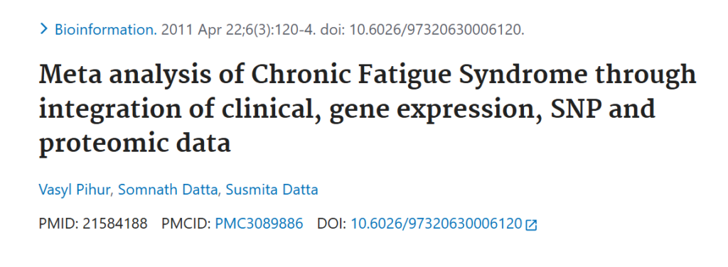
The obscure 2011 paper that provided a connection to ME/CFS.
included a 38-year-old woman with mysterious fatigue, which had started after a bout with infectious mononucleosis at age 16. Talk about serendipity.
Looking in her muscle tissue for evidence of the suspect gene, Hwang found abnormally high levels of WASF3 in the woman with fatigue (but not in the other family members).
He apparently latched onto ME/CFS when WASF3 popped up in an obscure 2011 ME/CFS study. That short paper – which used a novel gene-gene association technique – ended up focusing on WASF3. It stated that WASF3 “possibly regulates brain cytokines involved in the mechanism of fatigue through the p38 MAPK regulatory pathway”.
WASF3
WASF3 is best known for regulating the cell’s actin “skeleton” but can also disrupt mitochondrial functioning.
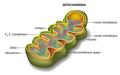
WASF3 can have profound effects on the mitochondria.
Hwang dug deeper and found that the fatigued woman had multiple mitochondrial abnormalities, including unusually prolonged recovery (phosphocreatine (PCr)) periods after exercise, a lower mitochondrial oxygen consumption rate (read energy production), a decreased ability to transfer electrons from complex III to complex IV in the electron transport chain of the mitochondria, and decreased cytochrome oxidase enzymes. No wonder the woman was so fatigued!
This made sense, as WASF3 can disrupt mitochondria by inhibiting part of the electron transport chain. Indeed, Hwang found that knocking down WASF3 levels in her cells improved mitochondrial function. He’d just discovered a new way to potentially explain the energy problems in ME/CFS.
Next, he created transgenic mice with high WASF3 levels in their skeletal muscles and stressed them with exercise. That resulted in higher blood lactate levels – a sign of mitochondrial distress, which is seen in ME/CFS – and lower glycogen levels. The mice also showed a significant reduction in endurance during a running test.
Nath’s Study Participants Take Center Stage
Next, Hwang brought in samples from ME/CFS patients in Nath’s intramural study. He and his colleagues found significantly increased WASF3 levels and dramatically reduced cytochrome oxidase and MTO1 levels. (MTO1 is a protein expressed in high-energy-demand tissues such as the muscles.)
So far, so good.
The Real Culprit Shows Up? (And the Viral Connection)
Looking upstream, Hwang found a dramatic reduction in an endoplasmic reticulum protein called BiP, which regulates WASF3 production. They apparently examined the endoplasmic reticulum (ER) because infections put the ER under considerable stress. Indeed, a marker of endoplasmic reticulum (ER) stress called PERK was high.
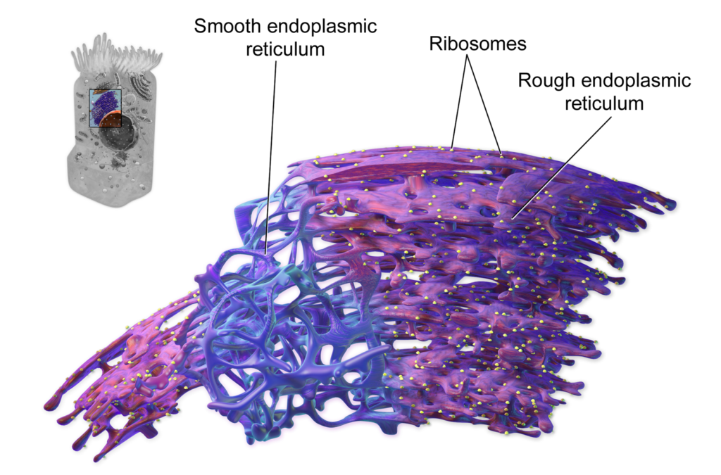
The key player? Stressed out endoplasmic reticulum? (Image from Blausen.com staff (2014). “Medical gallery of Blausen Medical 2014”. WikiJournal of Medicine )
The endoplasmic reticulum regulates protein folding. Proteins, which do the work of the cell, are extremely complex structures that must be folded correctly to work. The high ER stress levels suggested that too many improperly folded proteins were present in the ME/CFS patients.
It turns out that the endoplasmic reticulum (ER) plays an important role in cellular viral defense. Viruses attempt to counteract this by upregulating genes that impair its ability to properly fold proteins or remove misfolded ones.
The high WASF3 levels found in ME/CFS patients then appeared to result from an overstressed endoplasmic reticulum that could not properly regulate WASF3.
The authors concluded:
“WASF3, induced by ER stress…(provides) a molecular explanation for the energy deficiency symptoms of exercise intolerance and postexertional malaise in a patient with chronic fatigue.”
A Critically Important Process?
Hwang was clearly quite interested in the role mitochondria play in ME/CFS because two years later, in 2025, his group published an overview, “Mitochondrial Dysfunction in Myalgic Encephalomyelitis/Chronic Fatigue Syndrome“. The review, which highlighted exercise intolerance, suggested that the ER-WASF3 connection could be what we’re looking for: a “critically important cellular process” that affects many systems.
“ME/CFS could be a complex manifestation of multiple systems being affected by the derangement of a critically important cellular process that has wide-ranging effects depending on the characteristics of each tissue or organ.”
The authors noted that the mitochondria play a far larger role than just providing energy and cited over 20 studies suggesting the mitochondria have been affected in ME/CFS.
While substantial evidence suggests mitochondria are affected in ME/CFS, it’s not considered a mitochondrial disease because no genetic basis for the mitochondrial dysfunction is present. (Liz Worthey and Camille Birch argue that mitochondrial conditions can be “acquired”, that is, produced by non-genetic means over time in the body.)
The Big Shift
When the paper shifted to oxidative stress, something new emerged. The general understanding has been that because the mitochondria produce most of the free radicals, or reactive oxygen species, in the cell, damaged mitochondria in ME/CFS must be flooding our cells with these dangerous substances.
Hwang and Syed turned that idea on its head. Instead of being dangerous free-radical merchants, they portrayed mitochondria as containing the cell’s most “powerful antioxidant mechanism(s)”. (!)
The idea that the mitochondria may play an important antioxidant role has been around for over 30 years. It rests on the realization that while mitochondria do produce scads of potentially harmful reactive oxygen species, they also – at complex IV of the electron transport chain – turn most of those free radicals into water.
Mitochondria, in fact, contain a huge array of antioxidant factors (manganese superoxide dismutase (SOD2), glutathione and glutathione peroxidases, peroxiredoxins, mitochondrial thioredoxin (TRX2), thioredoxin reductase) designed to ensure that the vast majority of ROS are destroyed before they can cause damage.
More importantly, perhaps, because the mitochondria also take in free radicals from the cell and metabolize them to harmless substances, the mitochondria may actually play an essential role in keeping our cells free of damaging ROS (free radicals).
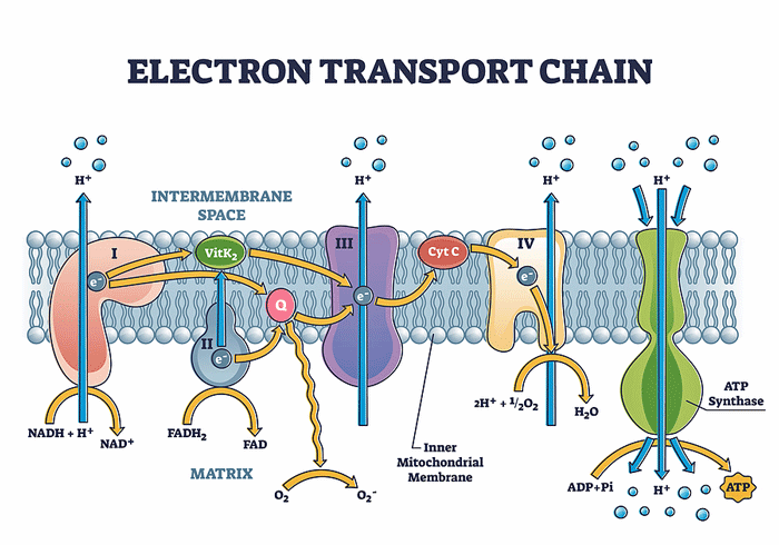
High WASF3 levels disrupt Complex IV – the part of the electron transport chain that produces antioxidants. (See the conversion to water (H20).)
In a 2022 “State of the Art Review”, “Mitochondria and oxygen homeostasis,” Hwang and Mori asserted that “insights gleaned from these basic mechanisms are likely to be important for understanding disease pathogenesis and developing strategies for maintaining health.”
The key point for ME/CFS, though, is that the mitochondria’s ability to keep cells healthy and clean depends entirely on having the energy needed to produce these antioxidants.
In their 2025 paper, Syed and Hwang proposed that mitochondria’s low energy status in ME/CFS means they can’t metabolize the ROS present, leading to the consistently high levels of oxidative stress found in this disease.
The real zinger, though, comes when WASF3 gets packed into the mitochondrial equation. It turns out that increased WASF3 levels disrupt the exact part of the electron transport chain (complex IV) responsible for cleaning up the reactive oxygen species in the mitochondria.
Inhibiting complex IV would decrease antioxidant and energy production, as well as increase reactive oxygen species levels.
A Potential Vicious Circle Emerges

A vicious circle (which does not require a pathogen) could occur.
Increased reactive oxygen species (ROS) levels could then loop back and damage the endoplasmic reticulum, creating a vicious circle: high ER stress, possibly caused by an infection, dysregulates WASF3. WASF3 damages the electron transport chain, reducing ATP production and increasing reactive oxygen species. Those reactive oxygen species further damage the ER, causing more WASF3 dysregulation, more mitochondrial damage, and on it goes. Syed and Hwang appear to be the first researchers to explicitly flesh out this process.
Once this self-reinforcing system, or vicious circle, is established, there’s no need for a pathogen to keep disrupting things.
Now we can see, at least in part, why Hwang is so fascinated by ME/CFS – the disease appears to perfectly fit their mitochondrial/major antioxidant thesis. WASF3 damages the core antioxidant process, which they believe gives mitochondria their superpower as the cell’s premier free-radical buster.
That’s not nearly the end of their hypothesis, though. Over time, Hwang and company have really worked this problem…
A Pathogen Fighter as Well?
They propose that this WASF3 upregulation is usually a short-lived adaptive change that helps the body fight off an infection. WASF3 does this by downregulating ATP production and upregulating glycolysis – the primary energy source immune cells use when they ramp up to fight an infection.
The potential problem in ME/CFS and related diseases occurs when this temporary adaptive change becomes chronic, and immune cells get stuck in anaerobic energy-production mode. (More on that below.)

Pathogens attempt to disrupt the ER.
Note that the increased reliance on glycolysis or anaerobic energy production fits what the ME/CFS and long-COVID exercise studies have found. Lactate increases when the cell can no longer produce energy aerobically and is forced to rely on the dirtier, much (much) less efficient, and less productive anaerobic energy pathways.
Hwang has produced a highly integrated and coherent hypothesis. If he is right, then exercise should cause WASF3 levels, ER stress markers, and reactive oxygen species (free radicals) in the muscles to skyrocket, and complex IV, ATP production, and antioxidant levels to decline.
Back to the “Critically Important Cellular Process”
Remember the “critically important cellular process” Syed and Hwang proposed could dysregulate systems across the body? Because WASF3 is found in many cells across the body (brain and retina, muscle, epithelial tissues, immune cells, and reproductive tissues), it could dysregulate many systems.
Immune System
Towards the end of the 2025 paper, they suggested that the mitochondrial insufficiency they’d found in the muscles could be affecting immune cells. Because WASF3 hasn’t been well studied in immune cells, they presented this as a possibility to be tested.
They focused on T and B cells, both of which have become a major area of interest in ME/CFS.
WASF3 could disrupt these cells in several ways. Its ability to regulate actin could impact signaling, migration, calcium entry, and interaction with antigen-presenting cells. Ultimately, it could impair their ability to interact with invaders.

T-cells may not have the energy to mount an effective attack against invaders.
Perhaps more importantly, WASF3 could affect energy production. While T-cells use glycolysis to ramp up and produce clones, they then rely on aerobically produced energy. Mature T-cells stuck in glycolytic mode by increased WASF3 levels would have trouble recognizing and killing infected cells, and would be inflammatory, too.
T-cell exhaustion seems like an expected outcome and offers another test: does stimulating T-cells increase WASF3 levels, impair aerobic respiration and increase glycolysis, cause calcium issues, etc.?
The insufficiency of the adaptive immune process (T and B cells) and the compensatory response of the highly inflammatory innate immune system in ME/CFS seems well established. By whacking the mitochondria in the T and B cells, WASF3 could conceivably account for that.
The problem would show up not necessarily in the initial response to pathogens but in a sustained response to pathogens.
Interestingly, because the JAK2/STAT3 pathway can also regulate WASF3 levels, JAK/STAT inhibitors – which are being assessed in long COVID – could be helpful. In fact, baricitinib, which is currently being tested in the huge (n=550) long-COVID trial, might be one of the better inhibitors to try.
Hypometabolic Muscles, Too?
“For example, in disorders associated with immune and exercise tolerance abnormalities such as ME/CFS, it could be speculated that the innate immune activation caused by exercise stress may contribute to the clinical symptom of post-exertional malaise.” The authors
The authors propose that during exercise, weakened mitochondria release a danger-associated molecular pattern (DAMP) called mtDNA into the cell interior (cytosol).

A muscle in a hypometabolic, antiviral state…(Really?)
That produces a location problem. mtDNA looks fine in the mitochondria, but once it enters the cell interior (cytosol), it looks like viral DNA to the immune system.
The innate immune system immediately goes on the attack. Energy production in muscle tissue drops, causing a prolonged metabolic shutdown. Reactive oxygen species (free radicals) levels skyrocket. Muscle repair mechanisms that should kick in after exercise get dramatically delayed.
The result: the muscles now exist in a hypometabolic, antiviral‑like state.
As the muscles spill cytokines and danger signals into the bloodstream, the vagus nerve activates nervous system circuits that tell the brain to produce the symptoms associated with “sickness behavior” to cause the patient to isolate themselves. The result: the big functioning hit and flu-like symptoms that pervade ME/CFS.
The difference is that, in this case, the symptoms are produced not by a virus but by a condition in the muscle cells (mtDNA) that mimics a viral attack.
Hence the authors suggestion:
“It could be revealing to investigate whether PEM, a key symptom of ME/CFS with neuroimmune features, is mediated in part by the mitochondrial innate immune signaling mechanism involved in skeletal muscle adaptation to exercise.
The Brain, Too?
And then there’s the brain. The authors wrote:
“Given the high energy demands of neurons for action potential generation and cell signaling, decreased mitochondrial respiration could affect brain function in unpredictable ways given the complexity of neuronal circuitries.”
and
“Because WASF3 is highly expressed in brain tissue, it is also tempting to speculate that disruption of WASF3 homeostasis could be involved in neuroinflammation as observed in ME/CFS.”
WASF3 is indeed found in many parts of the brain. Interestingly, it’s particularly abundant in the two brain regions which recent NIH studies suggest may exhibit reduced energy levels: the cingulate cortex and the locus coeruleus.
Because WASF3 is found in neurons and astrocytes, it could contribute to both reduced norepinephrine activity (neurons) and neuroinflammation (indirectly via astrocytes).

An energy-stressed brain?
Indeed, the WASF3 finding predicts that the NE-producing neurons would act as Goldstein and Aregawi found. They might start off normally, but as they became more stressed, they would quickly fail. WASF3, then, would also produce the increasing inability of ME/CFS patients to maintain a steady handgrip force – as the MRS brain findings suggested.
Ultimately, it could affect the effort/supplementary motor cortex/motor cortex/muscle recruitment issues recent papers suggest may be occurring.
A nice potential brain connection has emerged. One wonders if Hwang is working with Goldstein and/or Bedard at the NIH to explore it?
Connective Tissue Connection?
WASF3 may have one more trick up its sleeve. As noted earlier, WASF3 regulates actin, which makes up the “skeleton” of the cell, and is also involved in extracellular matrix (ECM) remodeling. Enter Rob Wust’s group, which found extensive ECM remodeling in muscle tissue that Slaghekke believes may be interfering with capillary blood flow to the muscles.
ECM remodeling, aka connective tissue remodeling, could be contributing to many issues in ME/CFS. Could WASF3 be contributing to this? This is also highly conjectural, but the potential connection seems to be there.
Endoplasmic Reticulum Stress: Take II
WASF3 could be doing a lot of damage, but Hwang’s findings suggest that endoplasmic reticulum stress is driving the high WASF3 levels. He was, after all, able to reduce WASF3 and restore mitochondrial functioning in the original patient’s cells with a drug that affects the endoplasmic reticulum.
In 2025, a small study (n=22) from Maureen Hanson’s lab, “Extracellular vesicle proteomics uncovers energy metabolism, complement system, and endoplasmic reticulum stress response dysregulation postexercise in males with myalgic encephalomyelitis/chronic fatigue syndrome”, brought ER stress to the fore.
Fortunately, the Glass/Hanson study used exercise as a stressor. Hwang’s study suggested that exercise should increase ER stress but could not test that. The Hanson group found that exercise dramatically increased ER stress – and that stress was strongly correlated with PEM symptoms.
The Hanson group’s study went beyond what Hwang achieved in his more limited study. BiP – the stress protein Hwang found dysregulated in muscle tissue – was dysregulated in the extracellular vesicles as well, but so were 9 other ER stress proteins. That suggested exercise had produced a broad dysregulation of the ER stress response. They also found a broad downregulation of proteins associated with aerobic energy production and other metabolic pathways. The authors wrote:
“Collectively, this highlights ER stress and protein folding as key therapeutic targets warranting further investigation.”
The fact that an independent lab exploring a different compartment of the body found similar evidence of ER stress was pretty compelling. The main issue – it was another small study…
Treatment Possibilities
If ER stress and WASF3 elevations are causing trouble in ME/CFS and related diseases, what to do about them? Hwang definitely has ideas. Whether he can test them is another question.

A clinical trial seemed to be on its way…
In 2023, Hwang stated he hoped to “embark on clinical studies” to improve energy levels in ME/CFS. In a 2023 LinkedIn post, one of Hwang’s co-authors, Jin Ma, reported the same thing (and posted a link to the Health Rising article :)). In Jan. 2025, in an NIH Advocacy call, Avindra Nath reported that Hwang was “going to do a small clinical trial”.
Everything seems to have stopped after that. They may have trouble finding the right drug.
Salubrinal – the drug he used to reverse WASF3 levels in patient one’s cells – is not FDA-approved. Interestingly, a 2024 paper from Nova Southeastern (home of Nancy Klimas’s lab) proposed using salubrinal to reduce ER stress and enhance cellular stress pathways in ME/CFS and long COVID.
Relyvrio seemed like a likely option until, in 2024, it failed a large ALS trial, prompting the drug company to withdraw it from the US and Canadian markets and the FDA to revoke its approval.
Almost a year later, Nath still reported that a small clinical trial was going to be done, indicating that Hwang had other options (sodium phenylbutyrate (Pheburane) and/or taurursodiol, guanabenz?).
However, an extensive ChatGPT search could not find references to an ongoing trial. Hwang still lists ME/CFS as a research focus on his NIH page, so he appears to still be involved, but no evidence indicates that a trial has, or is about to, take place.
Conclusion
WASF3/ER stress clearly has the potential to be a “critically important cellular process” that can disrupt many systems.

Where are we now? I will try and find out.
Is it? Could a basic mitochondrial problem be the key? Or is the WASF3 hypothesis another beautiful hypothesis, as Robert Phair has noted, that our very complex body ends up rejecting? Time will tell.
One nice thing is that Hwang appears to be operating in a potentially rich environment right now with NIH researchers looking at the muscles and the brain in potentially interconnected ways. Are they working together? We don’t know.
Note that we’re still very much stuck in a rather familiar place – we’re in intriguing results/small study mode! Small studies from Hwang, Goldstein, Berdard, Wust, Hanson’s group, Prusty, and many others are driving the field forward in potentially fruitful ways.
We’re just missing the next step – launch mode – which involves large studies that can validate and expand the smaller study results.
We’re not in a terrible place – we’re in a MUCH better place than we were five years ago – but thus far, major funders like the NIH haven’t been willing to step up, put on their big-boy pants, and fund some major grant projects. That’s when things will really start happening. (Let’s hope that ME/CFS advocates can get Congress to allocate $50 million for the ME/CFS roadmap.)
Because Hwang is an intramural researcher, it’s hard to know exactly what he’s working on. We know he has plenty of latitude to explore different options – and has used that latitude to dig deeply into WASF3 and ME/CFS, but what he’s doing now, how much funding he has, who he’s working with, whether a clinical trial is in the works, whether he’s able to bring in new patients, etc., is unclear. I will try to find out.





Cort, if Complex-IV is the site at which mitophagy is failing to occur, wouldn’t Rapamycin be the obvious drug? It might not downregulate WASF3 but it might disrupt the doom loop.
There’s at least two trials on the use of rapamycin in long covid or ME/CFS. Polybio, and Simmaron.
Does anyone know of the progress on these trials?
I’ve always been a bit skeptical on rapamycin, although I don’t really know why! It would be good to have some evidence either way on its potential
This being an adaptation reaction would explain why some people get ME/CFS from vaccines without a real infection?
I can’t help wondering if this could be a clue as to how medications such as Mournjaro may be assisting. I developed moderate ME-ICC (as per the International Consensus Criteria) in 2012, acute post viral. GLP-1 GLP meds are known to have anti-inflammatory actions but it is predominantly a metabolic drug.
I stumbled on it when being treated for sleep apnea via weight loss and was very surprised to feel ‘weller’ within the first few weeks of treatment (so not attributable to simple weight loss). This continued over time, to the point that I can now exercise (within limits) without post exertional symptoms. It’s astonishing & the first time since 2012 that I’ve been able to do this. Treatment was successful, I am at a healthy weight and sleep apnea is now mild and not causing distress.
Pacing is still essential, I still fit the moderate ME spectrum but quality of life is improved with better cognitive function, reduced OI, more energy & stamina, less pain, the sense that “I can do something” has expanded.
I went off it recently and started to decline so I’m back on it experimenting with microdosing. Seeking the dose that alleviates ME symptoms without affecting appetite.
As I read this blog, it seemed to resonate with my experience. Any thoughts?
I was in the Simmaron rapamycin trial in May of 2024 but dropped out after 4 weeks due to side effects. I think the trial must be over by now.
Well as far as I’m concerned it saved my life! I would say I was about 5% and now I’m at least 75 to 80%. I’ve been on rapamycin since December 2023.
That’s great. I hope the studies back up the positive, but certainly not universal, anecdotes such as yours. I presume side effects have been tolerable for you, given you have been taking it for 3 years?
Yes, I happen to be very lucky that I am a super responder. Everything else I tried made me worse. Crazy how we are all so very different in this. No, I don’t believe I’ve had any adverse side effects. It’s hard to tell of course because I haven’t felt really fully okay in years lol.
You are just what we need to identify – the super responders! I wish they could do a bunch of lab tests on you and figure out why you responded so well particularly since everything else made you worse. Good for you to keep trying.
Yes, thank you, I definitely was suicidal when I was on my back in the dark, unable to feed myself, no sound and no light 24/7. It is so hard to keep hope when you’re in that position. And when I kept trying things and they were making me worse it was even harder. I’m so glad I didn’t give up. And that’s my message to folks don’t give up you never know when there’s going to be a breakthrough. And you never know what’s going to help we’re all different unfortunately, but I know that there is something somewhere out there. We need to keep holding on to hope. Thank you for all that you do cort. Very grateful.
phase 2 is complete and published….people with flu onset responded, others did not…placibo controlled phase 3 getting started, expect to complete in 12 months..all in there on Simmaron website….https://rdcu.be/BGyiCZuevhja
Thanks! Will be interesting to see how the next phase goes, and also how the Polybio trial aligns (or doesn’t)
It appears that it could indirectly do so by improving autophagy/mitophagy – getting those damaged mitochondria out of there – which can be inflammatory – and improving mitochondrial efficiency, thereby reducing ER stress.
If ER stress is what’s causing WASF3 levels to rise it could theoretically be helpful. 🙂
I think people have to be careful because if its used too frequently it can suppress mitochondrial production – which is why I think most people are on the once a week plan (?)
It would be great if someone could test whether it affects WASF3 levels.
Thank u, Cort for all ur effort in reporting all this and past info.
Thanks!
Walitt told me by email that Hwangs trial is starting next year.
Relvyrio is not for sale in the US at the moment but it is very much available for clinical trials. There’s several underway using it. I believe the trial will probably apply relvyrio as salubrinal is not in widespread use.
Very nice, Jason! Thanks. I was hoping things were moving forward. 🙂
It’s nice if that is being trialled in addition to IVO-21!
I found this a very very interesting study when it came out a few years ago.
And I attended a very interesting Solve ME/CFS webinar last week featuring Dr Jay Chung of the NIH. Dr Chung is testing a substance called IVO-21 which acts on mitochondria, and he showed in the presentation that IVO-21 inhibits WASF3.
Maybe this is the trial, but Dr Chung took it forward rather than Dr Hwang?
I think there’s little doubt that mitochondria are significant in ME/CFS. But how?
I think it has been suggested that TUDCA can inhibit WASF3.
Matthias, do you happen to have a YouTube link for that seminar?
Maybe this is it?
https://m.youtube.com/watch?v=IBXYhRInY0o
Thanks! That webinar is on my list. So nice to see other options pop up 🙂
TUDCA is known to have effects on the ER so it’s worth looking into. It’s pretty safe to use and available if a little expensive. Likely popularity now because it might raise GLP1 so people are taking it to loose weight as well as liver detox. I took it when I first had Lyme and stopped because it didn’t seem to be doing anything. Maybe if I kept taking it it wouldn’t have progressed to CFS?
Given the following work on ER stress presented on Health Rising :
https://www.healthrising.org/blog/2023/10/21/ai-driven-chronic-fatigue-syndrome-clues/
it appears that TUDCA could also lower it but it seems that Dr Hwang prefers to try salubrinal and there may be a good reason for this
I can touch for tudca.works on me. One bile as well.serms the liver is involved.i also get good results with milk thistle
Vouch*
And many people, ive learned from natropathic physicians have,after the age of 50,experience low stomach acid.
A few doses of retained HCL has also helped.
without enough stomach acid it leaves the door open to pathogens
I remember this article and your recovery story. I tried all the supplements the way you had mentioned. Tuscan, NAC,q10 etc…Got some results but then didn’t continue. I tend to switch a lot.Went to Cyprus for a blood filtering treatments for me and my daughter. My sister has got the same illness.
There are patients which for them TUDCA was a game changer. Also it is possible that a personalized approach is needed
With AI assistance I landed on TUDCA as a potentially effective treatment for the ER stress glitch described in the study. TUDCA is a readily available OTC supplement (a simple bile salt helpful for people with liver and gallbladder issues). Needless to say, I’ve ordered some so I can do my own n = 1 trial. It will be delivered tomorrow.
Looking for patient reviews mentioning energy benefits, I found this on Amazon. The review was posted in 2022 and there has been no update but I think the information may be important, since this is a readily available, popular OTC supplement that will be very easy for people with ME/CFS, long Covid, etc. to try:
“I just started taking these for liver and gallbladder help and wanted to leave a quick preliminary review.”
“I will write a full review after I’ve been taking this for 30 days. Before I started taking these my stamina was pretty much at 0 and the fatigue in my legs was so bad that it hurt to walk and I would often lose control of my legs to the point of my knees buckling.”
“The first day I took one pill and noticed that after my afternoon nap, I was able to wake up quickly whereas ordinarily, it takes me more than 30 minutes to fully wake up and be able to stand. The next day I learned that the serving size was two pills so I took two at around 11am. By 7:30pm I had pain in the upper right quadrant and I felt very sick. Fever, sweats, nausea, the works. That lasted about an hour and then it went away completely.”
“At that point I wasn’t convinced it had anything to do with the TUDCA. I’m still not 100% sure. It could’ve been the meal I ate, which may have a little too much fat. The next day I took 2 more pills at 11am and about 8 hours later I started feeling a dull pain in the upper right quadrant again but this time there was no general sickness.”
“That strange pain grew into what felt like pressure and if I leaned against that part of my back, the pain got worse and the feeling of pressure grew and I eventually developed a headache from it. Even with that pain and pressure, I could feel my energy had gone through the roof and I was able to work out for nearly an hour with no fatigue at all. I haven’t been able to do that in well over a year.”
“I decided to lay off the pills for two days to let that pressure and pain clear up and they did. I started the pills again this morning with 1 pill only. More than 8 hours later there are no adverse reactions at all and my energy level is great. Not what it was the second day I took two pills but still much better than normal.”
“My legs feel about 80% stronger and there is no pain with the fatigue and no buckling knees. So far, I am thoroughly impressed. I’ve only taken 6 of these pills and I can already feel a big improvement. I have no idea what state my liver and gallbladder are in so I have no idea why I felt those adverse reactions.”
“I really just wanted to put this out there for anybody doing their own research on TUDCA without having seen a doctor first, like I did. So much of the research out there says there are no side effects with TUDCA but I obviously had some taking a full dose of 2 pills. With 1 pill though there are no bad side effects and I feel much stronger than I did before I started taking them.”
Hi jerry,
Thx for posting this.
Sounds hopeful , I will try.. brave souls that we are. Could you please keep me/us updated.
Thx
I will.
Thank you Cort.
While the finding is intriguing, the key questions now are: What infection—or other trigger—could be driving this process and linking muscle fatigue, PEM, immune abnormalities, and brain dysfunction?
And, most importantly, can the underlying culprit be identified and eliminated?
Dr Chung’s presentation suggested that infection is the usual trigger, but there are others. Regardless of the trigger, a doom loop is perpetuated, involving microglia and inflammation.
And he theorizes that IVO-21 can break that doom loop
Yes, there is a possibility that IVO-21 acts as a mitochondrial stabilizer. However, fatigue and “brain fog” may also be associated with disrupted neuronal function resulting from chronic inflammation and progressive deterioration, including changes in white matter that can impair nerve signaling.
If muscle weakness is related, at least in part, to nerve damage caused or exacerbated by overexertion, an important question is whether IVO-21, by stabilizing or restoring mitochondrial function, could help support the repair or recovery of damaged neural pathways. If so, that would represent a particularly significant breakthrough—regardless of the underlying trigger—not only for ME/CFS but potentially also for post-polio patients.
For now, however, we will have to wait and see whether these potential benefits are supported as research progresses beyond preclinical mouse models and into human studies.
Sorry I meant mitochondria rather than microglia
Why are some medical researchers suddenly leaving the US, the largest/wealthiest *?
“first world” country?
Harold.
*FROM CORT’S LAST BLOG:
“…The back-and-forth with the FDA over the use of a new technique to assess T-cells in the brain proved enormously complicated. At one point, they had to start over because the FDA disallowed a reagent they’d been using.
Last summer, he (Dr. Jared Younger) reported that initial tests on healthy controls indicated the new technique was successful and he was ready to begin testing people with ME/CFS. At some point, the radiochemist and radiologist suddenly departed for Canada, and the project collapsed.
Unless he can find another radiochemist, the project is over. (He mentioned Michelle James at Stanford (???). One of the heroes of the story has been MERUK, which funded an extraordinary project in the first place and has shown remarkable patience.”
”Why are some medical researchers suddenly leaving the US, the largest/wealthiest *? “first world” country ?
Are you asking that seriously ? I hope not !
Largest, wealthiest, first world. That was then and it is now.
The answer is very simple and rude: a damned fu… moron called Donald Trump screwing up all the scientific fields all over the USA.
So many researchers are actually leaving USA for Canadian Universities (and other countries) in all top notch health and engineering various fields to pursue their work in an environment not rulled by a gang of clowns at the White House.
Yes, that is a factor, but no it’s not that simple. The rudeness is gratuitous. While I am not fan of DJT, one wonders if “Trump Derangement Syndrome” is also too common in Canada — it’s a waste of energy and time, which could be spent on productive pursuits (e.g., advocating for reasonable public policy that actually improves the long-term health of North Americans).
PS.
TDS is also what sadistic leaning Trumpites thrive on; don’t give them the oxygen. The Democratic federal senatorial candidates in Texas and Michigan are a good examples.
No further “dialogue” is warranted.
Harold it IS that simple! They want to continue doing their research and DT has pulled a lot of money out of science at Federal and University level, which in some cases has interrupted decades of on-going research. I know researchers who have had grants canceled and had to cut grad students and projects. This wasn’t a one time thing, it’s still happening now. They are pulling funding from any program they can so scientists of all ages are accepting offers in countries all over the world who want their expertise and will pay for them to replicate their work overseas. Canada is actively soliciting these experts and right now, you’d be a fool not to go..
It’s an interesting question. The US is still easily the largest medical research funder in the world, but so many grants were cut during the first year that the NIH resorted to paying out 5-year grants in full (instead of by the year) in order to use up the funding available to it.
Now, the Trump administration is hammering Harvard for not bending the knee by reducing the number of grants it approves.
How reducing science funding makes America great again I have no idea…We’re not an industrial powerhouse anymore – intellectual prowess is our superpower. It just seems so counterproductive.
We’ll see about the attempt to put political appointees in charge of not just medical research grant approvals but science grants and many other types of funding.
That’s a kick to the nuts for researchers of all kinds. I don’t if it’s going to go through, though.
Cort, the WASF3 findings become even more interesting when placed downstream of the broader post-infectious ECM disturbance which is increasingly being recognised for playing a role in ME/CFS.
A plausible sequence would be:
1. Intermittent viral activity
2. Disturbed fibronectin production/turnover and ECM organisation
3. Abnormal integrin–talin / filamin–actin mechanotransduction
4. ER / Mitochondria ER Contact stress
5. Maladaptive Unfolded Protein Response signalling
6. WASF3 upregulation
7. Respiratory-supercomplex instability and loss of mitochondrial reserve.
This would position WASF3 not as the initiating defect, but as an intracellular effector of a much wider failure of cellular stress adaptation.
Fibronectin is particularly important because it sits at the interface between the extracellular matrix, integrins, actin architecture, membrane trafficking and organelle positioning – precisely the systems through which WASF3 operates.
If this is the case, disturbing fibronectin–integrin signalling experimentally, in relevant endothelial, muscle or immune cells, should prove to be sufficient to induce ER stress and WASF3.
Such an upstream connection remains hypothetical, but if demonstrated it could link the Prusty Lab’s fibronectin/ECM findings with Hwang’s WASF3 mechanism in a single virally initiated pathway.
What an interesting slant! It’s great to see another potential option and one that involves the ECM and potentially the blood vessels. I hope somebody can do that test…
I agree the WASF3 explanation probably works best as a downstream aspect of the illness.
I put a bit of probability on a sort of lipid/sterol dysregulation, perhaps caused by a persistent virus presence, that in turn leads to membrane issues, both extracellular, as in the endothelium and also intracellular, as in the ER. That could be more or less pronounced in different people, and in different cells in different people. (this is just one theory that can fit the limited evidence we have)
Two stories I’d love to tie together are Rob Wust’s necrotic muscle cells and Hwang’s ER stress. If exercise puts muscle cells into ER stress, then after a certain period perhaps 24h, the cell needs that stress to be resolved. If it is not resolved then the cell dies, either via apoptosis (controlled dismantling) or necrosis (messy explosion).
When I first read about the delay in ER stress leading to necrosis I immediately thought of delayed PEM.
Wow thank goodness for the gist. That was an intense blog.
Thanks again Cort for giving us hope for the future
Yes, an intense blog indeed! Exciting stuff, though. We’ll see how it turns out. 🙂
That is fascinating research. Many thanks to dr. Paul Hwang for couragously entering the field!
To me, it seems that both BiP and WASF3 are key indicators of what could be going wrong. Having ‘key points to look at’ is very important for further understanding our dissease. I also think it’s part of a complex defensive / inhibitory / Dauer reaction but unfortunately it seems to be a tricky point to try and intervene (I hope I am wrong!).
https://www.jbc.org/article/S0021-9258(20)35606-4/fulltext with title “The Molecular Chaperone GRP78 Contributes to Toll-like Receptor 3-mediated Innate Immune Response to Hepatitis C Virus in Hepatocytes”
citations:
“GRP78 expression level correlated with that of RANTES (regulated upon activation, normal T-cell expressed and secreted) and CXCL10, two inflammatory chemokines most frequently elevated in HCV-infected liver. Altogether, our data suggest that GRP78 contributes to TLR3-mediated, IRF3-dependent innate immune response to HCV in hepatocytes”
“GRP78 Depletion in Non-neoplastic Human Hepatocytes Impairs Expression of Antiviral Genes via the TLR3 Pathway”
My understanding in as plain as possible language:
A) decreased BiP (as seen in ME/CFS) increases WASF3 and that creates mitochondria to malfunction
B) mitochondria that malfunction create ‘mitochondrial debris’ that triggers a receptor called TLR3
C) activation of TLR3 creates a lot of antiviral responses leading to ‘sickness behavior’
D) a ‘simple’ way to decrease the effects of C) (decrease sickness symptoms) is to… decrease BiP since less Bip means less TLR3 activation means less ‘sickness behavior’
=> A) and D) conflict and lead to a ‘Mexican standoff’. Increase BiP (and likely decrease WASF3) too fast, and immune response flares up (risking more symptoms to lock in for a long time). When not increasing it, the current poor health status remains while preventing the risk for backfiring that increasing it could give.
So, IF it would work at all for any given subgroup, timing and dosing and dose over time might be key.
Great blog! Hit’s all the key points in my experience including the lactose issues. I haven’t taken TUDCA in 10 years after the first bottle did nothing for my Tick disease symptoms. Maybe I should try it again as I over did it last weekend and regretted it the next day. 😉
Done some digging. Found more than 10 drugs / natural substances that inhibit ER stress!
Might try one or two of the natural ones
“Collectively, this highlights ER stress and protein folding as key therapeutic targets warranting further investigation.”
Nice thought but ER stress and protein folding problems are probably just one of the many downstream processes triggered by an inadequate stress response. Trying to fix ER stress may therefore be as effective as fixing aging by treating the skin wrinkels that go along with it.
More interesting would be exploring the upstream processes:
a) the “missing link” between the dysfunctional stress response and the ER stress. What exactly happens in this blackbox? Viral reactivation? Overload of oxidative/”inflammatory” assault?
b) and what could be upstream of this? Barrier dysfunction exposing the brain (and other tissues) to all these goodies? Has anyone ever measured how the integrity of the blood-brain-barrier (or the endothelium in general) changes during PEM?
What I am trying to say: ER stress is just a link in the chain of events, the interesting part is the territory above…
Yes but I think there are multidirectional feedback loops at play, so I am not really sure if there is an ‘upstream’ or a downstream’! I am not sure it’s linear
“Nice thought but ER stress and protein folding problems are probably just one of the many downstream processes triggered by an inadequate stress response.”
I would not be surprised. Let’s get them validated in some decently sized studies and then start searching 🙂
Does WASF3 appear i Phair’s model?
Are WASF3 and Phair’s model each just “promising might be”? Sorry, my mind s a bit blurry today …
I don’t remember it appearing. Phair’s model if I remember it correctly does link the immune system to the mitochondria…if I remember correctly
Great. Now what?
Probably a whole lot of nothing like the last few decades.
They’re all so good at listing what’s messed up followed by nothing.
Great job
Cool, thanks
Yeah ,its getting rather PATHETIC
I think they are trying to figure out how they can get BIG PHARMA involved..$$$$$$$$$$$$$$$$$$$$
Another study implicating the brain / dopamine
https://fortune.com/2026/09/14/long-covid-brain-fog-symptoms-dopamine-nerve-terminals/
Bezisterim:
https://thesicktimes.org/2026/09/15/bezisterim-may-help-with-some-long-covid-symptoms-drugs-developer-reports-in-early-trial-results/
Doesn’t look earth shatteringly positive, but not bad either.
Main takeaway seems to be that patients with high symptom burden got benefit (ranging between small to moderate), while those with low symptom burden didn’t get any benefit.
Mind you, if I think of my daughter with a high fatigue burden, then I think we’d take a moderate benefit from treatment all day long, if it was relatively affordable
Dr Michael Peluso says, with regards to the study:
“There have been, at this point, a few dozen Long COVID trials of other agents, and really none of those have had results that are as consistent and as compelling as what I’m seeing here.”
Absolutely! Benefit is benefit, and most of us will take whatever we can get. Improvement in fatigue, cognition and PEM! Got my fingers crossed for this one.
Cort, maybe feed those poor dogs inside for a little while. Best of luck with the bear.
Never could quite understand why we give kids bears to sleep with.
The possible WASF3–mitochondria link is fascinating, especially the idea of a feedback loop involving ER stress and oxidative stress. It feels like an important avenue to investigate further, even if the mechanism is still far from settled.
It looks like Hwang is digging deeper into this. I’m trying to get an interview with him.
The proposed feedback loop between ER stress, WASF3 and mitochondrial dysfunction is especially intriguing. If larger studies confirm it, it could help connect several seemingly separate features of ME/CFS.
Yes, indeed!