


Geoff’s Narrations
The GIST
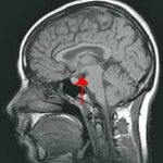
How timely Dr. Da Silva’s presentation on “Delineating clinical phenotypes and HPA-axis dysfunction in ME/CFS” at the 2025 IACFS/ME conference was. Jose Philipe Da Silva is a neuroscientist based at the University of Amsterdam. We’ve just heard from a slew of studies that pointed arrows straight at the brain, and now we have an ME/CFS brain autopsy report. (Thanks to the IACFS/ME for their support in virtually attending the conference.)

This is the third blog to emanate from the 2025 IACFS/ME Conference. More are coming.
Consider this – the Netherlands has done what the U.S. cannot – it started (in 2024) and is now delivering on an ME/CFS brain donor program. The NIH Neurobank is a huge brain donor program that encompasses ME/CFS. They were shocked at the interest shown by ME/CFS patients, and presumably have some ME/CFS brains, but what, if anything, has happened with them, we don’t know. )
 Health Rising’s Quickie Summer Donation Drive is On!
In the meantime, we have the Netherlands Brain Bank (NBB), a top-notch brain bank known for delivering high-quality, freshly harvested (terrible word) brains for research. According to the NBB website, researchers worldwide request brain tissue from the NBB each year.
These brain studies give us a unique slice (whoops, did it again) at ME/CFS. Thus far, autopsies have been done on 7 ME/CFS donors and more will be done. Each person had been diagnosed by an ME/CFS specialist, and the results were compared to matched controls who did not have ME/CFS.
It’s been almost seven years to the day since Health Rising last reported on autopsy reports in ME/CFS. A theme of dorsal root ganglionitis – damage to the neurons that transmit sensory signals to the spinal cord – prevailed.
This report looked at a different area of the brain and found something equally interesting.
THE GIST
-

Could inflammation in the hypothalamus be playing a key role?
How timely Dr. Da Silva’s presentation on “Delineating clinical phenotypes and HPA-axis dysfunction in ME/CFS” at the 2025 IACFS/ME conference was. We’ve just heard from a slew of studies that pointed arrows straight at the brain, and now we have an ME/CFS brain autopsy report.
- It’s been almost seven years to the day since Health Rising last reported on autopsy reports in ME/CFS. A theme of dorsal root ganglionitis – damage to the neurons that transmit sensory signals to the spinal cord – prevailed.
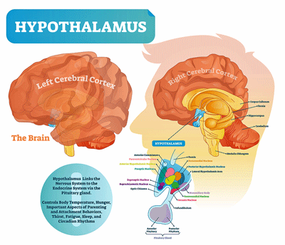
- Past brain autopsies have focused more on the brain stem, but this group, which specializes in the hypothalamus, started off at the HPA axis, which has been a focus of ME/CFS research almost from day one. Why? Because the HPA is the main stress-response and energy-regulation system in the body.
- The hypothalamus, located at the bottom of the brain, senses stress, blood sugar levels, inflammation, time of day, and more. It releases corticotropin-releasing hormone to the pituitary gland, which then produces cortisol, which has been called our body’s “stress and energy manager”.
- Cortisol mobilizes energy by raising blood sugar levels and helps us use fat and protein upon waking and during stressful periods. It also helps us stay alert and focused, and able to respond to threats or challenges and is an important anti-inflammatory, helps to constrict blood vessels, and affects memory, attention, motivation, and mood.
- The Dutch group counted the neurons in the hypothalamus, did a gene expression analysis of the pituitary, and assessed cortisol levels. With the lower cortisol levels, they expected to see increased levels of neurons in the hypothalamus in order to up the production of CRH. It was all quite straightforward, they thought.
- Something completely different, though, was going on. Instead of more CRH-producing neurons, they found almost no CRH producing neurons in the ME/CFS patients. That suggested that a general suppression of the hypothalamus had occurred, but the vasopressin and oxytocin neurons in the hypothalamus were either found at increased or normal levels. Only the CRH-producing neurons had been affected…
- A gene expression analysis found more evidence of HPA axis trouble: the pituitary receptors that were designed to respond to numerous neuropeptides were down-regulated.
- All in all, the HPA axis with the dramatic reduction of CRH-producing neurons in the hypothalamus, a shutdown of the pituitary, and lower cortisol levels found appeared to be pretty much of a wreck in these autopsied patients.
- Note that this may represent a very severe end-stage of ME/CFS, only seven autopsies were performed, and note that people with milder illnesses may have more functional CRH neurons left.
- Da Silva did not speculate on the cause. In 2020, though, Angus Mackay rather eerily conjectured – using a thought experiment – that the “stress integrator” in the hypothalamus called the paraventricular nucleus (PVN) may be playing a key role in ME/CFS. The PVN, it turns out, is the main producer of CRH.
- Mackay proposed that an inflamed PVN might overproduce CRH. While this study found low numbers of CRH-producing neurons, Mackay’s hyperactive PVN scenario could result in long-term damage and the eventual disappearance of those neurons.
- The situation seems all too familiar: it’s yet another version of the wired and tired / burnt out but stuck in threat mode problem that seems to pervade these diseases.
- The recent (as yet unpublished) norepinephrine finding featured in a recent blog might be related to this. Dense strands of noradrenergic (norepinephrine-producing) fibers from the locus coeruleus reach straight into the PVN, where they trigger CRH production. Hyperactive NE neurons could trigger CRH neurons in the hypothalamus, causing them to burn out eventually in the most severe patients.
- These findings suggest why adding cortisol in the form of low-dose hydrocortisone might sometimes have negative effects, if core portions of the cortisol-producing system are broken.
- Interestingly, Cortene’s CT38 hypothesis might fit. Cortene proposed using a brief pulse of a CRFR2 agonist to reset the HPA axis. The idea is that chronic activation of the CRFR2 receptors could suppress the HPA axis, thereby blunting cortisol production. Interestingly, a chronically activated CRF system could also contribute to the hyperactivity observed recently in the locus coeruleus.
- Interestingly, Cortene’s CT38 drug, if proven effective, might help by resetting the HPA axis and taking pressure off the hypothalamus.
- The autopsy data – which may come from very severe patients – could also fit a picture where chronic neuroinflammation in limbic/PVN/brainstem areas slowly erodes core stress-regulation regions there. In this scenario, the NE-producing neurons in the locus coerleus, the CRH-producing neurons in the hypothalamus, and the adrenals are all hit, and the two major stress response systems (HPA axis, autonomic nervous system) are clobbered.
- Treatments to tamp down neuroinflammation, calm the immune system, and turn off the danger response might help.
- We should expect more from the Dutch effort and the NIH’s RECOVER project already, get this, over 250 long- COVID patients in its autopsy bank and plans to include about 350. The possibilities for learning are immense.
Donation Drive Update

The connections appear to be piling up…
Thanks to the over 100 people who have contributed almost $10K to Health Rising’s year-end drive!
This blog demonstrates a couple of Health Rising’s strengths. One, a commitment to keep up with the latest research findings, and two, a commitment to integrate, if possible, past findings into current ones. In this blog, for instance, we saw that it’s possible – not proven – but possible that the low NE findings from an earlier blog could match up with the low CRH findings in the autopsy reports. It was also nice to see Cortene pop up again as a possibility. Finally, these findings suggest we have a lot to look forward to as the Recover Initiative gears up its massive long-COVID autopsy project.
If that floats your boat, please support us!
HEALTH RISING IS NOT A 501 (c) 3 NON-PROFIT
A Hypothalamic-Pituitary-Adrenal (HPA) Axis Focus
Past brain autopsies have focused more on the brain stem, but this group, which specializes in the hypothalamus, started off at the HPA axis, which has been a focus of ME/CFS research almost from day one. Why? Because the HPA is the main stress-response and energy-regulation system in the body.
The hypothalamus, located at the bottom of the brain, senses stress, blood sugar levels, inflammation, time of day, and more. It releases corticotropin-releasing hormone to the pituitary gland – which is found just below the brain – which then releases ACTH into the bloodstream. ACTH travels through the bloodstream all the way down to the adrenal glands (on top of the kidneys).
The end result is the production of cortisol, which has been called our body’s “stress and energy manager.” Cortisol mobilizes energy by raising blood sugar levels and helps us use fat and protein upon waking and during stressful periods.
It helps us stay alert and focused, and able to respond to threats or challenges. It’s also an important anti-inflammatory, helps constrict blood vessels, and affects memory, attention, motivation, and mood.
Studies have shown that morning salivary cortisol levels are low in ME/CFS. Akiko Iwasaki’s big long-COVID study rather shockingly found that it was low cortisol instead to T or B-cell, or an autoimmune process, that was “hugely predictive” for long COVID.
That was a bit of a shock. While HPA axis studies continued in ME/CFS, I don’t think anyone thought that low cortisol might be so predictive. The ever-increasing complexity of these studies and the data analytic techniques used means new insights are liable to pop out. (The Iwasaki group was critiqued for how they did the initial cortisol testing but redid them and got the same result.)
The Dutch group counted the neurons in the hypothalamus, did a gene expression analysis of the pituitary, and assessed cortisol levels. With the lower cortisol levels, they expected to see increased levels of neurons in the hypothalamus in order to up the production of CRH. It was all quite straightforward, they thought.
Producing CRH – The hypothalamus detects cortisol levels in the bloodstream via glucocorticoid receptors, which latch onto the cortisol. If too few GRs are activated, the brakes on cortisol production are taken off, and the hypothalamus produces more CRH. Given the low cortisol levels in ME/CFS, it only made sense that the hypothalamus would be trying to compensate by producing more CRH-producing neurons.
Surprise!
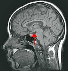
Except it wasn’t in the ME/CFS patients. Something completely different was going on. Instead of more CRH-producing neurons, they found almost no CRH-producing neurons in the ME/CFS patients. An image (embargoed) showed what was essentially a blank screen with a few faint scuff marks (the CRH producing neurons). Given that, it was not surprising to see dramatically lower CRH levels in the fibers projecting to the pituitary.
To their surprise, the researchers found very low levels of CRH-producing neurons in the hypothalamus of autopsied ME/CFS patients.
That suggested that a general suppression of the hypothalamus had occurred, but then they were stumped again. The vasopressin and oxytocin neurons in the hypothalamus were either found increased or at normal levels. Only the CRH-producing neurons had been affected…
Going down the HPA axis loop, they found much the same. A gene expression analysis found that pituitary receptors designed to respond to neuropeptides were down-regulated.
The pituitary responds to a lot of neuropeptides (CRH (corticotropin-releasing hormone) AVP / vasopressin, TRH (thyrotropin-releasing hormone), GnRH (gonadotropin-releasing hormone), GHRH (growth hormone–releasing Hormone), somatostatin, dopamine (from the hypothalamus)). The production of POMC, a “mother” peptide or prohormone that gets chopped into ACTH, was also downregulated. Decades ago, ME/CFS studies found that the pituitary in ME/CFS was not responding correctly to CRH.
Note that low CRH levels can affect more than cortisol. Low CRH levels could also affect arousal, “stress readiness”, produce feelings of being “jittery but weak” via autonomic nervous system dysfunction, increase pain, gut problems, poor sleep, and more. Reduced pituitary receptor activity could lead to increased pain, reduced libido, increased sensitivity to cold, etc.
All in all, the HPA axis with the dramatic reduction of CRH-producing neurons in the hypothalamus, a shutdown of the pituitary, and lower cortisol levels appeared to be pretty much of a wreck in these autopsied patients.
Since the final result of this pretty smashed-up HPA axis is to produce cortisol, the researchers looked for evidence of cortisol suppression, and found it. They are now looking for evidence of low plasma cortisol levels.
Note that this may represent a very severe end-stage of ME/CFS, that only seven autopsies were performed, and that people with milder illnesses may have more functional CRH neurons left.
Back to the Future?
The speaker noted that low cortisol levels can at least partially explain many of the core symptoms (fatigue, sleep, others) found in ME/CFS. So we appear to be back at the HPA axis.
Da Silva did not speculate on the cause. While this finding has not been published, and only 7 autopsies have been done, it’s worthwhile to ask what could be causing this strange HPA axis dysfunction and whether it fits with what else we know about ME/CFS? Also, what could the possible pituitary problems produce?
First, note that the hypothalamus has attracted considerable interest over time. Back in 2014, Dr. Bateman focused on inflammation in the thalamus, hypothalamus, and pituitary. In 2018, Theoharides proposed that mast cells in the hypothalamus were pouring out neuro-inflammatory factors that opened up the blood-brain barrier, whacked the mitochondria, and caused neuroinflammation at the same time.
In 2020, Angus Mackay rather eerily conjectured – using a thought experiment – that the “stress integrator” in the hypothalamus called the paraventricular nucleus (PVN) may be playing a key role in ME/CFS. The PVN, it turns out, is the main producer of CRH.
Mackay proposed that an inflamed PVN might overproduce CRH. While this study found low numbers of CRH-producing neurons, Mackay’s hyperactive PVN scenario could result in long-term damage and the eventual disappearance of those neurons.
The situation seems all too familiar: it’s yet another version of the wired and tired / burnt out but stuck in threat mode problem that seems to pervade these diseases.
But why would only CRH-producing neurons in the hypothalamus be affected? Perhaps because vasopressin and oxytocin-producing neurons at least in part occur in different parts of the PVN. Various hypotheses have been put forth regarding adrenal dysfunction, but in this scenario, the adrenal functioning might be impaired because they are “under-stressed”.
It shouldn’t go unnoticed that the recent (as yet unpublished) norepinephrine finding might be related to this. Dense strands of noradrenergic (norepinephrine-producing) fibers from the locus coeruleus reach straight into the PVN, where they trigger CRH production. The hyperdrive present in the NE neurons could produce hyperactive CRH neurons in the hypothalamus, which eventually burn out.
These findings suggest why adding cortisol in the form of low-dose hydrocortisone might sometimes have negative effects, if core portions of the cortisol-producing system are broken.
Interestingly, Cortene’s CT38 hypothesis might fit. Cortene proposed using a brief pulse of a CRFR2 agonist to reset the HPA axis. The idea is that chronic activation of the CRFR2 receptors could suppress the HPA axis, thereby blunting cortisol production. Interestingly, a chronically activated CRF system could also contribute to the hyperactivity observed recently in the locus coeruleus.*
The autopsy data – which may come from very severe patients – could also fit a picture where chronic neuroinflammation in limbic/PVN/brainstem areas slowly erodes core stress-regulation regions there. In this scenario, the NE-producing neurons in the locus coerleus, the CRH-producing neurons in the hypothalamus, and the adrenals all get hit, and the two major stress response systems (HPA axis, autonomic nervous system) get clobbered. Things are at their nadir when exhausted neurons begin to disappear.
The study found that some neurons remained, and it’s possible that others were too shrunken to appear. While neuron regeneration in the central nervous system can occur, it’s limited. Reducing inflammation, taking the pedal off the stress response, and using CT38 to reset the HPA axis could conceivably provide substantial compensation.
Treatment?
We always seem to end up talking about inflammation, which is actually good news, since fighting inflammation is such a big topic in the medical field. If neuroinflammation is driving this HPA axis disruption, several approaches could help.
Could inflammation in the hypothalamus be playing a key role?
We don’t appear to have any great neuroinflammation busters right now, but a number of treatments (minocycline, GLP-1 agonists, mast cell stabilizers, low-dose naltrexone, PEA, vagus nerve stimulation, cytokine blockers like etanercept) could help in that regard. The effects of Ibudilast, NLRP3 inhibitors, CNS BTK inhibitors, and TREM2 agonists on neuroinflammation are being assessed. By plumping up the prefrontal cortex, rTMScould take stress off locus coeruleus neurons. Baricitinib and other JAK inhibitors (e.g., REVERSE-LC) and drugs like bezisterim may indirectly help by calming the immune response.
Neuroplasticity practices may be able to tone down the danger response in some people, allowing the system to reset. I’ve heard reports that Bob Naviaux’s Suramin trial to turn off the danger response may be getting underway.
We’ve had some exciting reports from the IACFS/ME conference which appear to fit together well. Note that we are still awaiting publication of these studies, and the Dutch study was quite small – so time will tell. We should be hearing more from them as they assess more patients and explore more parts of the brain.
The big elephant in the autopsy room is the RECOVER Initiative which already has, get this, over 250 long-COVID patients to autopsy and plans to autopsy around 350.
*I am a board member of Cortene.
Donation Drive Update
Thanks to the over 100 people who have contributed almost $10K to Health Rising’s year-end drive!
This blog demonstrates a couple of Health Rising’s strengths. One, a commitment to keep up with the latest research findings, and two, a commitment to integrate, if possible, past findings into current ones. In this blog, for instance, we saw that it’s possible – not proven – but possible that the low NE findings from an earlier blog could match up with the low CRH findings in the autopsy reports. It was also nice to see Cortene pop up again as a possibility. Finally, these findings suggest we have a lot to look forward to as the Recover Initiative gears up its massive long-COVID autopsy project.
If that floats your boat, please support us!
HEALTH RISING IS NOT A 501 (c) 3 NON-PROFIT


Oh, interesting… when you Google symptoms of low cortisol, it’s basically all my daughter’s POTS symptoms.
She has intense sugar cravings- when she ‘needs’ sugar, there is a real desperation there, and says it’s the only thing that makes her feel better. It’s the first thing she wants in the morning when she wakes up 😬.
Symptoms of low cortisol:-
Extreme fatigue: Feeling constantly tired and weak, even after resting.
Weight loss: Unintentional loss of weight, often accompanied by a loss of appetite.
Muscle and joint pain: Generalized muscle weakness or pain, as well as joint aches.
Low blood pressure: Dizziness or fainting, particularly when standing up.
Digestive issues: Nausea, vomiting, and abdominal pain.
Cravings: A strong craving for salty foods because the body struggles to regulate sodium levels.
Low blood sugar: Sweating and other symptoms related to hypoglycemia.
Mood changes: Depression, irritability, or other mood disturbances.
raw cornstarch in the night reduces the period with lack of glucose.. raw cornstarch is digested very slowly giving regular supply of glucose. We have some similar issues. oxaloacetate helped a lot with glucose and other issues.. our drs suspect gluconeogenesis is affected at krebs cycle level.. 2.5 litre of ORS is helping gain weight and regulate glucose through the day.. we get abdominal pain with lots of interventions that are beneficial at lower dose.. we think body has limited energy available with no buffers and when interventions increase energy need it reduces peripheral energy usage by reducing motility etc..
Before I got me/CFS my doctor was treating my adrenal problem and he told me the problem originated in my pituitary. Then I read about the HPA axis and thought, that sounds like it could be me. So there’s that question that we always have-cause or effect? Did the HPA axis problem cause or contribute to me/CFS in these 7 or did the me/CFS cause the HPA axis problem.
I wonder if EBV has damaged some of these glands? In Australia they call “Mono” “Glandular Fever”….
My MECFS came from EBV. I might have been able to avoid it if I had been able to rest when I got mono, but my doctor thought it was mental illness despite that not making any sense at all. I only found out I had mono after she referred me to a psychiatrist who ordered the lab test after my consultation. He told me I didn’t need him because it was mono. He was right.
My cognitive function can’t read and understand much. Any suggestions / opinions appreciated: Have had ME/CFS since 1975 (undiagnosed, of course, the first 14 yrs.). Told by top expert must’ve started as an encephalytic event. Also told not to donate blood. Wanting to contribute to scientific study, for decades I’ve been registered in my state with two universities (one funded by NIH) re post-mortem brain donation. Came up with zero when I tried emailing some applicable professors re brain banks specific for ME/CFS. Now will use info on HealthRising to contact NIH. Anyone have further advice for my quest?
Комментарий *Do you consider oxidative stress leading to the formation of oxysterols as a possible cause of ME/CFS?
DHCR7, LIPA, NPC1, NPC2, SC5D, DHCR24, CH25H, CYP46A1
Have you analysed the listed genes, mutations in which lead to the oxidation of certain metabolites into oxysterols?
Is there an increased frequency of mutations in these genes in your ME/CFS donor group?
Cortef saved my life. I’ve been on it 10 years. I’m monitored closely for side effects, and have weight gain as the only one. Hypertension is problematic, but varies widely and doesn’t correlate well with dose. It currently seems linked more closely to hydration status. Recent addition of Zepbound 2.5 mg is significantly reducing my hunger which is the direct cause of the weight gain. I’ve already lost 10 of those extra pounds. While I’m still only at 50% of my pre POTS/ME illness activities, it’s enough for a decent quality of life. Cortisol labs through the years have been confusing, and I’ll leave it at that. Every doctor tells me I should stop the steroids, but offers no alternative. Before Cortef I required a wheelchair outside the home, so I rarely left, and at times became bedbound and intermittently comatose. Doctors wrote many nasty comments in my chart through the years which have greatly negatively impacted my care due to this doctor described “addiction to steroids”. A couple of years ago, while hospitalized for something else, they took me off Cortef “to restart my adrenals” (go figure) and I landed on a vent. Thanks guys.
This is all fascinating. I see neuroplasticity training can have an impact. Ketamine therapy is supposed to bolster and stimulate neuroplasticity healing pathways. Are any researchers looking at this? Has anyone published on it? I know a clinician who uses ketamine therapy for chronic pain and fibromyalgia and he does research too. Do you know of an article I could provide him which might convince him to do a study on these infusions with CFS/ME patients?
Please can you make the gist shorter and easier to understand? Please don’t just cut and paste long wordy sentences full of medical terminology from the main article, please could you summarise it into something people with ME can actually digest. If you don’t have the time, you could plug the whole article into AI and ask it to provide a short summary at an eighth-grade reading level. As it stands, the gist for this article is unreadable for me. I have moderate ME.
I will try to do better. 🙂 It’s the last thing I do which means I’m often pretty wiped out but I will try to simplify it better.
At last. From early childhood I was not able to get tan. It was very slight. All my family members were nicely tanned. I even when I tried to increase sun exposure was pale. Once I came back from summer vacations from Spain and my friends didn’t want to believe me I was in Spain. I did not give a lot of thought to that. I just accepted that. It’s just how my skin reacts.
But this year I started to connect the dots. And I have measured my ACTH. It was border low. End I had to drive far to test it. So the stress was high.
I am sure that my hypothalamus is affected.
I suffered from POTS from early childhood but it was diagnosed in my 30s.
Until 12 I my health was quite normal, only allergie..
Later, I developed endometriosis, Hashimoto and pernicious anemia and allergies. I read that low level of ACTH can higher the risk of autoimmune diseases.
So I am impressed how slow my ME was progressing. I am moderate/severe now and I am 50.
CORT !!!
As you must realize, I am both your ally and your foil (see: Shakespeare).
This article is THE ONE. The one in which: by virtue of your science degree and accumen, experience, and logical-yet-humble brain hit on selected elements that have slowly accumulated in
my understanding
over 5 years to build : the CRUX of CFS/ME causation-therapeutic targets.
It’s about: endothelial health, blood pressure, failure of blood pressure,
because of
the way pandemic flus have culled many endothelial cells, causing rigidity,
and now
the way that Covid finally was witnessed as seen in this blog as a DIRECT ATTACKER upon ACE2 receptors,
with all those nasty sequelae and “longhaul-ness’es, once again infecting smooth muscle cells by the same receptor as the flus always do: a7 nAchr. (more to follow on that)
Please do not ignore my support of today’s article as possibly frivolous, but instead, please review. Narrow your focus? It has had to be perhaps too broad for too long?
(I enjoyed your brain jokes here, in fact I’m planning to serve brain slices (mine) on rosemary-currant baked crackers at my next soiree) (humour ! : I am not that sociable 😉
The most important things I read here in this current blog on brain autopsies have been EXCELLENT of you to point out. I know that you always put your fullest into each Gist or review, but I sensed today that your mind was especially energized during this analysis. Please review.
Vasopressin is EXTREMELY important when you consider CFS symptoms or subsets including:
POTS: vasopressin release results from Renin release by the kidneys and works with Angiotensin. Angiotensin tenses… in response to the heart pressure (pumping) and also releases when the constrictor is converted to a dilator. (Ang II…ACEII)
Oxytocin: is a vasoconstrictor. Just when the brain needs increased blood pressure to compensate for the huge shunt of blood to the legs and core, something fails to constrict the vessels to the brain as required.
You said: maybe not ENOUGH response to stress?
I think the most helpful thing for all of us to do right now, in light of the findings you have elucidated here, is to Zoom out.
Which failure can account for ALL of the downstream symptoms, or at least triggers
seen in
(a) long-haul infection sequelae
and in
(b) Long covid outcomes, as seen in these autopsies?
ANSWER: Renin-Angiotensin-Aldosterone-System
N.B.
The Renal (Kidney) system is sat upon by the Adrenal gland system, on both sides of the back.
The Adrenal system COMMANDS exertion, but also relaxation.
If either tension or relaxation fails, this will impact upon:
i) ENOUGH blood getting to the area needed most (shunting)
ii) the DELIVERABLES in blood getting to/not getting to the area needed most.
I think that after 5 years on Healthrising, it is safe to say that there is
INSUFFICIENT PERFUSION of rich blood to the tissues on the other side of the Capillaries. This can thank low bpv (low blood-pressure variability) for its advent.
When insufficient perfusion occurs… conFUSION occurs.
(sorry… jumped ahead there).
“occurs…” there is a lack of
A) oxygen
B) glucose
C) removal of byproducts of Aerobic respiration (water, C02)
What is the likely result? HYPOXIA
After Hypoxia comes the fall
(hypoxia)
Ischaemic Cascade
of symptoms.
including: reperfusion injury
and eventually, during PEM:
Apoptosis from excessive calcium levels in cell fluid.
For me it is clear that the effect of microbial infections such as:
serious bacterial infection (triggering fever and Anaerobic immune response)
flu pandemic reaction
Covid cytokine storm
that the number ONE victim via dysregulation (dyshomeostasis, dysequilibrium)
is :
Blood pressure homeostasis.
The blood is the
SUPPLY CHAIN.
The blood is: SYTEMIC.
The damage to smooth muscle cells and responses is
SYSTEMIC EXERTION INTOLERANCE DISORDER !
And NOW, with this Dutch study result,
we can reverse-engineer HOW.
I forgot my main kicker (logical point) :
Yes !!
there actually IS
a brain-
— inflammation buster:
RESVERATROL.
Present in the skins of red grapes,
but also in a few other foods.
It not only heals the xonulin:occludin breakdown beyween tight junction cells of the BBB, sealing that up again,
but it also decreases priduction of inflammatory cytokines at this bbb border. Its a tough read, but I can share the research article…
How does this aligns with Manuel Ruiz autoimmune hypophysitis theory and what do you think about it?
I find that Ruiz EXPLAINS this HPA Axis condition.
And after 5 years of reading Healthrising,
I find that ASIA
Autoimmune/inflammatory syndrome induced by adjuvants
is the only phenomenon that bringa under its umbrella all of
CFS, FIBRO, IBS, MS and all of the CFS subsets.
https://www.sciencedirect.com/science/article/pii/S1568997223000216
Please share the article?
Oops. Sorry, I see the link now. 🤦🏼♀️
I am completely well after taking low dose Aripiprazole for 3 years. I had ME for 40 years. It seems that this medication may work on the HPA axis.
Wow thank you so much for this article Cort. That’s is terrifying yet perfectly matched why being severe feels like a whole different ballgame
It is a little terrifying isn’t it? We should learn more about whose brains were autopsied.
I think all the studies sum up a big circle. I’m very sad that those banks don’t consider taking the entire body of decreased patients, we all know muscle biopsies show massive changes in mitochondria and by only looking at the brain, or the body, I think they are missing the full context of how those things could all interact so badly.
Also I want to point out, that many of of have tested cortisone, have brain MRI and so on and have been told all is well, because doctors actually don’t understand and check for at which times probes were taken, don’t see the odd stuff on the MRI and so on….
I think we all living beings would be the perfect cohort, of our DRs would actually take our claims serious and look twice.
Thanks, Zoe. I relate totally to what you are saying. I have also had great sugar cravings especially following exertion. At one time grabbing a piece of cake (forbidden by various health experts) out of desperation, saved me from an impending melt-down. this was a discovery for me.
My earlier, wise doctor had me tested for cortisol, morning and night. Morning was lower than night, which explains my near inability to get up, (I don’t do mornings) and feeling better in the evening when it is higher. such observations are ignored by doctors and even some researchers. Thanks, Cort, this study provides validation for these experiences.
My teen daughter is experiencing the same. Very low am cortisol and higher in the evening. Stays up until 3 or 4 am. Could sleep in until 1pm the next day or later. She’s completely lost the ability to attend school or take online classes. Learning, in this manner, takes too much energy.
I am at a complete loss. I have not found one single practitioner to guide us. I am thankful to have found a few who want to help, but they don’t have the depth of knowledge or time in an appointment to even begin to address this complicated problem.
I’d love some new ideas to float out there. I’d also love to find a practitioner who could offer us suggestions.
Been a night owl my entire life.i dream ONLY when past 7A.M.
All night long I’m trying my darkest to get to sleep but can’t…and if I do get some night sleep its only between 11pm-3a.m….from 3a.m To 7am I may as well go play cards
What HAS helped lately is going to bed earlier as ive developed a head cold. In an attempt to hope this doesn’t enter my chest, ive been heading to bed between 7pm-8pm…ive gained 1.5 hrs of sleep.i normally only get 3 hrs sleep..now 5 hrs
I’ve had sleep problems all
my life: prescribed first sedative aged nine years old.
Am again finding it hard to get to sleep and to stay asleep , now aged 83 with FMS/CFS for 23 years.
Using an Apple watch 10 sleep tracker shows highly fragmented sleep, rapidly alternating between different sleep / wake levels (and it generally errs on side of identifying “core sleep” when actually I am AWAKE).
This has been happening particularly since, a month or so ago, I stopped taking a variety of supplements — including Magnesium and 5HTP (L-5-Hydroxytryptophan): with a drop from an average 50 mins of “deep” sleep (0-4Hz electrical oscillations Delta sleep) to under 30 mins per night.
So today I started to take the supps again, & expect to start sleeping a lot better again, as a result of the Magnesium and 5HTP.
I have the same pattern and occasionally the same craving when I overexert myself physically.
My doctor first insisted I had mixed up my cortisol samples because they were so opposite of what he expected. After reviewing some other labs he realized that the samples were listed correctly even though they were so abnormal.
Some long covid and me/cfs research shows that vagus nerve affects the hypothalamus. Could this cause this impact?
Bingo! I asked Gemini AI about this. It stated that
So here we have another way to put more stress on the PVN – potentially causing neurons in it to become depleted or even possibly burn out. ‘
Plus, because the PVN communicates with the vagus nerve, a whacked out PVN could contribute to vagus nerve problems.
Thanks for the question 🙂
We have been tracking this pathway to determine if we should be treating borderline cortisol in stimulation test. We also have kynurenine and serotonin issues and after UPenn serotonin paper pointed out that low peripheral serotonin affects hypothalamus through vagal affront pathway we decided to hold back on low dose cortisol. https://pubmed.ncbi.nlm.nih.gov/37848036/
From UPenn serotonin paper linked above.
“Circulating serotonin does not cross the blood-brain barrier15 but can influence the brain via afferent sensory neurons.52 To explore the impact of peripheral serotonin on sensory neurons, we measured neuronal activation in sensory terminals of the nucleus tractus solitarii (NTS) in the brainstem. Novelty exposure led to an increase in cFos+ cells in the NTS, but this response was abrogated upon poly(I:C) treatment (Figures 7M and 7N), suggesting that serotonin depletion causes cognitive impairment through reduced sensory neuron activity. Consistently, restoration of peripheral serotonin levels using 5-HTP rescued cognition in poly(I:C)-treated mice (Figures 7O and S7O), and so did the TRPV1 agonist capsaicin, a strong stimulant of sensory neurons (Figure 7O). Of note, capsaicin treatment did not affect peripheral serotonin levels (Figure S7O), and neither capsaicin nor 5-HTP treatment ameliorated poly(I:C)-induced ISG responses in the brain (Figure S7P), highlighting that restoration of sensory input from the periphery is able to rescue cognition despite serotonin deficiency or ongoing neuroinflammation. Peripheral serotonin reduction alone, as in the case of platelet depletion, did not trigger inflammation in the brain (Figure S7Q).
TRPV1+ sensory neurons can be broadly categorized as vagal and spinal cord afferents. To distinguish between both possibilities, we chemogenetically activated Phox2b-expressing neurons, which are restricted to the vagus nerve. Indeed, stimulation of Phox2b neurons during poly(I:C) treatment restored activation of hippocampal neurons and the formation of short-term memories (Figures 7P–7R and S7R). To determine the mechanism by which serotonin influences the activity of vagal neurons, we used an in vitro system in which we cultured neurons from nodose ganglia and exposed them to serotonin. Vagal neurons robustly responded to serotonin treatment, as evidenced by rapid calcium influx (Figure 7S), suggesting a possible direct effect of peripheral serotonin on the vagus nerve. Single-cell transcriptomics data52 showed high and selective expression of the serotonin receptor 5-HT3 on vagal neurons (Figure S7S). To determine whether serotonin signaling via 5-HT3 receptors was sufficient to restore cognition during viral inflammation, we used the pharmacological 5-HT3 receptor agonist meta-Chlorophenylbiguanide (m-CPBG). Indeed, m-CPBG treatment normalized both novelty responses of hippocampal neurons and performance in the novel object recognition paradigm (Figures 7T and 7U). Taken together, these findings suggest that serotonin reduction dampens vagal signaling and thereby impairs cognitive function.”
How does this aligns with Manuel Ruiz autoimmune hypophysitis theory and what do you think about it?
Whoa….autoimmune hypophysitis certainly seems like it could fit.
“Autoimmune hypophysitis can lead to deficiencies in one or more pituitary hormones”
We did a blog on Ruiz at one point. It’s amazing how quickly I forget these things…
https://www.healthrising.org/blog/2024/11/14/exhausted_immune-t-cells-chronic-fatigue-syndrome/
So, do you think that may be what is behind ME/CFS?
For that to be the case, everybody should have problems with the hypothalamus, right?
This is interesting. I got diabetes insipidus in my last couple of trimesters, drinking 10-11 liters a day, and visiting the restroom frequently, both day and night. It passed after giving birth, but slowly crept back after a few years.
Had it for a couple of years again, drinking 7-8 liters a day. I got a doctor that helped me manage it with Desmopressin (synthetic vasopressin), and I added daily electrolytes all day, and got better at avoiding PEM by following heartrate.
Now, a couple of years later with these treatments, I’m well again (from this particular issue).
I have always craved a LOT of salt.
I found a few study while back showing some ME/CFS-patients might do well with Desmopressin due to low bloodvolume.
For ME/CFS: https://pubmed.ncbi.nlm.nih.gov/27401397/
For POTS: https://www.researchgate.net/publication/224914133_Desmopressin_acutely_decreases_tachycardia_and_improves_symptoms_in_the_postural_tachycardia_syndrome
This one: https://www.sciencedirect.com/science/article/abs/pii/S0006322398002327
And this one: “Conclusions:
Our findings suggest that deficiency of vasopressin secretion is a fundamental measurable part of the disease mechanisms, which may underlie a number of symptoms in ME/CFS, including the common complaint of orthostatic intolerance.”
https://www.neurology.org/doi/10.1212/WNL.0000000000205761
*Selectively* missing (dead?, to an extend of being near absent) CRH neurons in a part of the brain but fairly normal amounts of similar neurons still present sort of rings like something similar to Parkinson Disease. There it are dopamine neurons in the motor cortex that are selectively killed.
I hope this finding doesn’t hold, as reversing advanced Parkinson is near impossible up to today. The missing neurons are not keen on comming back. If we would have something equivallent with CRH neurons :-(.
Anyhow, Parkinson Dissease has features of autoimmune disorder, while it isn’t an autoimmune disorder by itself. See https://pubmed.ncbi.nlm.nih.gov/33131704/ saying “Recently, several notable data have highlighted various immune alterations underlying that PD is associated to autoimmune features and could be considered as an autoimmune disease. In this short article, we briefly review key elements participating to this emerging viewpoint.”
Hi Cort,
Thank you so much for this amazing article!
Could one keyword – baritcinib- be misspelled? Baricitimib (Olumiant) is one of the 4 interventions NIH has chosen for clinical trials, along with Low dose naltrexone (LDN), stellate ganglion block, and GLP-1s such as Zepbound.
It will be important in the future that people researching JAK inhibitors such as Olumiant to find this article.
Thanks Cheryl! I have butchered the spelling of that drug many times. I think it’s fixed now. 🙂
A superb article. The subject matter astounding. Exactly M.E. A breakthrough i am thinking in research.
Tragic that the Trump dictatorship is forcing such research out of America, possibly never to return
Yes, definitley my POTS connections an extension of this , and so much more hellish symptoms thses last forty years.
I recently emailed with Jarred Younger who said he will be teaching next year instead of fully concentrating on research because his funding is being held up.
States need to take the lead now. My Maryland state delegate, Del. Greg Wims, has pre-filed a bill to support the formation of an Initiative in Maryland to support Long COVID R&D. I’m aiming for initial funding levels at $25M annually. The bill will be introduced when the state Assembly opens Jan. 13, 2026. It will have a bill number then. The bill’s fate will largely be decided by March 23, 2026. Sen. Nancy King has pledged to cross-file in the Senate. We are hoping to support Maryland researchers (particularly HIV researchers) who have been disenfranchised by the current federal administration by redirecting their skills to Long COVID (and related). I’m happy to provide a draft of the bill if you’d like to model a similar program there in your own home state.
Hi Cheryl.
I would love to see the bill if you would be willing to share it with me. I am a ME/CFS/Long COVID/Rare disease researcher at UAB and have worked with Jarred. Would love to see if that might work here in Alabama also.
Thanks!
Liz
Manuel Ruiz has suggested Korean red ginseng for help with low cortisol. It may not be strong enough for everyone, but it does help me.
I am wondering if acetylcholine has anything to do with this.
cholinergic hypothalamic input stimulates CRF gene expression in the PVN and CRF secretion into the portal circulation under physiological conditions.
https://academic.oup.com/endo/article-abstract/136/11/4858/2497561
It may be worthwhile to try Korean Ginseng.
https://pmc.ncbi.nlm.nih.gov/articles/PMC5879065/
Is norepinephrine part of this picture?
https://pmc.ncbi.nlm.nih.gov/articles/PMC4538596/
Many of us with myalgic encephalomyelitis suffer from it due to heavy metals—especially mercury, as in my case—and mercury is one of the poisons that most severely damages the hypothalamus and the HPA axis:
Relationship between mercury and CRH
Evidence from animal and cell studies shows that mercury can:
✔ Reduce the viability of hypothalamic neurons
→ Fewer functional neurons = possible decrease in CRH production.
✔ Alter the regulation of the HPA axis
By affecting stress homeostasis, mercury can:
Decrease CRH expression
Or, in cases of toxic stress, abnormally increase it
✔ Interfere with the gene expression of regulatory hormones
Including genes related to CRH and its receptors.
✔ Cause neuroinflammation
Inflammation in the hypothalamus can alter the function of neurons that produce CRH.
📌 In summary
Yes. Mercury poisoning can affect the production and regulation of CRH, either by reducing the function of the neurons that produce it or by altering the dynamics of the stress axis.
I would not be surprised at all. When I was eating a lot of fish my limbs starting tingling and falling asleep and hair analysis showed higher than normal levels of mercury. I think I’m probably abnormally sensitive to it.
Nah, not 7 years since HealthRising last reported on ME/CFS brain autopsy:
Prusty’s “Tissue specific signature of HHV-6 infection in ME/CFS” https://pubmed.ncbi.nlm.nih.gov/36589231/ was 3 years ago and you did report 🙂 https://www.healthrising.org/blog/2022/12/15/prusty-hhv-6-brains-chronic-fatigue-syndrome/
In case you are wondering what Dr. Prusty is working on now, visit his website https://www.prustylab.org/groundbreaking-contributions
He is still focusing on reactivation of herpes viruses in ME/CFS and Long Covid.
Interesting that he had to go to Latvia to do this work.
Thank you Betty :-)! I’m glad Prusty remains committed to the topic of ME/CFS. I include his discovery that cell-based latent viruses can actively influence other cells into my personal current “best guess” ME/CFS model (epigenetic changes triggered by a range of immune stressors; possibly involving latent viruses causing epigenetic dysregulation either in a key class of cells and/or influencing activity of other cells; leading to cascade dysregulation of the body).
I saw an older interview from when he had just started in Latvia https://www.rsu.lv/en/news/pioneering-virology-research-tenured-professor-bhupesh-prustys-impact-rsu where he said that he had previous cooperation with Riga university. To me it looks like Riga gave him the opportunity to work independently more than Würzburg could.
It’s simple…..ask Amy Proal (pry it out of her lips lol) what her experimental treatment was that pulled her out?
Millions of people’s lives have been shattered
Usually I do not get things done, but sometimes I feel that I really need to finish something urgently – to the point that I feel acute stress. Then I get some energy to do it, but it seems like I work on adrenaline at these occasions. I work in a haste and my temper is close to anger. If the cortisol-releasing system is shut off, maybe I “cheat” by using the adrenaline system instead. Maybe the research findings support explain my experiences.
Thank you for sharing this. I’ve passed it on to several people.
I also appreciate your honest disclosure.
When I went looking for info about C-38, I came across this commentary about a study done by your company – 2021 results.
https://mecfsscience.org/intime-the-results-of-cortenes-ct38-trial/
I had three endocrinologists turn me away because that stupidly inaccurate blood cortisol test was normal. But three saliva 24-hr cortisol tests this year were severely abnormal, which they all discounted. Finally a ME/CFS doctor started me on low dose hydrocortisone, and what do you know, one of the biggest improvements since getting Long Covid, POTS, and ME/CFS from my March 2020 Covid infection. I have a long ways to go, but this is a big deal.
For how long do you need to take hydrocortisone? Doctors suspect Addisons (low dhea, cortisol and high acth). Im worried because hydro is bad for you right for long time? And in this article they say that hydro is also bad. Can you give some more info and how much it helped and which stmptoms?
It does work for some people!
Thanks for the reply! But what about the body dependce and long term issues of hydrocortisone? Does it make the HPA axis more damaged? Like prescribed in your article. Does this article also imply its Addison? Or is this adrenal damage different?
It’s such a low dose – anything under 15-20mg daily is not going to cause a problem according to my doctor. I’m on 5mg BID. And it’s just barely getting me into the acceptable range, so I’m still cortisol deficient. But feeling relative improvement.
Thank you Cort for this (and all of the other) great posts. I have low morning cortisol and this explains why low dose hydrocortisone didn’t help. You also mention JAK inhibitors. I have started Jakafi (ruxolitinib) 3 months ago – I have essential thrombocythemia and hematologist and immunologist thought that it might help with the chronic inflammation and in turn with ME/CFS. I cannot find anything on that particular JAK inhibitor and ME/CFS and was curious if the effects depend on the specific type of the drug or if in general all JAK inhibitors have the same effect on ME/CFS.
Perfect timing for me on this subject, I suspected an overlap of me/cfs/pem and hpa axis suppression symptoms. For me I believe the cause cfs flow disruptions from tandem stenosis. I plan to resolve the compressions and pray for a resolution of symptoms.
Thanks !
Do you mean IJV stenosis? I have this as well (bilateral) and have been wondering if it could be causing -or contributing to- my me/cfs.
No I’m talking about cervical and lumbar stenosis cfs flow. Google “cervical stenosis fatigue”
Good Luck
Oh, got it – thanks for clarifying. Hoping you’ll get some symptom relief with treatment. Best of luck.
Thanks for the great write-up. It’s eye-opening to see evidence that the brain’s stress response system may be seriously impaired in ME/CFS patients. I actually had a cortisol blood test done today through INIM (after my recent first visit). Curious if you think findings like this lend more support to nervous-system–focused approaches, like meditation.
For sure, I think they suggest that neuroplasticity and mind/body approaches that take pressure off the stress response can be helpful in some people.
Little downside to trying it I guess; might as well ) Thanks!
Cort, how can someone access the actual presentation? Can I pay the conference fee after-the-fact to access it? Has da Silva’s findings been published in any journals? I have a few grumpy endocrinologists that I need to send it to, along with your summary. Thank you 🙏🏻
It’s not available anymore even to people who paid for the conference. I think they took it off the air a week or so ago. His results have not to my knowledge been published. The paper will help a lot.
“Studies have shown that morning salivary cortisol levels are low in ME/CFS. Akiko Iwasaki’s big long-COVID study rather shockingly found that it was low cortisol instead to T or B-cell, or an autoimmune process, that was “hugely predictive” for long COVID.”
Cort, can you elaborate on this statement? And I’ll just share a couple of possibly related experiences, as well as ask a question about RU486 (mifepristone), as well as give some of my anecdotal Vitamin D experience.
As you will remember, I had either ME/CFS a/o chronic Lyme along with the hormonal changes of late perimenopause (which I think were aggravated by oral Progesterone supplementation) for a period of about 5 years. I, fortunately, even after testing negative on ELISA tests several times, popped a perfect bullseye rash on the inside of my left arm one day after doing yard work (no memory or evidence of tick bite). Interestingly enough, I was positive for Lyme, bartonella, babesia on standard WB and other tests, treated with abx and recovered. My EBV titres were off the charts showing active or reactivated infection during this time. My LLMD told me that EBV and Lyme cross-reacted serologically…and that if my titres came down with abx treatment, we were dealing with Lyme, not EBV. However, given the reactivation of EBV (or Lyme? or both?) seen in LongC, I’m not sure the explanation is that simple. Long story short, I was treated with abx and recovered. Also, interestingly enough, I suffered through several trials of high dose ergocalciferol, my storage form D low at the time and the prevailing wisdom was to replace. That was a disaster..I became hypercalcemic, thyroid antibodies went through the roof and I truly believe I went through HE, given the impact on the CNS. I have since discovered, that while my storage form D is low, the active form is usually elevated…sometimes greatly so. Whatever is going on, with me at least, causes an overconversion of storage D to active. And with D being so inextricably linked with cortisol levels, I have to wonder if there isn’t some connection.
My first strident symptom from the 2nd Pfizer BNT vax I received (omicron bivalent), was an all over itchy rash..then adrenaline dumps in the afternoons. I then promptly got probable COVID and have been ill ever since, although do have some periods of feeling almost recovered. Interestingly enough, I have also had very low iron levels and had to have an infusion in July, probably will require another in January. Iron replacement seemed to at least coincide with cessation of the afternoon adrenaline dumps.
I had a very good ENT/savvy thyroid doc who had me do a LabCorps 4 pt saliva cortisol. I was scraping the bottom of the barrel, but still within normal range EXCEPT during the afternoons when the dumps occurred.
Although I can have some brutal days here and there, I find I am somewhat functional from about mid-morning on..Mornings can be hell on earth which makes me wonder (and I want to do another 4-pt saliva cortisol test) if my own case doesn’t support the low morning cortisol theory.
Finally, years ago, same doc as mentioned above was sure I either had Cushing’s OR a pheo. He did a LOT of testing, we came up empty handed, but did have me read up quite a big on “resetting” HPA axis a/o cortisol receptors. At the time, there was a lot of focus on RU-486, mifepristone, for doing that..particularly in women who had post partum mental health issues OR even in people with psychosis. A lot of good data and reading out there if you’re interested. But, I’ve heard Iwasaki mention the drug more than once as a possible LongHaul treatment.
Welcome your thoughts. As always, thanks for the blog.
I have incredibly low morning cortisol and everything in this report fits with my symptoms! I’ve said this so many times but my body reacts so badly to any daily stressors, like just living. A raised voice, a thought triggers me and the jittery weak feeling is me to a T! I also have reactive hypoglycemia, can’t sleep and have issues with mast cells. I believe my worsening symptoms happened when my female hormones plummeted. I also believe there’s a connection with neurodivergent people. Dr chandy (a GP with B12 knowledge) knew of the connection to cortisol as he mentioned it to me and had me check my cortisol when I told him of my M.E diagnoses. I know the HPA axis holds the answers!
Do you consider oxidative stress leading to the formation of oxysterols as a possible cause of ME/CFS?
DHCR7, LIPA, NPC1, NPC2, SC5D, DHCR24, CH25H, CYP46A1
Have you analysed the listed genes, mutations in which lead to the oxidation of certain metabolites into oxysterols?
Is there an increased frequency of mutations in these genes in your ME/CFS donor group?
LDN has been helpful, the adderall I take for ADHD helps me safely get through a day when I have to leave my apartment, but the absolute best I have ever felt was when I took 10 mg of prednisone. I understand why I can’t take it regularly but it’s one of the only medications I have ever considered “seeking” because I felt FANTASTIC.