


Geoff’s Narration
The GIST
Personal Update
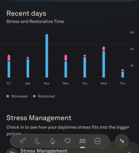
I’ve been on extended rest for about a week, and it shows – remarkably high restorative times (for me) from the Oura ring.
A Big Crash – I always feel compelled to explain what happened when the blogs suddenly stop for an extended time. This time it was a self-inflicted injury – which is a good thing, really. I am usually very good in hot temperatures, but not wanting to move camp, which itself takes considerable energy, I tried to stick it out for too long in the hot desert.
Strange symptoms started appearing, and finally, I got the message and got out of there. (Then I really crashed (lol)).
That was two weeks ago, and I’ve been on extended rest since then. (See high restorative times in the Oura ring to the right). I’m slowly improving and hopefully will be back to baseline sooner rather than later.

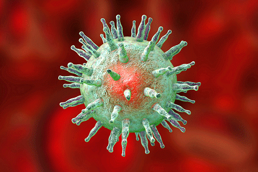
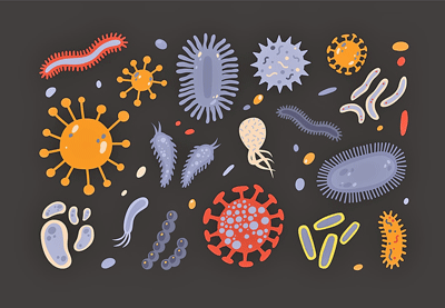
Bye-Bye “Microclots”: Hello Microclot Complexes
Dr. Pretorius has identified 4 different kinds of microclots in long COVID – setting the stage for precision blood clot therapies.
Resia Pretorius demonstrated just how rapidly the long COVID field has moved with her presentation on blood clots. First came the microclots, but in what seems like a giant leap forward, Pretorius and her team now believe they’ve identified 4 distinct microclot complexes (cell-debris–seeded complexes, NET-containing immune complexes, misfolded fibrinogen, and amyloidogenic aggregates on intact cells) in long COVID and ME/CFS.
Interestingly, post-vaccination syndrome microclots differ from Long COVID microclots. Plus the microclots in people with pre-COVID POTS, Long COVID, and Long COVID POTS appear similar but differ in their chemistry. So now we have microclot subsets!
The most fascinating part of her presentation, though, focused on the idea that one problem – called a phosphatidylserine “flip” – is producing all the different microclot issues. Phosphatidylserine (PS) “flips” when it moves from the inner side of the cell membrane to the outer side of the cell. Once there, it triggers clot formation.
When endothelial cells, red blood cells, tissues, etc., are injured, they release extracellular vesicles, apoptotic bodies, membrane fragments, mitochondria-containing debris, or cellular remnants loaded with PS. Depending on what kind of cell/cellular fragment is present, different kinds of microclots are found.
These suggest that targeting the phosphatidylserine “flip” could eliminate the microclot problem entirely. It’s entirely possible, though, that things happening “upstream” of the flip (persistent antigen, platelet hyperreactivity, endothelial injury, complement activation, autoantibodies, hypoxia, oxidative stress, mast-cell activation, impaired fibrinolysis) – are causing the flip and should be addressed first.
The GIST
- The blogs have stopped for the time being because I’ve been on an extended rest since I overdid it in the heat. See the blog for more.
- The second blog on PolyBio’s 2026 Spring Symposium starts off with a very promising finding: Resia Pretorius has found four different kind of microclots in long COVID – each of which would respond to a different kind of treatment. This is precision medicine for microclots!
- This fits nicely with another finding that “netosis” – a process by which neutrophils blow themselves up and produce “nets” that trap clots and cellular debris is occurring in long COVID. These nets can damage the blood vessels, and, in fact, blood vessel damage was worse in LC patients with higher NET levels.
- The presenters’ hypothesis that the coronavirus or remnants of it were constantly leaking into the gut or the blood vessels mirrored the finding from another study. That study found that healing the gut and preventing it from leaking reduced coronavirus levels in the blood and improved symptoms.
- The idea that the coronavirus, or another virus such as EBV, simply needs to be bottled up better is intriguing.
- Looking back at a cerebrospinal presentation covered in the last blog, I asked how an infection could cause such dramatic damage as seen in craniocervical instability (CCI). When CCI occurs, the skull slams down on the brainstem, causing damage.
- The body, though, has all sorts of ways to prevent this from happening. For one, the brain floats on a cushion of cerebrospinal fluid. It is also protected by thick membranes and is held in place by ligaments and muscles.
- One interesting possibility comes back to blood flow. Because cerebrospinal fluid cushions the skull, the virus could affect it by compromising the blood-brain barrier, damaging blood vessels, and dysregulating the pressure/flow systems that regulate CSF. Time will tell, but it’s yet another way that compromised blood flows may be impacting these diseases.
- There’s a lot of excitement about the role that chronically activated and exhaustged T-cells may be playing in ME/CFS. A ton of work on them is being done in long COVID as well.
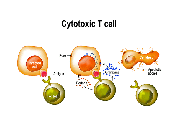
- T-cells are amongst the most important immune cells we have. One of the two big players in the adaptive, or later, immune response, they’re responsible for eliminating cells that pathogens have infected. Once they’ve identified the pathogen, they produce massive numbers of specifically targeted clones that then destroy those cells and clean the pathogen from the body.
The Chime ME/CFS study will be looking deep in the gut tissues for evidence of enteroviruses.
There is so much interest in T-cells in general that it’s almost impossible to keep up with the number of T-cell drugs available. That suggests that if a specific T-cell problem is shown to play a major role in these diseases, a treatment may very well be available.
Two presentations found that the T-cells in long COVID had been activated by three pathogens: the coronavirus, Epstein-Barr virus, and cytomegalovirus. Interestingly, the activation appeared to increase over time.
This finding suggests several treatment options may be helpful: antivirals to remove the viruses, immune modulators to calm the T-cells, and/or to strengthen them.
A pilot trial of the Furium microtesla device resulted in improvements in several aspects of cognition (including attention) and emotional well-being. These devices use very small magnetic pulses to strengthen the mitochondria. This device was used to reduce neuroinflammation.
This device is not yet available in the U.S., but David Putrino, the leader of the study, was jazzed enough about the results to launch a major trial that he hopes will lead to FDA approval.
Finally, the might UCSF LIINC long COVID project is now studying ME/CFS as well! Besides employing whole-body scanning of T-cells (there they are again) and metagenomics, the project will dig deep into gut tissues to see whether enteroviruses, as Dr. Chia has proposed, are present in ME/CFS. This project is led by researchers who are highly skilled in detecting pathogens in the tissue. After many years of promoting the idea that enteroviruses are involved in ME/CFS, Dr. Chia will finally get his due.
Health Rising’s Donation Drive Update

A big thanks to everyone who has contributed
We met our goal 🙂
Pretorius’s group has identified three different intervention approaches — restoring fibrinolysis, targeting platelet-monocyte and NET aggregates, and addressing upstream chemistry with glycation inhibitors or antioxidants — that they believe could impact microclots, and are currently testing them in a clinical trial.
If successful, we could have precise treatments that address the different kinds of microclots present in patients with long COVID or ME/CFS. Pretorius’s progress shows how far one dedicated and persistent research group can move a field.
Check out a PolyBio interview with Dr. Pretorius
Caught in NETS?
More evidence of netosisis – where neutrophils explode themselves into create nets that mop up blood clots and cellular debris – is present in long COVID. These nets can damage the blood vessel walls.
Health Rising recently covered several studies that found evidence that NETosis – a dramatic process in which neutrophils essentially explode in order to form nets that capture cellular debris and clots – was present in the microvascular blood vessels in long COVID (and perhaps ME/CFS). Remarkably, one study found that neutrophils from long COVID patients did not require any outside triggers to go on their suicide spree.
A 94-person study from Lael Yonker seconded that finding. Neutrophils exposed to long COVID plasma exhibited high rates of NETosis. The highest NETosis rates were seen in patients with detectable coronavirus spike protein.
However, more analyses indicated that the spike protein alone wasn’t the problem; instead, immune complexes (antibodies plus the spike) were driving the neutrophils to blow themselves up. We’ve seen evidence before that it’s the immune complexes – not the spike protein itself that are the big deal.
Not surprisingly, given the NETs’ ability to damage endothelial cells lining blood vessels, long COVID patients showed evidence of endothelial injury.
Yonker proposed that SARS‑CoV‑2 or its remnants persisting in the gut and/or blood vessels chronically leak into the circulation. The immune reaction that results – including these NETS – further damages the blood vessels. This suggests that neutrophils may play a major role in long COVID, and potentially opens the door for a variety of treatment possibilities.
This fits in nicely with another Symposium presentation suggesting that larazotide may be able to help by healing the gut barrier (larazotide) and preventing the coronavirus (or EBV or another virus?) or other toxic factors from spilling into the blood.
Other drugs that reduce immune complex formation (FcyR/complement), inhibit NET formation (DNase‑based approaches), or protect the endothelium (antiplatelet/anticoagulant regimens) might be called for.
Craniocervical Instability Pt. II
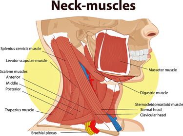
Four sets of neck muscles help to stabilize the skull
Getting back to Murakami’s presentation on craniocervical instability. One question concerns how a viral infection could create a condition as dramatic as craniocervical instability.
Stabilizing the brain so that it doesn’t smash down on the spinal cord is something the body clearly takes very seriously. Most of the stabilizing actually comes from the cerebrospinal fluid, which cushions the head so that it kind of floats above the spinal cord. Allowing the head to float in a liquid greatly reduces its downward thrust.
Three layers of tough membranes covering the brain and the spinal cord provide further support, and four ligaments keep the skull from moving in ways that could kink or compress the crucial junction between the spine and the skull. Four sets of neck muscles also keep the skull centered properly. (Deconditioning could contribute to weak neck muscles.)
A SARS-CoV-2 infection might particularly affect people who already had weakened ligaments or joint hypermobility but were largely asymptomatic because they were able to compensate by using their muscles, their healthy autonomic system, and abundant cerebrospinal fluid flow. The virus may also unleash mast cells, which attack ligaments that were perhaps already weakened and were helping to hold the skull in place. If COVID-19 produces extensive bed rest, that could weaken the muscular “guy wires” in the neck that help to hold the skull in place.
Because cerebrospinal fluid cushions the skull, the virus could affect it by compromising the blood-brain barrier, damaging blood vessels, and dysregulating the pressure/flow systems that regulate CSF.
If the same blood flow problems found in the body exist in the brain – and it looks like they do – that may be all that’s needed. Because one of the major waste removal systems in the brain – the glymphatic system – runs in the perivascular spaces found right next to the blood vessels, anything that affects blood vessel functioning could impair the brain’s ability to get rid of its waste products.
That, of course, suggests finding ways to return the blood flows to normal could be helpful in many ways.
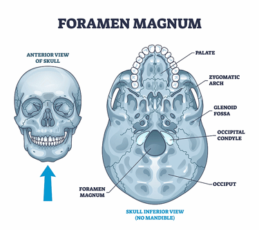
Increased cerebral spinal fluid pressure – which appears to be common on ME/CFS – can impact the foramen magnum – the small opening at the bottom of the skull through which the spinal cord, arteries. ligaments and nerves pass through
Low cerebrospinal fluid (CSF) levels or pressures can cause the brain to sag downward, producing intracranial hypotension (low CSF pressure). High CSF pressures, which studies suggest are common in these diseases, can cause the small hole (foramen magnum) through which a variety of structures (lower medulla, upper spinal cord, arteries, meninges, ligaments, membranes, nerve roots) make their way through to the brain, to become compressed and produce a wide variety of problems.
The virus can also produce CCI-like symptoms by producing neuroinflammation in the brainstem and/or vagus nerve, by restricting venous outflows of blood from the jugular vein, by producing Chiari-like crowding of the cerebrospinal fluid, via POTS/dysautonomia, vestibular dysfunction, small fiber neuropathy, cervical muscle spasms, and MCAS-related inflammation/swelling.
Immune Functioning
The T-cells Are Back!
Actually, they’ve never left. Health Rising just reported on the exciting T-cell work in ME/CFS being carried out by Selin, Kumar, and Kohlbruger in Boston. Two presentations in the Polybio Symposium feature T-cells. Over the past couple of months, more than 10 long COVID studies have featured T-cells.
Since it looks like T-cells are here to stay in the long COVID/ME/CFS space, let’s take a brief look at what these powerhouses of the adaptive immune system do and why so many researchers are interested in them.
A Short T-cell Interlude
T-cells are responsible for finding and destroying pathogen-infected cells.
T-cells are a major target in the drug space. From T-cell activation inhibitors, immune checkpoint inhibitors, intracellular signaling inhibitors, immunosuppressants, cytokine pathway modulators, CAR‑T products, T‑cell engagers, TCR‑based therapies, and Treg‑targeters, probably hundreds of different T-cell-affecting drugs exist. In fact, the T-cell space is so dynamic, and so many T-cell-affecting drugs are being assessed, that any attempt to come up with a solid number is likely to be quickly outdated.
One would think that if specific T-cell problems are validated in these diseases, drugs targeting them would be available.
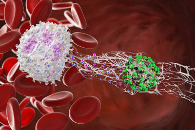
T and B cells are the mainstays of the adaptive immune response, which is responsible for eliminating infection once and for all. When activated – a process which takes enormous energy – both send out armies of clones specifically designed to find the pathogen and kill it
B-cells hunt extracellular pathogen hunters (free-floating pathogens not inside a cell). They produce clones that bind to the pathogen, preventing it from entering cells and replicating. As they’re doing that, they place a tag on it which is like catnip to macrophages and neutrophils, which then rush in and engulf (eat up) the pathogen.
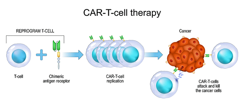
CAR-T-cell therapy is just one of the many T-cell therapies present. CAR-T weaponizes T-cells by aiming them at specific targets such as autoreactive B-cells. If autoimmunity is present, it’s a possibility.
T-cells, on the other hand, hunt for intracellular pathogens – pathogens found inside cells. They do this by examining MHC molecules on the cell surface, which cells produce to communicate their state to the immune system. If a pathogen is present, these molecules exhibit a piece of the pathogen – an antigen – on the outside of the cell, for all to see.
If a cytotoxic or killer T-cell determines that a pathogen is present, it kills the cell itself by boring a hole into it and dropping in a suicide program. Note, though, that pathogens have developed several ways to prevent MHC complexes from appearing on the cell surface. If that happens, the cell can still be destroyed by cytotoxic T-cells, which detect the low levels of MHC complexes on the cell, suspect a pathogen is present, and then move in and kill the cell in exactly the same way that T-cells do.
Coronavirus / EBV Activated T-cells Take I
In Nadia Roan’s presentation, “A type of cell-killing immune cell stays abnormally active in Long COVID — especially in women — which may help explain why Long COVID disproportionately affects women“, she looked at T-cells from a different angle and ended with a familiar result.
T-cells specifically activated by the Epstein-Barr virus were found.
First, she used mass cytometry to identify T-cells associated with a variety of pathogens (SARS-CoV-2-, EBV-, CMV-, and influenza-specific CD8 T cells) to determine their state. The recovered COVID-19 participants’ T-cells had low levels of exhaustion markers (their T-cells were not activated and were ready to pounce on the next invader), whereas the T-cells in long COVID patients were highly activated and showed evidence of exhaustion.
Note that the longer these killer T-cells are chronically activated, the more likely they are to trigger an autoimmune reaction and/or damage the blood vessels, nerves, produce fibrosis, etc.
An analysis found clusters of T-cells had become activated by SARS-CoV-2, EBV, and/or CMV (but not the flu). Each of these pathogen-specific clusters showed increased cytotoxic T-cell CD29⁺/Granzyme B⁺ frequencies, indicating that even years after the initial exposure, these T cells were still on high alert and primed to act. In fact, the level of activation even seemed to increase over time, perhaps indicating that further bits of SARS-CoV-2 and/or herpesviruses were continuing to escape. This was more prominently seen in women.
Impaired cytotoxic NK cell exhaustion was found in ME/CFS many years ago. So now we appear to have problems with both of the cells (cytotoxic T and NK cells) responsible for going after intracellular pathogens. If that’s true, it’s no wonder that EBV reactivation, in some form, remains a problem, or that the innate immune system is highly activated, trying to fill the gap.
Validating this finding could open a wide variety of treatment approaches which might a) target EBV via antivirals or monoclonal antibodies, b) seek to calm the T-cells, c) seek to revive the exhausted T-cells,
Coronavirus / EBV Activated T-cells Take II
Mark Painter ·of the J. Craig Venter Institute, provided more evidence of T-cell exhaustion caused by coronavirus (or remnants of it) and/or EBV in his “Virus-specific CD8 T cells as biosensors of antigen persistence” talk.
Painter assessed cytotoxic T-cells that have been specifically primed to attack SARS-CoV-2, EBV, CMV, and the flu. The basic question was – are they still reacting to the virus? If they are, that suggests the virus (or bits of it) is still present and driving the T-cell activation.
More evidence of cytotoxic T-cell exhaustion was found.
Lo and behold, he found that the cytotoxic T-cells in about a third of long COVID patients were still reacting to the virus, were in an activated/exhausted state, had trouble producing cytokines, and proliferating. These and past findings suggest that the coronavirus is tweaking the immune system in a substantial subset of long COVID patients, but certainly not in all.
He also found signs of EBV reactivation in a subset of long COVID patients. No signs of CMV or flu-activated T-cells were found.
These EBV findings track well with a recent German study, which found that cytotoxic T-cells lost the ability to effectively rein in EBV early in a COVID-19 infection.
They also align perfectly with Selin-Kumar-Kohlbuger’s pilot ME/CFS work, which found that T-cells in ME/CFS are activated by a pathogen or an autoimmune response. They’re now on the hunt to identify the specific T-cell triggers in ME/CFS.
MicroTesla Therapy to the Fore?
(Elon Musk supporters or non-supporters and everyone in between, please note that this MicroTesla device has nothing to do with Musk’s Tesla enterprise.)
Now for a completely different treatment approach. The Furium microtesla device used in this long COVID study is gentle (check), can be used at home (double-check), and appears to have no side effects (double-double-check). It’s being trialed by David Putrino at Mt Sinai, who seems to be checking out just about any treatment that makes sense.
So far so good. Magnetic devices have already proved themselves to various degrees in disease, but this one is decidedly different. It produces low‑energy, low‑frequency magnetic fields (hence the “micro-Tesla”) that are ~100,000× weaker than those used in rTMS devices being trialed for fibromyalgia.
The mictoTesla device gives the brain’s mitochondria a gentle nudge to combat neuroinflammation.
The magnetic fields are far too weak to cause neurons to fire. Instead, the goal is to engage hormetic mechanisms. These treatments give the brain’s mitochondria a gentle nudge, prompting them to rebound, produce more mitochondria, clean up damaged mitochondria, and exert an anti-inflammatory effect.
These devices are aimed at the head. The goal is to reduce neuroinflammation. A 2025 paper, “Transcranial microtesla magnetic fields suppress neuroinflammation and neuronal oxidative stress burden” appearing in Science, found that in various disease animal models, this electromagnetic therapy had “robust anti-inflammatory, antioxidant, and neuroprotective effects”,
It was a small trial, just 30 persons with long COVID, but it was triple‑blinded, randomized, and sham‑controlled. The participants used the device for 15 minutes every three days for four weeks.
People with the active device showed improvements in visual attention, processing speed, attention/inhibition, and emotional well‑being. That processing speed and attention improvements are biggies, as delayed processing speed (“what did you just say?”) and attention (“what was I doing again?”) are prime cognitive issues in these diseases. Better emotional well-being is an obvious help as well.
This device is being trialed in other neuroinflammatory diseases, but this is the first time it’s been tested in long COVID. It’s not yet available on the US market, and it should be noted that this was a pilot trial, but Putrino was excited enough about the results to report that a multi‑site pivotal trial for FDA approval was underway. I was unable to find a website associated with this product.
The Mighty UCSF LIINC Project Takes on ME/CFS
“research on enterovirus persistence as a driver of ME/CFS has stalled over the past decade due to a lack of teams in the field with the infrastructure needed to collect tissue samples from patients with the condition or to identify viruses in such samples using cutting-edge methods.” Chime
Saving the best (for ME/CFS patients) for last, Michael Peluso, UCSF professor and co-founder of the mighty LIINC long COVID project, reported that, thanks to PolyBio, the LIINC project is now taking on ME/CFS (yah!).
The Chime ME?CFS study will be looking deep in the tissues for evidence of enteroviruses.
It looks like Dr. John Chia, after pounding the enterovirus drum for decades, is finally going to get his study. Despite the fact that enteroviral RNA has been found in the brains, muscles, and guts of ME/CFS patients, the last ME/CFS enteroviral study (by Chia) I could find was in 2010.
In 2021, though, O’Neal and Hanson’s ME/CFS enterovirus review asserted that “further studies of appropriate biological samples with the latest molecular methods are urgently needed.”
Enter three experts (Mike Peluso, Steven Deeks, Tim Henrich) with deep experience in ferreting out viruses (HIV) that hide deep within tissues. They will use the same ultrasensitive methods (single-molecule assays, peptide arrays, bacteriophage display, and transcriptomics) they’ve used in HIV and are using in long COVID to search for enteroviruses in ME/CFS.
In the “Chronic Infections and Inflammation in Myalgic Encephalomyelitis (CHIIME): study, preCOVID ME/CFS patients will receive intensive in-house visits that will include providing a wide range of biospecimens (all biobanked), including gut biopsies (enteroviruses), brain imaging, neurocognitive tests, and metagenomic analyses.
Plus, an advanced whole-body PET (positron emission tomography) will use a special tracer they’ve developed to map the location of the activated T cells throughout the bodies and brains of the ME/CFS study participants. (There are those T-cells again!).
When I emailed Peluso several months ago, he said the CHIME ME/CFS study was immediately filled.
Conclusion
The four big things that popped out for me in this Symposium were the creation of the Long COVID Cure Initiative, the tissue findings, the pathogen/T-cell findings, and, of course, as a long-term ME/CFS patient, the CHIME project.
The arterial plaque, retinal, gut, lymph, spinal, and brainstem findings suggest that, as Amy Proal asserted, the tissues may be where we’ll find the “most signal”.
We saw more evidence that persistent pathogens are playing a role in long COVID.
If there’s one thing that PolyBio has been focused on, it’s been viral persistence; i.e., the idea that the coronavirus and/or EBV are continuing to tweak the immune systems of long COVID patients.
With two presentations finding evidence of T-cell exhaustion due to chronic responses to the coronavirus and/or EBV, T-cells are clearly going to continue to spark strong interest.
It appears that the immune system is activated but is unable to clear the remnants of SARS-CoV-2 infection and/or tamp down EBV in a significant subset of long COVID patients.
Two other presentations suggested that the problem may not be the pathogen itself, but its ability to escape into the bloodstream from the gut or to trigger immune cells to attack the blood vessels. This suggests that gut lining support/immune modulation may be helpful.
Finally, LIINC’s CHIME project is a breath of fresh air for a field that is making progress but has struggled to gain the traction one would have thought it would after long COVID showed up on the scene.
Health Rising’s Donation Drive Update
A big thanks to everyone who has contributed
We met our goal 🙂


Cort, bless you for taking the time to write this very interesting and inspiring blog despite the crash that you experienced recently (I hope your recovery is continuing).
It’s really encouraging to read of these developments. Almost makes me think that an effective treatment could be arriving in the next year or two. Is that too optimistic?
I don’t know. Here’s what I in my laymen’s brain think – I think new possibilities like the Furium or larozatide will continue to pop up and they will be helpful. Getting them validated will be particularly important – which is why it’s so important to have people like Putrino in the field who can take on bigger trials.
Getting to the heart of these diseases – which will require biologically identifying subsets and matching them with treatments – will take more time. PrecisionLife seems like an outlier here – they believe they’re already able to do that using genetics. They are focused on genetics which clearly plays a role and it’s not the whole story. I’m looking forward to see what they produce over the next year or two. I would suspect that they would introduce new treatments that are beneficial if not the end all and be all. For me, I would love to improve by 20%
I don’t know what everyone is doing. It’s possible that some group is right now identifying what the T-cells in each person are reacting to in long COVID (pathogen, autoimmune process) and will be able to identify drugs that will work.
Maybe Pretorius’s group will have a breakthrough in precise microclot treatments, or that some of the big long COVID immune drug trials that are underway will identify specific subsets. It’s possible that RECOVER identifies a subset of long COVID patients that REALLY benefit from IVIG, or a Patterson maraviroc/prevastatin subset, or a gut subset that responds to larozitide shows up, etc. The Chime study could show – whoa! there’s lots of enterovirus where it’s not supposed to be in a subset of ME/CFS patients. That would open things up.
I could see a study showing up over the next year which suggests that hey, this treatment looks really effective. If that happens it will probably take another bigger study to validate the results and that will take time but people who have access to a physician who is willing to give the drug a try would get a shot at it quickly.
In the end, I think big projects like PolyBio’s Long COVID Cure project and the OMF’s Bioquest project that methodically look for and identify biomarkers are the best things going. They will take several years to produce results but they have the ability to transform these fields and really get things moving.
There’s no telling how each of these efforts will go but it’s the accumulation of things that will tell the tale. I think we should be patient and recognize that these diseases are very complex AND there are a lot more pans in the fire now and new possibilities are going to continue to show up.
We shall see!
Larazotide is available in the US as a (pricy) supplement. I ordered it after seeing the positive results of the MIS-C study in children. Hasn’t yet arrived but one of many interventions I’m trying simultaneously.
Good luck!
It seems like we’ve been going in circles for 30 years. Personally, I no longer believe that the cause of ME will ever be found. Nor do I have any indication or hope to think that a working medicine will come. We are psychosomatic sufferers at all times.
Its a connective tissue disease
Everything is downstream of that.
If we can edit the connective tissue. We can beat this
Certainly in terms of the immune system it seems like it has been going in circles for 30 years. One of the reasons why I don’t think the immune system is the primary driver. More likely the brain, which has been much harder to research, although getting better
Hypermobility is the critical driver. Its our tissue type.
It leads to the brain issues. But the brain issues are secondary. Its all downstream reactions if being born with weak collagen
The tissue, across the body, depending where affected is more prone to virus, endothelial damage, hyperactivity on glial cells, mast cells.
Ebv and other pathogens find it easy to hide abd reactivate, blood reacts differently.
The tissue type diagnoses is not fit for purpose. Jen brea was told she had m.e. then cranio cervical instability. But she passed the beighton scale eds test. But she had some defect on the collagen in her neck.
So we can call tgis sub clinical eds that overtime becomes a pathology.
The integrity of the blood brain barrier and the gut wall as well as the endothelial wall is vital for health.
If the collagen is weak, then you will develop problems more easily.
What does the organism do after years of any kind if stressor? It shuts down and switches to a dauer state.
Ground zero is the tissue, everything else is a downstream individual description of pathology that develops. Some people might be almost entirely mast cell related, others autoimmune.
We need stem cell therapy and regenerative medicine.
It feels to me were going round in circles because we’re not trying to stop the hub of the damage at source.
Problems in the brain. Sure. But why?
The fact that hypermobility shows up on cfs time abd time again should make it a massive area of study
‘But Why?’
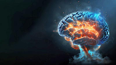
Chronic neuroinflammation.
Neuroinflammation can explain all symptoms.
Such as muscle issues / PEM:
https://medicalxpress.com/news/2024-07-scientists-block-muscle-fatigue-covid.html
Yes but why? What is neuro inflammation.
It comes down to tissue type .
And sadly its far more complex than just in the body. Affected tissue can be anywhere in tge body.
We are not all made of the same stuff. And even those if us that are have it expressed in different places in the body.
Omf has been looking at distribution of affected tissues in the body. The variations will describe what kind of m.e. you have.
Neuroinflamation is for sure a big component for many, but you have to ask why? What is neuro inflammation.
Why are some people more prone to it than others. What creates resilience.
Its the strength of your tissue
Whenever anyone is sure the cause is *one* thing, you can be pretty sure they’re wrong.
How about some epistemic humility, fellas?
Because we’re going round in circles and in this heterogeneous illmess the underlying ground zero seems to be how tissue expresses itself under insult of any kind.
I hear what youre saying, abd im open to anything, but at the same time, connective tissue comes up in all these different manifestations of m.e.
In the OMF s deep dive on whitney they concluded that eds of some kind was a neccesary part of the illness.
To me that gives hope and a frame work.
There’s so many people doing different things, which is great, but I think theres enough evidence to suggest this is a tissue based disease at heart and id love to see a concerted effort on that. Thats why im so adamant. From my virw it looks like a total non trainer.
https://pmc.ncbi.nlm.nih.gov/articles/PMC12437428/
There are a lot of very recent papers noticing these crossovers that the rccx theory has elicited.
Check the study.
This is one study amongst many noticing the presence of eds.
The omf deep dice concluding similar. As we see, hypersensitivity, inflammation etc etc all occur
I also wonder if the different EDS genetic variants may provide us with further “subsets”?
I think almost certainly. I also think there will be subclinical variants. Jen brea didn’t traditionally have eds, but she did in her neck.
I really think its in its infancy. It can ‘ describe’ say more benign variants such as ibs, where theres only those collagen expressions in the stomach and its mild.
I think k if we can get a handle on how to restore or strengthen or change these collagen defects, a lot of these things we think of as m.e. such as leaky bbb , leaky gut, pots, autoimunnity, sensitivities due to a leaky gut, weak mitochondrial membrane and immune system will not occur. Theyre all downstream disease processes of the original lack of collagen integrity. And as you point out , the different expressions of m.e. will likely correlate to cfs subsets.
Obviously, anything that helps us is very welcome short-term but I think we are a ‘ type’ of human.
There was a time when I was functionally well. Why cant we get back to that state?
I am one of the people who has had an episode of a post viral ME/CFS accompanied by FM, from which I completely recovered. Ten years ago, after twelve months of mild ME/CFS I returned to full health and in fact following recovery, in my early fifties, was fitter and stronger than ever.
Three years ago I caught a cold virus and it took into a severe state of ME/CFS (interestingly no FM this time). I have progressed now, out of the severe state to a moderate state.
I remain convinced that under the “correct circumstances” (whatever they might be) some of us have inherently “something” (whatever that might be!) that allows our bodies to self-correct and overcome ME/CFS. I’m not in any way suggesting this is true of everyone/every subset. However, this concept is all I have to hold onto as I hope that my body can somehow regain purchase.
My WGS showed EDS variants….
Hmm. What do you have, Liam? ME/CFS? Long Covid? Both? How long have you been ill? I have always thought it was unfortunate that Cort mixed Long Covid in with ME/CFS on Health Rising. I have both and they are not the same. Long Covid is much worse.
This is to ling white clouds.
I think, without being able to prove it forensically, that people with eds constellations have a certain predisposition to viral attack. Like you say, the conditions have to be right.
Do you know Ron davies had myocarditis as a kid.
So did my mum.
Both their sons have developed severe m.e. although, mercifully for me, ive not gone to the places whitney had.
Ive also had periods of recovery similar to yours, although ive never felt ‘ stronger than ever’ as you did.
I think the healing cycles that naviaux talks about are on a hair trigger to shut down to protect us, as we are more vulnerable organisms.
Its such a complex interplay obviously but eds is involved in autoimmunity cell integrity, hormone production, cell overactivation etc etcthat I cabt help but think its the root cause
I don’t know, but the immune system is so powerful….It’s actually a major driver of many neurological and neurodegenerative diseases. With some it’s simply chronic inflammation, with others such as MS, immune cells are attacking tissues.
When we talking about neuroinflammation that’s inflammation which is produced by the immune system.
It’ll be fascinating to see what Jarred Younger finds with his immune cell brain invasion study…
I think its going to be both 🙂
When is Younger’s study out?
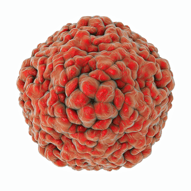
An interesting Japanese study on long covid has been released and shows HHV-6Bb reactivation in the brain in a large subset. And that in that subset, a dementia drug offers promise.
I am interested to see how the study on Bezisterim gies.
Goes
I don’t know, but I will by the 20th. We have a Zoom interview planned. I found this interesting study . I wonder if the ME/CFS field has kind of missed the boat with HHV-6. If I remember correctly, Dr. Klimas highlighted HHV-6, and my test results showed HHV-6 reactivation not EBV.
” Detection of salivary EBV and HHV-6 DNA was highest early in the morning….However, salivary HHV-6 DNA levels were positively associated with a greater aggregated LC propensity score, as well as anxiety and depression scores.
Interpretation: The observed correlation between salivary HHV-6 shedding and symptom severity suggests HHV-6 may contribute to post-acute disease, though mechanisms remain unclear.
While our study did not identify a relationship between salivary EBV shedding and LC, EBV may still play a role at earlier time points in the disease course, or in compartments not sampled here. These findings highlight the potential importance of HHV-6 in LC pathophysiology and underscore the need for longitudinal, multi-compartment studies of herpesvirus reactivation in LC.”
https://pubmed.ncbi.nlm.nih.gov/42238391/
I also saw this one which suggested the pathogens could impacting autonomic nervous system functioning. Interesting that HHV-6 and HSV-1 were in there but not EBV (they tested for it), and that so much ANS disruption was found in Lyme disease.
Logistic regression analysis revealed that, beyond classical hemodynamic parameters, antibody status served as a significant predictor of HUTT outcomes, with specific associations identified for HSV-1, HHV-6, Coxiella burnetii, Toxoplasma gondii, and Borrelia spp. Multinomial regression further indicated that negative IgG antibodies, particularly to HSV-1 and VZV, predicted Lyme disease group membership. These findings support the hypothesis that ANS dysfunction in post-infectious syndromes may be driven by persistent or prior infections, highlighting the need for integrative diagnostic approaches.
https://pmc.ncbi.nlm.nih.gov/articles/PMC12656751/
Here’s an article on the Japanese study. In essence, viral infection in the brain leads to reactivated HHV6, this then impacts on acetylcholine. Possibility that a dementia drug that acts on acetylcholine might help. Has anyone tried Huperzine A? That acts on acetylcholine.
I am very interested in this sort of research as I think the brain is central.
https://mainichi.jp/english/articles/20260623/p2a/00m/0li/018000c
Prusty has heavily researched HHV6?
And Polybio are trialling an antiviral that acts on HHV6
If you look at understanding and treating ME/CFS like it’s some treasure buried deep in a mountain I would say we’re not going in circles but instead are digging deeper and deeper into the mountain.
We’ve been talking about pathogens and ME/CFS for as long as I can remember, for instance, but only recently does it look like it’s possible to identify the pathogen/autoimmune process that’s exhausting the T-cells (and potentially destabilizing the immune system). We’ve been talking about enteroviruses for 30 years – and now we have a study that will be able to say, yes or no, if they’re actually present.
Dozens of drugs could become possibilities if an autoimmune subset, for instance, is identified.
We used to talk about microclots like they were one thing – and now we find there are four different kinds of microclots – each of which requires a specific treatment.
No reason to give up now! 🙂
I admire your optimism. But perhaps the map to the hidden treasure deep in the mountains is wrong. But giving up, no. It is just going to take a very long time, and I don’t think our generation will live to see the treasure found.
I hope you’re feeling a bit better soon. How hot was it where you stayed? 40 degrees Celsius. It’s very hot in Europe too. I always collapse when the weather cools down.
It was 95-105 degrees (40 C = 104 F) over about 3 weeks or so. I had gotten through about a week of 100+ earlier and I’ve always been able to handle heat before. I don’t think I’ve been in such extended heat for a long time. Lesson learned!
Research into Long COVID continues to evolve as scientists investigate the biological mechanisms that may explain why some individuals experience persistent symptoms long after an initial infection. One area of discussion at the PolyBio Long COVID Symposium focused on the possible roles of lingering pathogens, abnormal blood clotting, and neutrophil extracellular traps (NETs) in the development and persistence of the condition.
Researchers are exploring whether viral remnants or other persistent biological triggers may contribute to ongoing immune activation in some patients. While evidence is still emerging, these hypotheses aim to explain why symptoms such as fatigue, cognitive difficulties, shortness of breath, and exercise intolerance can continue for months or even years after the acute illness has resolved. https://spinbetters.org/
The reason the chronic latent virus hypothesis is only now resurfacing after 40 years is that the required technology and funding simply didn’t exist back then to prove atypical herpesvirus reactivation profiles. Decades ago, the prevailing infectological dogma dictated that if no active virions were detectable in the blood, reactivation could be ruled out. Anyone challenging this dogma was treated like a fool.
Two things changed everything: first, the development of high-resolution immunological technologies over the last several years; second, the historic $1.15 billion in NIH funding for Long COVID and ME/CFS research—successfully lobbied for by patient organizations like Solve M.E. This massive influx of capital finally drew elite, mainstream immunologists like Akiko Iwasaki and many others into the field, fundamentally shifting the paradigm.
Hi Cort, I remember Jennifer Brea was such a case who started out as post-infectious ME and developed CCI.
She wrote a number of blogs about it 6/7 years ago, see https://jenbrea.medium.com/how-infection-can-damage-the-cervical-spine-d43d3dac5734 and https://jenbrea.medium.com/onset-part-i-viral-onset-2a431c149800 .
I think it had something to do with connective tissue, with infections compromising connective tissue integrity. Have a look at the blogs if you like.
Googleing “how infections cause CCI” might also yield newer reviews of the topic, such as this one: https://www.youtube.com/watch?v=SNsqylGrWPk “Infectious or Environmental Causes of CCI: A Review of What we Know” (3 yrs ago).
Thanks – I had forgotten about that blog. I would think connective tissue damage would be key. All you need is a vulnerability there or a reaction to a pathogen that goes awry and decides to attack the ligaments holding the skull in place. Or maybe impaired blood flows play a role?
Maybe – just brainstorming here – if you stop the attack, the ligaments could repair themselves (?) or you could find a way to facilitate that.
My belief is that eventually, we will need gene editing to strengthen the tissue.
Think about all the crucial areas in the body, the gut, the BBB integrity is crucial to stop inflammation or autoimmunity, or pathogen entry.
Also the tissue integrity, because its weaker, usually creates immune hyperactivity. Over activation of glial cells. Overstimulating of mast cells which cannibalise tissue.
That kind of invasion potential into the body creates a space for all body shutdown and switch to a more damaging energy supply.
Im sure there will be loads of modalities we can use on the way but tgis is almost certainly an issue with connective tissue integrity.
I think we all remember a time before we were ill.
We need to find a way to reverse our way back over that event horizon
When I did my WGS with S……. .com there was a pop up note with my results which asked me, on account of my EDS variants, if I would be willing to have my WGS sent through and be part of a study with the Norris Lab. I don’t know anything about this organisation but I did link into their study, and I wait with interest to see their results.
PS The Norris Study onboarding questionnaire was a small handful of questions which included specific questions about if I have ever had ME/CFS FM. I assume they are looking into the links between the conditions.
For the vaccinated victims and long Covid patients. Concerning reports.
https://www.mdpi.com/2073-4409/15/11/978
https://panagispolykretis.substack.com/p/the-vaccine-induced-immune-reaction
COVID-19, Spike Protein and “Turbo Cancers”
Published case reports and social media reports have reported that exposure to the spike
protein, particularly following mRNA vaccination for COVID-19, is associated with “turbo
cancers” (hyperprogressive disease). (60-69) These include new cancers that are highly
malignant, often in young patients and rare cell types/locations, as well as tumor recurrences in
patients after remission. It has also been proposed that long COVID-19 can predispose
recovered patients to develop cancer and accelerate cancer progression. (70) The U.S.
Department of Defense Medical Epidemiology Database (DMED) reported a 664% increase in
malignant neoplasms following the deployment of COVID-19 mRNA vaccination in the military
(until this data was erroneously removed). This information is now supported by an analysis of
the VAERS database which has demonstrated a strong “safety signal” for many cancers but
particularly for breast, colon, liver lung and kidney cancer. (71) Death trends for cancers in the
USA (ICD codes C00-D48) for individuals aged 15-44 demonstrated a rise which started in 2020
(1.7%) and accelerated substantially in 2021 (5.6%) and 2022 (7.9%). (72) A similar trend has
been reported in the UK.(73) It is likely that this trend has continued into 2023 and 2024. It
should be noted that cancer is largely a disease of the aged and the increase in cancers deaths
in this young cohort of patients is alarming. A recent Japanese study demonstrated a significant
increase in age-adjusted mortality rates from cancer as a whole, as well as some specific types
of cancer, namely leukemia, as well as ovarian, prostate, oropharyngeal, pancreatic, and breast
cancers.(74) In this study some excess cancer mortalities were observed in 2021 after mass
vaccination with the first and second vaccine doses, and significant excess mortalities were
observed for all cancers after mass vaccination with the third dose in 2022. The increasing risk
of cancer with the increasing number of “shots” may be related to increasing immuno-
suppression likely caused by immunological imprinting (antigenic sin).(75, 76) In addition, the
systemic toxicity of spike protein (spikopathy) increases with the number of vaccines.(77)
It has been suggested that SARS-CoV-2 converts normal cells into cancer cells by modulating
central metabolic pathways or hampering genomic integrity mechanisms, consequently
inhibiting the apoptotic machinery and/or enhancing cell proliferation. (70, 78) It is likely that
decreased CD8+ cells with impaired immune surveillance plays a major role in turbo cancers.
This may explain the explosion of turbo cancers after repeated “boosters” rather than the
primary vaccine series. Angues and Bustos have suggested a multi-hit hypothesis to explain the
increased risk of cancer associated with COVID mRNA vaccination.(79) The specific pathogenic
mechanisms by which SARS-CoV-2 and/or the spike protein leads to increased tumorigenesis
have not been well studied, however, several possible mechanisms exist. The spike protein
damages mitochondria and alters mitochondrial function; this may play a central role in cancer
cell development and propagation. (80-83) SARS-CoV-2 results in dysregulated innate and
adaptive immunity. Depletion of CD8+ and natural killer cells reduces immune surveillance and CANCER CARE (Version 2.2) 2024-10-1 Page 26
alters the tumor microenvironment to promote tumor proliferation and metastases. (84) The
retinoblastoma protein (pRB) is a tumor suppressor protein that prevents excessive cell growth
by inhibiting cell cycle until a cell is ready to divide. The non-structural protein 15 (Nsp15) of
coronaviruses induces the nuclear export and ubiquitination of pRB leading to its degradation
via proteasomes. (85) A second potential oncogenic mechanism has been hypothesized for
SARS-CoV-2 consisting of the degradation of the tumor suppressor protein p53 mediated by
NSP 2 and Nsp3. (86) The open reading frame 8 (ORF8) protein of SARS-CoV-2 interacts with
p62, the main autophagic cargo receptor, thereby inhibiting autophagy. (87) Jiang et al
reported that spike protein localizes in the nucleus and inhibits DNA damage repair by impeding
key DNA repair protein BRCA1 and 53BP1 recruitment to the damage site. (88) Spike protein
impairs type I IFN signaling increasing the risk of cancer as type I IFN signaling suppresses
proliferation of cancer cells by arresting the cell cycle, in part through upregulation of p53 and
various cyclin-dependent kinase inhibitors. (89, 90) Metabolic reprogramming is a distinctive
feature of SARS-CoV-2, and this may play a role in tumorigenesis. Metabolic reprogramming
includes amino acid and lipid metabolism, carbohydrate, and energy metabolism, and immune-
related pathways. (70) More recently Simian Virus 40 (SV40) DNA plasmids have been isolated
in the vials of the COVID-19 vaccines (social media reports). SV40 is a known oncogenic virus.
(91) IgG4 Antibodies are induced by repeated COVID-19 vaccination.(92, 93) IgG4 plays an
essential role in cancer immune evasion. (94-96) IgG4 induces tolerogenic M2-like macrophages
and correlates with disease progression in colon cancer. (97) In addition, Zang et al have
demonstrated that the combination of glutathione and IgG4 promotes tumor growth through
the effect of the Fc-Fc reaction between IgG4 and other tissue-bound IgG subtypes resulting in
local immunosuppression. (98)
In a patient with cancer, it may be difficult to establish a causal role with SARS-CoV-2/spike
protein. However, the tumor can be stained for spike protein, establishing this causal
association. As these “turbo” cancers are frequently highly malignant, an aggressive treatment
is suggested including the guidance offered in this monograph.
👆that from the independent medical alliance
Check out their site.
They actually care about your and my health.They have lots of life saving information
Good to see you back, Cort. I was wondering what happened, & while I’m sorry you crashed, I’m glad you know why & are able to do something about it.
Also, this was an interesting new blog post.
Thanks! Low autonomic drive appears to be the problem. The solution (hopefully) lots of rest, small, often liquid meals, Trioral, no caffeine or stimulants, no THC gummies for sleep, and better sleep overall.
Interesting symposium, I agree!
I hope those things do help. You’re always missed when you’re gone (& not just for the posts, though of course, those are definitely appreciated & very helpful).
Thanks – I am slowly getting better. I’ve had to break camp several time and drive and that has set me back. Hopefully, I can stay where I am now for longer and get back to baseline 🙂
Hi, Cort, Where can I find out more about your exhaustion from “overdoing things in the heat”? I have noticed a strong increase in CFS symptoms since it got hot here in Florida (and I walk in the heat an hour each day). I’d like to hear about your experience and how you’re doing/coping. I hope you feel better soon.
Thanks, LC. Like I said I usually do well in the heat and, in fact, had done OK while taking it easy when the temps hit around 105 degrees while camping outside of Phoenix. I was tentative about that – but it a short term thing – and I was a little surprised at how well I did. This time I spent too much time camping out in high heat. Lesson learned!
I will say that using Trioral salts really helped.
Things slowly built up and I was not aware that I was getting in trouble. The new symptoms I experienced were increased tingling in my ankles, nausea after eating just anything, intermittent cold sweats on my face, and poor sleep (about 5 hours/night). When I saw that the temps were going to get even worse I left for the mountains.
Packing up and leaving and re-establishing camp takes enormous amount of energy. I thought I would feel better once I got up there but then really crashed. It didn’t help that I had break camp again and move elsewhere – another hit. Plus I was at 7500-8500 ft high – which puts more stress on the autonomic nervous system. That’s my resting heart rate on the Oura ring dropped dramatically.
I asked AI about all this – normal heart rate (until I hit the mountains), HRV mostly OK, no temperature increase at night, no problems breathing – just a lot of fatigue, cold sweats during even mild exertion, nausea when I ate almost anything, and the increased tingling in my ankles.
The verdict – was low autonomic drive. No heat exhaustion – just a depleted autonomic nervous system that was not properly shunting blood to my gut when I ate (nausea) and to my face (cold icy sweats) The prescription – checked with Theresa Dowell, my ME/CFS doctor – keep with the pyridostigmine bromide (parasympathetic nervous system enhancer), eat small meals (mostly soups and juices), do mild exercise and stretching, no caffeine or stimulants, and rest.
AI also suggested that I not take clonzepam or THC gummies for sleep (sympathetic nervous system enhancer), do gentle deep breaths – and none of the other breathing exercises.
Since then I overexerted myself (too much walking), and had to break camp again suddenly twice (fire in the area, musty camping site) and drive long distances. Each time produced another relapse. Now, it appears that I have a good site, and will be able to stay here for awhile, and hopefully recover. I’ve dramatically upped my meditation – generally doing 1 1/2 hours + in the morning and it seems to help.
Wow…so many challenges but you’re a real trooper still getting out there and traveling. You know your body and you’re fortunate to have a CFS/ME doctor.
I always thought the desert southwest dry heat was like an oven, but I’m visiting DC now from Florida and in Florida 85 degrees is unbearable with our humidity. But I walked in 105 degrees in DC yesterday and it was okay because the humidity was low.
You’re fortunate to be in a dry climate. Keep on keeping on.
Hi Cort,
It’s been a while, hoping you’ve been able to stay put and recover.
Thanks. Slowly getting better. 🙂
On neutrophils and blood vessels: I always read the blood vessel studies with particular interest because my own disease trigger was the aftermath of an autoimmune vascular disease. The specific mechanism of autoimmune damage in my disease was via an antibody that specifically attacks neutrophils.
And the talk of neutrophils here reminded me of an intriguing study I read recently that found the majority of people with my condition go on to meet the criteria for ME/CFS even when in remission from the autoimmune disease itself.
Even more interesting is that there are two possible subtypes of antibodies and one is significantly more likely to cause fatigue — Google tells me that the more fatiguing type (anti-MPO) does the kind of damage that creates Nets, while the explanation of the less-fatiguing type (anti-PR3) says nothing about Nets.
That’s the best I can understand or explain as someone without any understanding of biology, but I found the overlap to be very interesting! Here’s an article describing the fatigue study if anyone is interested: https://ancavasculitisnews.com/news/most-aav-patients-have-fatigue-severe-enough-be-disorder/
I am not sure how to get his information out so I will post it here.
I have struggled with ME for 33 years. I recently went to a functional medicine physician who screened me for vector borne illnesses (tick, fleas, lice carry these illnesses). I came up positive for Bartonella.
In the 30 years I have had this illness, no one ever screened me for anything except lyme disease.
Turns out a recent study has shown that 50% of ME/ CFIDS pts screened positive for Bartonella or Babesia.
These are potentially treatable illnesses.
My Dr prescribed the IGENIX lab to screen for these. People with ME/CFIDS may want to inquire about these tests.
Here is the recent report.
Study uncovers hidden Bartonella and Babesia infections in ME/CFS patients
https://www.lymedisease.org/bartonella-babesia-me-cfs/
Thanks for passing that on!